Introduction
Upper limb injuries are among the most common types of injuries people experience, often resulting from sports, accidents, or repetitive activities. These injuries can affect any part of the arm—shoulder, elbow, wrist, or hand—impairing your ability to perform everyday tasks. Timely and effective treatment is key to reducing pain, preventing complications, and restoring function. Whether you're dealing with a fracture, dislocation, or strain, understanding the injury and how to treat it is the first step toward recovery.
Anatomy of the Upper Limb: Why Injuries Matter
The upper limb consists of several interconnected structures, each playing a critical role in our ability to move and perform tasks.
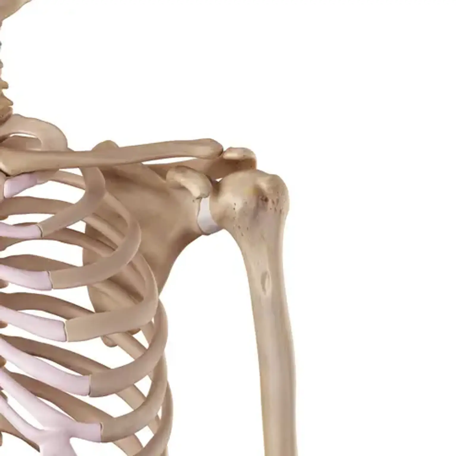
Shoulder: A ball-and-socket joint that allows for a wide range of motion.
Elbow: A hinge joint that allows the forearm to move toward and away from the body.
Wrist and Hand: Complex structures with multiple small bones, muscles, and ligaments that help with gripping, fine motor movements, and flexibility.
Each part of the upper limb is vulnerable to different injuries. Whether it's a rotator cuff tear in the shoulder or a wrist sprain from a fall, the damage to these structures can range from mild discomfort to severe disability, affecting daily life significantly.
Types of Upper Limb Injuries
Upper limb injuries vary widely, from minor sprains to more severe fractures and dislocations. Here are the most common types: