Ureteral Reimplantation Surgery
Last updated date: 13-May-2023
Originally Written in English
Ureteral Reimplantation Surgery
Overview
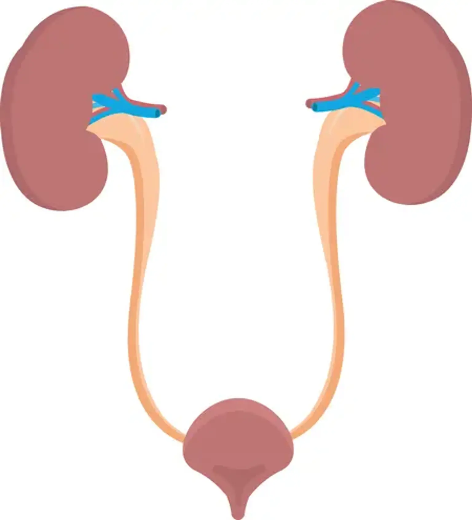
The ureter normally enters the bladder, which is formed of muscle, in such a way that urine may enter the bladder but not return up to the kidney. When the ureter enters the bladder improperly, the bladder's muscular backing does not completely cover the ureter, allowing urine to flow back toward the kidney. This is known as vesicoureteral reflux. Ureteral reimplantation is a surgical procedure that changes the location of these tubes where they enter the bladder wall, which aids in the treatment of reflexes. One (unilateral) or both (bilateral) ureters may require repair.
What is Ureteral Reimplantation?
Ureteral reimplantation is used to treat reflux, which occurs when urine from the bladder flows back up into the kidneys via the tubes that link the kidneys to the bladder.
When these tubes, known as the ureters, are functioning properly, urine goes only one way out of the kidneys and into the bladder, where it may exit the body. The ureters link to the bladder via a tunnel that works as a valve to prevent urine from backing up.
A ureter can sometimes become disconnected from the bladder wall. Reflux occurs when there is insufficient tunnel at the connecting site. If left unchecked, reflux can cause kidney scarring and irreversible kidney damage. When reflux is not expected to resolve on its own or is causing kidney damage, surgery is required. Reflux surgery involves constructing a new tunnel into the bladder to change the way the ureter attaches to the bladder. The ureter is "reimplanted" by the doctor to reconnect it to the bladder.
Relevant Anatomy
The ureters are muscular channels that convey urine from the kidneys to the bladder. In adults, the ureter is around 25-30 cm long and follows a S curve down the retroperitoneum. The renal pelvis is at the ureter's proximal end, while the bladder is at the distal end. The ureter originates at the level of the renal artery and vein, which are located posterior to these tissues. This ureteropelvic junction is normally located at the second lumbar vertebra on the left, with the right being slightly lower.
Why is it done?
A disorder known as vesicoureteral reflux (VUR) occurs when the valves where the ureters meet the bladder are unable to seal correctly, enabling urine to reflux back up into the ureters from the bladder, sometimes reaching as far back as the kidneys.
VUR is more frequent in infants and young children than in adults, and it increases the risk of recurring urine infections and kidney damage. Ureteral reimplantation surgery can assist to fix the problem by relocating the locations at which the ureters enter the bladder so that they are covered by bladder muscle, which can shut the ureters and prevent urine from backing up.
Procedure
When your kid returns from the operating and recovery rooms, he or she will be wearing one or more of the following tubes:
- An intravenous (IV) tube. A little plastic tube is inserted into a vein in your child's arm. It is used to administer fluids and medications to your child until they are able to drink.
- A urinary catheter. A tiny tube is inserted into your child's bladder to drain the pee. The sort of catheter your child is using is determined by the type of operation. A urethral catheter is inserted into the bladder via the urethra, which is the tube that transports urine from the bladder to the outside of the body. A suprapubic catheter is inserted into your child's bladder through a small hole in his or her abdomen (belly).
- A drain. This is a little rubber tube that is inserted into your child's stomach to drain any excess fluid that may have accumulated during surgery.
- A stent. This is a tube that drains urine from the ureter above the location of the procedure. The stent will be inserted through a tiny incision in the abdomen.
- An internal stent. This is a tiny tube that may be left inside the body and removed six weeks later. It aids in the drainage of urine from the kidney to the bladder. This aids in the healing of the surgical site.
In order to know what to expect following the procedure, ask your doctor what sorts of drainage tubes your kid will most likely require. The doctor or nurse can explain how long each tube will be needed for your kid.
About Your Anaesthetic
Normally, no food or drink is permitted for six hours before to surgery. Chewing gum and sweets are examples of this. Prior to surgery, specific instructions on 'nil by mouth' needs are provided. If the youngster sucks a sweet or consumes even a small amount of food, the procedure will be delayed or even cancelled.
The procedure is performed under general anesthesia. Patients are unconscious throughout the procedure and have no recollection of it. Gas is administered to young children via a tiny mask, which puts the kid to sleep and eliminates the necessity for a needle insertion while the child is awake. However, as soon as the youngster falls asleep, a drip is inserted for safety. It is recommended that a parent be present at the start of the anesthesia. Once sleeping, a Caudal Block will be administered.
A needle is placed into the back, and medicine is given to numb the surgical site. This guarantees that the kid requires less pain medicine during the anesthesia, and that after they wake up, the caudal continues to act for several hours, blocking the nerve supply to the surgical region and preventing pain.
What is the follow-up after surgery?
Depending on their healing, children are frequently discharged on the second to fourth day after surgery. If your kid has a drain or catheter, please schedule a removal appointment one week after surgery. Schedule an appointment four to six weeks following the procedure if there is no drain. At the time of the follow-up appointment, children commonly receive an ultrasound. During this time, you may be given Ditropan and Tylenol or Tylenol with codeine to reduce spasms before your next appointment.
It is critical that your youngster continue to take the low-dose antibiotics. The antibiotics prescribed at the time of hospital discharge should be finished by the time you return, and your kid should continue taking his or her pre-operative maintenance antibiotic on a regular basis.
Your child will be scheduled for a kidney ultrasound one month after surgery. This test determines whether or not there is a blockage at the surgical site. It does not tell us whether the reflux has been addressed; only a cystogram can accomplish that. Your youngster should have a voiding cystogram (VCUG) four to six months after surgery. If both of these tests come back normal and the reflux has gone away, your kid can stop taking the low-dose maintenance antibiotics. Your child should return to us in a year for a blood pressure check and another ultrasound to examine the kidneys.
Will my child have problems urinating after surgery?
It is usual for children to have bladder spasms or intermittent cramping, urinary frequency, urinary incontinence, and loss of tiny volumes of blood-tinged urine following this kind of surgery. If the symptoms worsen, a drug known as Ditropan (oxybutynin) may be recommended. It will not remove all spasms, but it should reduce their severity. Putting your child in a small tub of warm water or laying a moist, warm washcloth on the perineum (the skin between the anus and the genitals) may help make your child feel more at ease.
Older children may be concerned if they lose control of their urine, especially if the urine is stained with blood. Until the problem is resolved, your child might wear light mini-pads in his or her underpants. Urinary frequency and bloody urine may persist in some youngsters for two to three weeks. This is typical. Assure your youngster that control will be restored after the bladder recovers.
- Do the medications have side effects?
Ditropan (oxybutynin) may result in flushed cheeks, heated skin, a dry mouth, and a reduction in appetite. A lack of appetite is not uncommon, but you should provide your child water on a regular basis — a few sips every 15 minutes or so — to ensure appropriate urine production. Smoothies produced from blended fruit and yogurt are high in vitamins and are typically well tolerated. To persuade your child to drink fluids, you may need to be patient and persistent.
While in the hospital, your kid may be given Morphine, Droperidol, or Demerol. For nausea, vomiting, and discomfort, droperidol or compazine may be prescribed. These drugs may cause drowsiness in your kid. Some youngsters react to pain relievers by becoming too enthusiastic and agitated, or by breaking out in a rash. Inform the nurse if this occurs, and the medicine will be modified.
The medicine will be changed to Tylenol with codeine before discharge (Tyco). This is available in tablet and liquid form. Because the codeine in this drug causes constipation in some children, it is critical to urge your kid to be as active as possible and to offer lots of liquids, fruit, and vegetables. You can gradually begin to relieve your child's discomfort with Tylenol or Children's Motrin. Within a few days to a week of discharge, your kid should begin to feel more like himself or herself.
How should I care for the wound?
- One day following surgery, remove the gauze dressing from the wound. Tiny STERIS StripsTM (small white strips that assist seal the borders of the wound) are frequently placed beneath the incision to stabilize it. These are usually left in place for 1-2 weeks. They can then be removed when they naturally peel away from the skin.
- The incision will be red, somewhat elevated, and swollen. This is typical. The sutures are behind the skin and will naturally disintegrate. They are not required to be removed.
- It is normal for small amounts of dried blood to be present
- After the bandages are removed, there may be adhesive left on the skin. Glue remover from your local drugstore or supermarket might assist in removing the adhesive.
How much drainage is normal?
It is normal to have swelling and bruises around the incision. Although a limited amount of fluid or red leakage is normal, if the incision is pouring blood, apply pressure and notify your doctor.
When can my child take a bath or shower?
- For the first day following surgery, your youngster can take a sponge bath. They can bathe or wash normally 48 hours (2 days) following surgery.
- Simply pat the incision dry with a towel.
- Take only brief showers or sponge baths while a stent tube is in place until it is removed. The tube is sewn in place and taped to the skin.
What can I expect after surgery?
The anesthesiologist and surgeon will prescribe and discuss the medications that will be given to your kid to keep him or her comfortable. During surgery, a catheter (thin tube) will be put into your child's bladder to empty pee. Your child's urine may be crimson or pink in color, with a few blood clots. The urine will progressively turn a clear golden color. Blood in the urine is frequent and may appear and disappear depending on your child's activity and hydration level. It is not alarming; however, if your kid has trouble emptying the bladder owing to big clots, please tell your surgeon.
Bladder spasms might occur, particularly in the first few days following surgery. Bladder spasms are contractions of the bladder wall induced by one or more of the following factors:
- Healing of the bladder wall incision
- Catheter irritating the wall of the bladder
- kinked or blocked catheter (urine not able to drain out of the bladder)
- Constipation
Bladder spasms, like muscular spasms, can be excruciatingly painful. They appear and disappear often and generally last less than a minute. Medication to relax the bladder will alleviate the pain.
Following surgery, your kid will be given an intravenous (IV) line to ensure enough fluid intake. It can also be used to provide medications like pain relievers and antibiotics. When the IV and IV medications are no longer required, the doctor will make the decision.
While on IV pain medications, a pulse oximeter may be used to check your child's oxygen saturation.
Conclusion
Ureteral reimplantation is a procedure that repairs the tubes that link the bladder to the kidneys. The procedure alters the position of the tubes where they connect to the bladder, preventing urine from backing up into the kidneys.