Urethral cancer

Last updated date: 16-May-2023

Originally Written in English

Urethral cancer

Overview

Urethral cancer is a condition in which malignant (cancer) cells originate in the urethral tissues. The urethra is a tube that transports urine from the bladder to the exterior of the body. The urethra is around 112 inches long in women and is located directly above the vagina. ‏
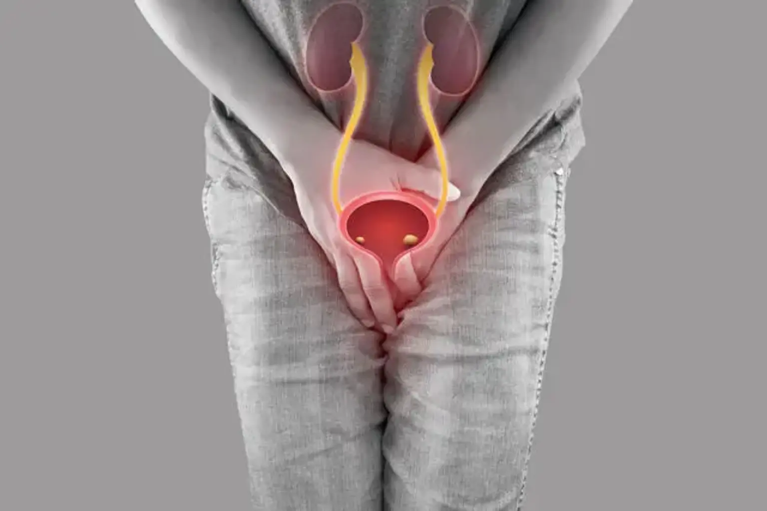
 

Urethral cancer definition

Urethral cancer definition

Primary urethral cancer (PUC) is an exceedingly uncommon disease that accounts for less than 1% of all cancers. It is more common among the elderly, males, and African Americans.

Because of the rarity of this cancer, practically all information on its treatment and outcomes are collected from retrospective, single-center case studies. The disease's rarity precludes prospective trials to establish the best treatment results. Furthermore, the urethral anatomical changes between men and women modify the therapy choices accessible. The location of the tumor's genesis, as well as the histology of the tumor, can all have an impact on treatment choices and prognosis. As a result, urethral cancer treatment is generally tailored to the person.

Because of the changes in the urethra between males and females, anatomic and histologic aspects are important in urethral cancer. The male urethra is 21 cm long on average and is split into anterior and posterior components, whereas the female urethra is 4 cm long and does not require subdivisions.

The histologic pattern of the urethral mucous membrane evolves distally from transitional epithelium to squamous epithelium in both the male and female urethra. Histologically, urethral cancer is classified as squamous cell cancer (SCC), urothelial carcinoma (also known as transitional cell carcinoma [TCC]), or adenocarcinoma based on the presence of mucosal cells (AC). Urothelial carcinoma is the most frequent histology in males, followed by SCC and AC; AC is the most prevalent histology in women, followed by SCC and urothelial carcinoma.

The labia, vagina, and bladder neck are the most common sites of tumor invasion in females. The vascular spaces of the corpora and periurethral tissues; deep tissues of the perineum; urogenital diaphragm; prostate; and the penile and scrotal skin are the most common sites of extension in males, where PUC can produce abscesses and fistulae.

Early discovery, as with most cancers, provides the highest chance of treatment. Most tumors are confined at first, with regional metastases to nodal locations found in up to 30% of cases in both genders. Distant metastases at presentation are uncommon (0–6%), but can arise in up to 40% of individuals with recurrent illness.

The importance of multimodal treatment in the management of PUC, particularly in advanced illness, has been widely shown in the literature. However, the optimal mix of chemotherapy, radiation, and surgery remains uncertain.


Epidemiology

Primary urethral cancer was found to occur at a rate of 4.3 per million in males and 1.5 per million in females in around 10% of the US population. The incidence rises with age; in the population aged 75-84 years, the male incidence is 32 per million and the female incidence is 9.5 per million. Primary urethral cancer was discovered to be twice as frequent in African Americans as in whites, and nearly three times more common in males than females.

Urethral cancer has been documented in people ranging in age from 13 to 90 years old, suggesting that it can strike at any age. However, it is most typically detected in the seventh decade of life.


Anatomy

Urethral cancer Anatomy

The urethra is a mucous membrane supported by a stroma of connective tissue, elastic fibers, and smooth muscle underneath the mucous membrane. The male urethra is 21 cm long on average, whereas the female urethra is 4 cm long on average.

The kind of epithelium of this mucosa changes depending on location in the male urethra. The urethral meatus and fossa navicularis are made up of stratified squamous epithelial cells. The urethra's penile, bulbar, and membranous parts have pseudostratified and stratified columnar epithelium, whereas the prostatic urethra has transitional-cell epithelium.

Furthermore, the Littré submucosal glands connect with the urethra. The glanular (meatus, fossa navicularis) and penile sections of the anterior urethra are drained by the inguinal nodes. The posterior urethra, on the other hand, empties into the pelvic nodes (bulbous, membranous, and prostatic).

The corpus spongiosum, which sits between the corpora cavernosum, surrounds the male urethra. Because each corpus is covered by a similar fascial sheath, urethral tumors can spread straight into surrounding tissues and circulatory areas (Buck).

The female urethra is significantly shorter and has a less complicated histology. The distal two-thirds are made up of stratified squamous epithelium, whereas the proximal one-third is made up of transitional cells. Skene glands are found in the urethral meatus's submucosa and are continuous with the urethra. Pseudostratified and stratified columnar epithelium is seen in these formations. The female urethra drains into the superficial or deep inguinal nodes, whereas the proximal two-thirds drain into the pelvic nodes (external iliac, internal iliac, obturator).


Etiology 

The cause of urethral carcinoma is unknown. Although cigarette smoking, aromatic amine exposure, and painkiller addiction have been linked to transitional-cell carcinoma of the bladder, no similar link has been found with urethral cancer. Patients having a history of bladder cancer, on the other hand, are at an elevated risk of urethral cancer.

In certain studies, human papillomavirus (HPV) infection has been linked to approximately one-third of occurrences of urethral cancer. PUC risk factors in males include urethral stricture (25–76%), sexually transmitted infections (24–50%), and trauma (7 percent ). Chronic irritation (including HPV infection), diverticula, sexual activity, and delivery are all linked to the development of PUC in females.

Chronic inflammation as a cause of urethral cancer is a hotly debated topic. According to one study, 88 percent of male urethral cancer patients had a history of stricture; however, another study showed the association in just 16 percent of patients. This is confirmed further by the high prevalence of primary urethral cancer in the bulbomembranous urethra, which is also the most common site of urethral strictures.

Arsenic use has been linked to an elevated risk of primary urethral cancer in rare cases.


Pathophysiology

Because urethral cancer is uncommon, detailed pathophysiologic aspects are unclear. Chronic inflammation, infection, or irritation of the urethra, on the other hand, is considered to precede the development of urethral cancer. Rapid urethral mucosal cell turnover predisposes to the development of dysplasia and neoplasia.

Inflammation, infection, and irritation may potentially impair the urethral mucosal cells' normal DNA repair processes. The tumor frequently invades deeply and spreads to nearby structures. Because urethral cancer is frequently identified late, decisive therapy such as surgery and radiation may be ineffective.


Diagnosis

Urethral Cancer Diagnosis

Tests that examine the urethra and bladder are used to diagnose urethral cancer.

The following tests and procedures may be used:

  • Physical exam and health history: An examination of the body to look for general indicators of health, including the detection of disease-related symptoms such as tumors or anything else that appears strange. A history of the patient's health habits, as well as previous diseases and treatments, will be collected.
  • Pelvic exam: A vaginal, cervix, uterus, fallopian tubes, ovaries, and rectum examination. The doctor or nurse inserts a speculum into the vagina and examines the vagina and cervix for symptoms of illness. The doctor or nurse also inserts one or two lubricated, gloved fingers of one hand into the vagina and places the other hand over the lower abdomen to feel the uterus and ovaries' size, shape, and location. A greased, gloved finger is also inserted into the rectum by the doctor or nurse to feel for lumps or abnormal regions.
  • Digital rectal exam: An examination of the rectum. A greased, gloved finger is inserted into the bottom section of the rectum to feel for lumps or anything else that appears strange.
  • Urine cytology: A laboratory test in which a sample of urine is checked under a microscope for abnormal cells.
  • Urinalysis: A test to determine the color of urine as well as its contents, which include sugar, protein, blood, and white blood cells. If white blood cells (a symptom of infection) are discovered, a urine culture is routinely performed to determine the kind of infection.
  • Blood chemistry studies: A method in which a blood sample is examined to determine the levels of specific compounds produced into the blood by the body's organs and tissues. A chemical in an unusual (higher or lower than normal) concentration might be a symptom of sickness.
  • CT scan (CAT scan): A technique that involves taking a number of detailed photographs of places within the body, such as the pelvis and abdomen, from various angles. A computer coupled to an x-ray machine creates the images. To make the organs or tissues show up more clearly, a dye may be injected into a vein or ingested. This is also known as computed tomography, computerized tomography, or computerized axial tomography.
  • Ureteroscopy: A technique that examines the ureter and renal pelvis for abnormalities. A ureteroscope is a narrow, tube-like tool with a viewing light and lens. The ureteroscope is introduced into the bladder, ureter, and renal pelvis through the urethra. A instrument may be placed via the ureteroscope to collect tissue samples for examination under a microscope for disease symptoms.


Management

Urethral Cancer Management

Because of the disease's rarity and a lack of high-quality data, no significant agreement on treatment options for primary urethral cancer (PUC) has been achieved. Large multicentric studies have been reported, shedding light on PUC management. The significant variety of treatment regimens and research populations, on the other hand, restricts the interpretation of the results. 

urethral cancer patients benefit from centralized treatment in terms of clinical results. The availability of practice pattern modifications such as multimodal therapy, aggressive surgery, and regional lymphadenectomy appears to contribute to the reported higher results in high-volume hospitals.

The main therapeutic premise is that the patient must understand the possible risks and advantages of the various techniques. Following that, clinicians may decide whether to proceed with drastic surgery in a shared decision-making process with patients. Alternatively, based on a risk-benefit analysis, the patient's medical team may suggest that dramatic action is largely contraindicated.

Following correct staging, the urologist should have a thorough discussion with the patient about the degree and severity of the illness. The necessity of reconstruction, urine diversion, social and familial support, and physical therapy cannot be overstated.


Medical Care

The treatment for urethral cancer depends on the stage and location of the tumor. If the illness is invasive and covers more than half of the penile urethra, radiation may be an option for treating unresectable lesions. Tumors in the bulbocavernosus urethra, as well as those in the prostatic urethra, can be treated with radiotherapy and chemotherapy. Treatment for advanced-stage illness includes major surgery, chemotherapy, and adjuvant radiation. In situations with severe metastases, systemic treatment is the only choice.


Radiation therapy

Radiation therapy can be used as main therapy, in conjunction with chemotherapy and/or surgery, or as adjuvant treatment for local recurrence following surgery in the treatment of urethral cancer. Radiation treatment might be external beam, brachytherapy, or a mix of the two. Definitive radiation is occasionally used for advanced-stage malignancies, but because monotherapy of big tumors has demonstrated poor tumor control, it is more commonly used in combination with surgery or chemotherapy.

Although the use of radiation in the treatment of urethral cancer has proven positive oncologic results, with reported 5-year survival rates of up to 41%, over half of the patients suffer from treatment-related side effects such as stenosis, fistulas, bladder bleeding, or necrosis. Fistula formation is more common in cases of big tumors penetrating the vagina, bladder, or rectum. Severe complication rates for definitive radiotherapy are in the range of 16% to 20%. Toxicity rates rise when dosages above 65-70 Gy. In an effort to reduce local morbidity from radiation, intensity-modulated radiation treatment has become more prevalent.


Chemotherapy

The chemotherapeutic literature for urethral cancer is limited to retrospective, single-center case series or case reports. Over the years, a diverse range of medicines, either alone or in combination, have been described, and their usage has mostly been extrapolated from experience with other urinary tract cancers.

Chemotherapy treatments for PUC are determined mostly on the underlying histology. Thus, cisplatin, gemcitabine, and ifosfamide are recommended for squamous cell carcinoma; 5-fluorouracil (5-FU), gemcitabine, and cisplatin-based regimens for adenocarcinoma; and MVAC (methotrexate, vinblastine, doxorubicin, and cisplatin) for urothelial malignancies.

Nonetheless, determining an appropriate therapeutic regimen for PUC is challenging, especially given the possibility of histologic overlap. Platinum-based chemotherapy regimens are the most widely employed in the treatment of PUC. In one study of 44 patients, the overall response rate after platinum-containing chemotherapy was 72%, with a median overall survival of 31.7 months for the entire cohort.

In patients with advanced disease, neoadjuvant chemotherapy with or without adjuvant systemic treatment enhanced 3-year overall survival and progression-free survival much more than adjuvant therapy alone. Based on the available evidence, a combination of chemotherapy and other treatment modalities appears to be optimal for improving patient survival.


Surgical Care

Surgical excision remains the gold standard as a primary form of therapy for both male and female patients with non-metastatic urethral cancer, offering a survival benefit. The scope of surgery is determined by the tumor's placement inside the urethra and its clinical stage. Given the disease's extremely aggressive nature, extensive surgery is often recommended to increase survival.

In highly chosen patients with superficial illness, minimally invasive urethra-sparing procedures are gaining acceptability. This less forceful method protects body image and cosmesis, as well as sexual and reproductive function; but, in these circumstances, vigorous, attentive, and regular follow-up is required.

Prior to decisive surgery, accurate tumor staging is critical, especially if considerable reconstruction is anticipated. The patient should have been to the operating room at least once for a transurethral biopsy and anesthesia-induced evaluation. Based on these findings, an imaging modality like MRI or CT scanning should be used to forecast the degree of local invasion.


Surgery in male patients

The following four modalities of surgical management in male urethral cancer:

  • Urethra-sparing management
  • Partial penectomy
  • Radical penectomy
  • Pelvic lymphadenectomy and en bloc resection including penectomy and cystoprostatectomy with the removal of the anterior pubis


Partial penectomy

The malignant tumor is excised with 2-cm margins during a partial penectomy. This therapeutic option is only available for infiltrative lesions of the distal penile urethra. If an infiltrating tumor is found in the proximal part of the penile urethra, a complete penectomy is recommended. Only if the nodes are palpable is ilioinguinal node dissection conducted. In contrast to penile cancer, there is no discernible advantage to preventive groin dissection.


Prognosis

Estimated 5-year survival rates for 165 patients treated for primary urethral carcinoma were as follows:

  • Local recurrence-free survival: 51%
  • Disease-specific survival: 48%
  • Overall survival: 41%

Distal malignancies of the male urethra have much higher survival rates than proximal tumors; the cure rate can exceed 90% due to early identification due to more obvious symptoms. Proximal neoplasms are often more invasive and aggressive upon presentation, necessitating extensive surgery that includes excision of the penis, urethra, scrotum, and pubic bone as well as radical cystoprostatectomy. The disease-free survival rate for these patients is reported to be between 33% and 45%.


Postoperative care

General postoperative precautions that are paramount to reducing complications include the following:

  • Hemodynamic support with intravenous fluids, both crystalloid and colloid
  • Intravenous antibiotics
  • Incentive spirometry and aggressive pulmonary toilet
  • Deep venous thrombosis prophylaxis


Strict monitoring of 24-hour intake and output from all drains is required in order to manage fluid status correctly and evaluate whether spontaneous diuresis is progressing. Based on these findings, diuretic medications may be indicated.

Stoma nurse care and education are required, especially when the patient is returned home, because they will most likely need to record their output at first. Initial instruction in stomal appliance maintenance and/or intermittent catheterization gives the patient much-needed autonomy and contributes to the development of a healthy and proactive self-image. If the patient is unable to fulfill the high demands of these treatments at first, visiting nurse support may be required.


Complications

In patients treated with radiation therapy, the overall risk of complication is roughly 20%.  Complications include the following:

  • Urethral stricture
  • Radiation cystitis/urethritis
  • Bowel irritation
  • Fibrosis
  • Infection
  • Bleeding
  • Fistula formation 


Patients undergoing urethrectomy or partial penectomy have a decreased chance of urethral stricture formation or the development of urethral fistulae, but these concerns should be discussed with the patient prior to surgery. Urine incontinence can occur as a consequence of overactive bladder and high urgency, or as a result of injury to the external sphincter, which can cause stress incontinence or proceed to complete urinary incontinence.

The return of a tumor can cause erosion or abscess of the penile, scrotal, and perineal skin. Necrotic tissue at these areas can cause poor wound healing as well as the formation of fistulae and abscesses, which can lead to sepsis.

Complications of radical cystoprostatectomy include bowel obstruction, infection, and leakage, particularly as a result of the use of intestinal or colonic conduits for urine diversion.


Conclusion 

Urethral cancer

Urethral carcinoma is the most uncommon kind of urological cancer. This kind affects just 1 or 2 persons out of every 100 cancer patients. It affects males more than women. Some persons with urethral cancer have no symptoms, but the majority do. This article will provide you with additional information about urethral cancer, how it is diagnosed, and treatment choices.