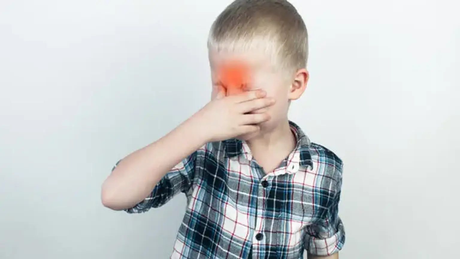
Uveitis

Last updated date: 14-Apr-2023

Originally Written in English

Uveitis

Overview

Uveitis is described as inflammation of the uveal tract, which has anterior and posterior components. The iris and ciliary body make up the anterior tract, whereas the choroid makes up the posterior tract. As a result, uveitis is inflammation of any of these components, as well as other adjacent tissues such as the sclera, retina, and optic nerve. Uveitis is frequently idiopathic, although it can also be caused by genetic, traumatic, immunological, or viral factors.

Diagnosis and therapy of the underlying condition are critical not only for treating the sickness, but also for preserving eyesight and perhaps revealing underlying systemic problems.

If left untreated or improperly managed, acute inflammation can progress to chronic, sight-threatening inflammation, stressing the need of the primary eye care practitioner in managing these patients carefully and efficiently.


Uveitis in Children

Uveitis in Children

Pediatric uveitis is an inflammatory eye condition that can cause vision loss and ocular problems. It is most commonly linked with juvenile idiopathic arthritis (JIA), although it can also be found in other autoimmune diseases such as Behcet's disease or sarcoidosis. Idiopathic uveitis, or uveitis that does not have a systemic cause, is at least as frequent as JIA-associated uveitis. The most frequent symptom is anterior uveitis, which is classified by anatomic location. Ocular problems have been documented in up to 50% of children, vision loss (VA of 20/50 or worse) in 25–40%, and legal blindness (VA of 20/200 or worse) in up to 25%. A chronic course characterized by remissions and relapses is typical, and many children require long-term immunosuppressive medication. 

It might be acute, subacute, or chronic. In the United States, there are an estimated 14–17 cases per 100,000 people, with 38,000 new cases verified each year.  Uveitis accounts for 10% (30,000 new instances each year) of legal blindness in the United States, and it can afflict people of all ages, though it is most frequent in people in their third and fourth decades. Under some conditions, the body creates a natural inflammatory response as a self-healing mechanism.

Excessive infiltration of neutrophils, macrophages, and lymphocytes may cause irreversible damage to the ocular tissue in chronic or untreated situations. Infectious, noninfectious, and masquerade illnesses can all cause anterior uveitis.

The primary eye care practitioner is frequently the first health care physician to diagnose and treat anterior uveitis. Understanding the immunoinflammatory cascade is required to successfully manage and cure ocular inflammation. This pathway is made up of two major components: the innate immune system and the adaptive immune system.


Epidemiology

Uveitis may affect persons of all ages and can vary greatly depending on the patient's geographic area and age. In a research conducted between 2006 and 2007, the incidence of uveitis was 24.9 occurrences per 100,000 people. Anterior uveitis is the most frequent kind, accounting for around half of all uveitis occurrences, whereas posterior uveitis is the least common. Uncontrolled inflammation found in untreated uveitis, as well as consequences connected to this uncontrolled inflammation, are believed to account for around 10% of blindness in the United States.


Uveitis affects around 10–20 percent of children with JIA. Those who have positive antinuclear antibodies (ANA), are young (6 years old) at the time of their arthritis diagnosis, have oligoarticular or polyarticular RF negative JIA, and are early in their disease history (4 years) are deemed high risk. Current screening standards indicate that these youngsters be monitored every 3–4 months. oligoarticular JIA category and younger age at JIA diagnosis increased risks of uveitis in 287 children with JIA, 18% with uveitis. Furthermore, 86 percent developed uveitis during the first four years following JIA diagnosis, highlighting the significance of regular monitoring throughout this interval.


Etiology 

Etiology of Uveitis

Uveitis is usually idiopathic, however it has been linked to traumatic, inflammatory, and viral causes. Patients may appear with concomitant systemic symptoms or infectious illnesses, indicating that the pathogenesis is not limited to the eye. Idiopathic uveitis accounts for 48 to 70% of all uveitis cases.

HLA-B27-related entities, juvenile idiopathic arthritis, inflammatory bowel disease, sarcoidosis, Behcet's disease (BD), or tubulointerstitial nephritis are all systemic inflammatory illnesses that are usually linked with anterior uveitis (TINU).

A short interval between arthritis and uveitis diagnosis, a young age at uveitis start, male gender, uveitis identified anterior to arthritis, and the presence of vision loss or problems during the first ophthalmology exam are all risk factors.

Viruses (HSV, VZV, CMV), bacteria (endophthalmitis, syphilis, TB, etc.), and parasites/worms are examples of infectious causes (toxoplasmosis, Lyme Disease, toxocara, Bartonella sp. or other atypical infections).


Pathophysiology of Uveitis in children

In general, the pathophysiology of uveitis in children is not well known. Trauma to the eye has been postulated to induce cell damage or death, resulting in the production of inflammatory cytokines and post-traumatic uveitis. Inflammatory disorders induce uveitis, which is hypothesized to be caused by molecular mimicry, in which an infectious agent cross-reacts with ocular-specific antigens. CD4 Th1 cells are responsible for vision-threatening inflammation.

Only activated lymphocytes are normally allowed to cross the blood-retina barrier, reducing the sensitization of naive T cells to ocular proteins. 


Uveitis symptoms in children

Uveitis symptoms in children

Children may complain of vision problems or impaired vision from time to time. Your child's eyes may appear red or hazy. However, because these symptoms normally appear gradually, severe eye injury might occur before any visual impairments are discovered. Your child requires regular eye exams with a pediatric ophthalmologist to detect eye abnormalities and prevent them from causing harm.

A thorough medical, familial, and ophthalmic history (particularly surgical) is required for diagnosis. A thorough system evaluation may also aid in the identification of a systemic illness with ocular signs. Symptoms will vary based on the affected tissue. Acute anterior uveitis can be unilateral or bilateral, with symptoms including discomfort, impaired vision, photophobia, and circumlimbal injection (ciliary flush).

Due to posterior synechia (iris adhesions to the anterior lens capsule) development in anterior uveitis, the afflicted pupil may be constricted or irregular in shape as compared to the unaffected eye. Intermediate uveitis does not normally cause discomfort but might produce impaired vision and floaters, whereas posterior uveitis frequently causes vision loss and/or dysphotopsias. Panuveitis is typically characterized by a mix of all of these symptoms due to many parts of the eye being damaged at the same time.

Visual acuity tests, slit lamp biomicroscopy, intraocular pressure measurements, and a dilated eye exam should all be included in physical exams. Pinhole visual acuity is especially useful when the patient has forgotten their glasses at home or is suspected of having an uncorrected refractive defect. This is readily achieved by piercing a small hole through a styrofoam cup and having the patient read the visual acuity chart through that single hole. Due to optic nerve involvement, an afferent pupillary abnormality may also aid in the differential diagnosis.

Local or widespread ciliary flush may be noticed on slit lamp examination. Cells and flare are also expected in the anterior chamber. The term "cell" refers to a collection of white blood cells in the anterior chamber; the cells may be so dense that they have settled to form a white, dependent, hypopyon (layered white blood cells within the anterior chamber), indicating severe inflammation that should be evaluated by an ophthalmologist as soon as possible.

A fluorescein angiography (an eye dye-based imaging method) might detect vascular ferning (vascular leakage). In certain situations, inflammation can spread into the anterior chamber, making it difficult to distinguish between anterior and intermediate uveitis.

Posterior uveitis is characterized by retinal and choroid lesions, which may include localized chorioretinal spots, retinal whitening, retinal detachments, retinal vasculitis, and optic nerve edema. Ocular history, such as a recent intraocular operation, raises the possibility of retinal hemorrhages and vasculitis being caused by an infectious etiology.


Most Common Key Signs/Symptoms:

  • Anterior uveitis:

Symptoms: red, painful eye

Signs: anterior chamber cell and flare, posterior synechiae, keratic precipitates


  • Intermediate uveitis:

Symptoms: worsening floaters, decreased vision

Signs: vitreous cell and haze, snowballs, pars plana snowbank, cystoid macular edema


  • Posterior Uveitis:

Symptoms: worsening vision, visual field changes

Signs: chorioretinal lesions, retinal whitening, vascular sheathing


  • Panuveitis:

Symptoms: red, painful eye; severely depressed visual acuity; floaters

Signs: anterior chamber cell and flare, vitreous cell and haze, chorioretinal lesions, retinal whitening


Diagnosis

Diagnosis of Uveitis

A laboratory workup is typically not required for the initial episode of anterior uveitis, but recurrence should warrant additional assessment. Because of the relationship with systemic disorders and the greater risk of permanent vision loss, intermediate, posterior, and panuveitis necessitate additional assessment with differential diagnosis-guided laboratory evaluation. If the patient has recurrent anterior uveitis, more testing may be required to rule out an underlying autoimmune or infectious condition.

Multiple sclerosis neuroimaging should be evaluated in the appropriate demographic population. Laboratory selection should be guided by the findings and other concomitant systemic symptoms. Syphilis is the "great masquerader" in the body, as it is everywhere else, and should be investigated in all cases of uveitis. 

Your child's ophthalmologist will use a special lens or light to inspect the interior of your child's eye and may prescribe additional tests to assist make a diagnosis. Among these tests are:

  • Blood tests
  • Skin tests
  • X-rays
  • Eye imaging, such as optical coherence tomography (OCT), a non-invasive imaging test that uses light waves take cross-section pictures of the retina


Management

Management of Uveitis

Uveitis is a dangerous eye infection that can result in scarring of your child's eye. It must be addressed as quickly as feasible.

Steroids and topical cycloplegics are used to treat inflammation and discomfort. Any subsequent treatments are dependent on the linked processes. Anti-viral drugs, for example, are required in herpetic uveitis, but bactrim can be utilized in toxoplasmic chorioretinitis.

Antimetabolite, biologic, and other immunosuppressive drugs (e.g., methotrexate, azathioprine, mycophenolate, cyclosporine, adalimumab, and infliximab) are frequently required for chronic, non-infectious cases, particularly when accompanied with systemic inflammatory illnesses. These agents, however, should only be prescribed by physicians who have been particularly educated in their usage and are familiar with their side effect profile.

The most prevalent kind of uveitis, anterior uveitis, is treated with topical corticosteroids and cycloplegics. To avoid rebound inflammation, steroids should be reduced based on clinical response. Follow-up should be established to monitor for resolution of inflammation and to monitor IOP due to the possibility of a sudden increase in IOP with continued topical steroid treatment.

Cycloplegics, such as atropine or cyclogyl, are used to reduce discomfort by lowering ciliary body spasms and to avoid and/or lyse posterior synechiae (adhesions of the iris to the lens capsule). These drugs are normally used twice daily until the inflammation has subsided or has diminished greatly before being discontinued.

Treatment for intermediate, posterior, and panuveitis is even more complicated and should be led by ophthalmologists, especially uveitis experts, whenever feasible.


Differential Diagnosis

Differential Diagnosis for eye

There are numerous different reasons of a painful, red eye with visual changes/loss that are commonly encountered in routine care and the emergency department. When examining an eye complaint, it is critical to have a broad differential in mind because there might be substantial overlap with the patient's symptoms. To identify between the following reasons of a red, sore eye, particular abilities must be required in each circumstance. Visual acuity tests, IOP measures, slit lamp biomicroscopy, and fluoroscein staining are examples of these specialized talents.

Conjunctivitis can manifest as a painful red eye accompanied by photophobia. Bacterial conjunctivitis can cause purulent discharge, which uveitis does not. Patients with viral or allergic conjunctivitis commonly have a serous discharge, although they can also be dry. The presence of palpebral conjunctival follicles and the absence of anterior chamber inflammation aid in the diagnosis of conjunctivitis. Chemosis (conjunctival edema) is also seen in conjunctivitis but not in uveitis.

Keratitis is a severe infection of the cornea. It is most usually observed in individuals who have a history of contact lens use and manifests with discomfort, photophobia, and perilimbic injection. The cornea in keratitis is frequently obscured, if not completely opaque (i.e. corneal ulcer with ring infiltrate), which is not seen in uveitis. Also, in keratitis, the pupil is normally normal in shape, although it may be constricted in anterior uveitis due to posterior synechiea development.

Acute angle closure glaucoma can cause a painful red eye as well as a change in vision. The patient may complain of a unilateral headache and may feel nauseated, even vomiting. The afflicted pupil is frequently mid-dilated and light receptive. The IOP will be greatly elevated in acute angle closure glaucoma, as it is in the majority of cases with uveitis.

However, high IOP is found with herpes infections in the setting of unilateral anterior uveitis, hence a slit lamp examination especially searching for anterior chamber cell and keratic precipitates should be conducted in each instance to correctly diagnose and treat the patient. The cornea may also become hazy as a result of the rapid rise in IOP in angle closure glaucoma.

A corneal abrasion frequently results in acute eye discomfort, photophobia, and weeping. A history of trauma to the afflicted eye is frequently endorsed by the patient. A drop of proparacaine at the moment of examination will relieve the pain almost completely, if not totally. This sensitivity to topical anesthetic is not a characteristic of any kind of uveitis. Fluorescein examination will reveal the abrasion as a brightly stained corneal lesion that, although infectious, will not show stromal infiltrate/whitening.

A panuveitis, such as that found in herpes infections and endophthalmitis, produces significant inflammation, resulting in a red, painful eye. In the anterior chamber, a hypopyn may be discovered. This discovery, as previously mentioned, should be cause for concern. A dilated eye exam is necessary to detect retinal whitening in acute retinal necrosis and substantial vitritis in endophthalmitis, and as such, an ophthalmology consultation is most likely required.

If any of these illnesses is suspected, an urgent ophthalmology appointment should be scheduled because both are eye emergencies, with earlier treatment yielding better outcomes.


Most common causes of a red, painful eye:

  • Anterior uveitis
  • Keratitis
  • Corneal abrasion
  • Acute angle closure glaucoma
  • Conjunctivitis
  • Scleritis


Prognosis of uveitis in children 

Prognosis of uveitis

In most circumstances, uveitis has an excellent prognosis if detected early and treated properly. While the majority of patients will suffer an eye problem, effective therapy and surgery, if necessary, reduce the likelihood of irreversible vision loss. Because of the severe morbidity and mortality associated with some specific systemic disorders that can cause uveitis, determining the underlying etiology of uveitis is also critical.


Complications

Uveitis complications

Ocular problems can cause vision loss in a considerable proportion of children with mild to severe uveitis. In 2003, 123 children with anterior, middle, posterior, and panuveitis were assessed in the Netherlands115 for specific causes of vision loss and factors associated with poor visual prognosis. Uveitis was seen anteriorly in 36% of cases, intermediately in 24%, posteriorly in 19%, and panuveitis in 21% of cases.

It was chronic in 83% of cases and bilateral in 73%. In 76 % of cases, uveitis complications occurred. The most common problems were cataract (35%), followed by glaucoma (19%). 

Cataracts, posterior synechiae, epiretinal membrane (ERM), cystoid macular edema (CME), band keratopathy, hypotony, glaucoma, and optic nerve edema are among the consequences. While topical steroids can be used to treat anterior chamber inflammation, intraocular, periocular, or oral steroids should never be used unless infectious etiologies have been ruled out by careful examination and laboratory assessment due to the danger of deteriorating illness and visual prognosis.


Follow-up care

Children and adolescents with uveitis may be seen by an ophthalmologist, a rheumatologist, or both. When your child's eye inflammation is active, she may need to be evaluated every other week. Once the inflammation has been controlled, she may be asked to return for follow-up sessions every three to six months to ensure that her condition has not changed.


Conclusion 

Uveitis

Uveitis is one of the most prevalent kinds of ocular inflammation encountered by primary eye care practitioners. A number of etiologies can induce anterior uveitis, including viral, non-infectious, and masquerade illnesses. 

If uveitis is suspected, immediate ophthalmology referral/consultation is required. Corticosteroids and cycloplegics should be used as soon as possible for anterior uveitis. Other forms of steroids (oral, periocular, or intraocular) should never be used until infectious etiologies have been thoroughly eliminated. The patient will need to be followed up on for additional ocular exams and IOP measurements. 

In addition to presenting signs and symptoms of the disease, short-term and long-term therapy of uveitis should involve examination of location, duration, pathology, and laterality. A comprehensive assessment of systems, a thorough examination, and laboratory tests may help the practitioner reduce the list of probable causes of uveitis. This is critical because once a list of diagnoses is established, a tailored strategy to therapy can be pursued. Early detection and treatment of children at high risk of uveitis may improve visual results.