Vascular Malformations
Overview
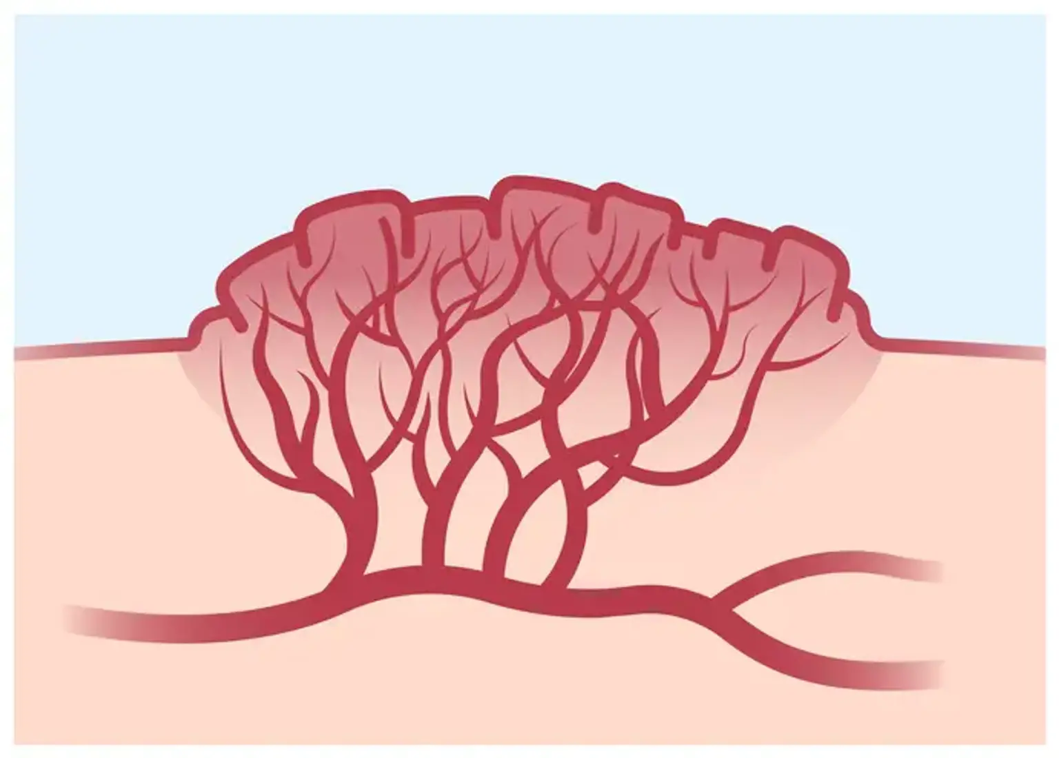
Blood vessel abnormalities, whether congenital or acquired, can affect arteries, veins, capillaries, lymphatics, and combinations of these blood vessels. Venous malformations and arteriovenous malformations are examples of vascular malformations, or arteriovenous malformations (AVMs).
What are Vascular Malformations?
Vascular malformations are a type of birthmark or growth made up of blood vessels that can create functional or aesthetic difficulties. While many of these blood artery anomalies are visible at birth, some do not appear until later in life, throughout childhood and adolescence.
Vascular abnormalities can occur anywhere in the body, from the head to the toes. They may manifest as a soft tissue mass, discomfort, swelling, and/or discoloration of the skin. Some vascular abnormalities occur on the face, neck, or near the brain or spinal cord. Others seem to be birthmarks or red blotches.
Adults seldom acquire vascular abnormalities as a result of trauma or another occurrence. Treatment may be required for vascular abnormalities that cause discomfort, limit function such as vision or activity, or cause bleeding or other difficulties. Vascular anomalies are uncommon. They occur in around 1% of all births. Venous malformations are the most frequent kind, affecting 1 in every 5,000 to 10,000 persons.
There are various varieties of vascular malformations, which are called by the type of blood vessel that is most commonly afflicted. Hemangiomas are the most frequent blood vessel abnormalities that affect infants in their first year of life, and the majority of them improve during infancy. The following are examples of vascular malformations:
- Capillary vascular malformations (port-wine stains)
- Spider angiomas
- Venous malformations
- Arteriovenous malformations
- Lymphatic malformations
- Pyogenic granulomas (lobular capillary hemangiomas)
- Angiofibromas (formerly called adenoma sebaceum, in patients with tuberous sclerosis)
- Glomangiomas
- Hemangiomas and pigmented skin lesions
What Causes Vascular Malformations?
The majority of vascular malformations are caused by the aberrant development of vascular and/or lymphatic channels in utero. They are commonly present at birth (congenital)
A vascular malformation might occur after an accident in which there is undiagnosed damage to the vessels. A venous malformation may become visible for the first time due to an accident or hormonal changes during puberty or pregnancy. In a limited percentage of cases, people inherit genetic abnormalities (mutations) that increase the likelihood of vascular malformations. These genetic reasons are currently being studied by researchers.
Capillary vascular malformations (port-wine stains)
Port wine stains (also known as capillary malformations) are birthmarks that are permanently red or blue in color. They are fairly frequent, affecting around three out of every thousand newborns. Some port wine stains are little, while others are rather extensive.
Port wine stains can form everywhere on the body, although they are most common on the face, neck, arms, legs, and scalp. They will increase with the infant and become darker as the youngster grows older. They might cause people to feel self-conscious or lose confidence.
Birthmarks are neither communicable or inherited, and there are usually no significant complications. There is a rare risk of epilepsy or vision difficulties if the port wine stain is on the forehead, eyelid, or cheek. If it affects the body or the limbs, the afflicted limb may become somewhat bigger than the opposite side.
Signs and symptoms of port wine stains
- The birthmark is a flat region that ranges in color from red to blue. With age, the color generally darkens to a purple hue. After a number of years, they may grow thick and bumpy.
- Their bodily regions range in size from tiny to enormous. They will not expand to cover a bigger area of your child's body, but they will increase in proportion to your child as they get older.
- They seldom cross across the midline (an imaginary, vertical line drawn down the centre of the body). This indicates that a port wine stain can be discovered on either the left or right side of the body, but not both.
Stains from port wine are present from birth. If your child gets a birthmark later in life, it will be a different type of birthmark.
When to see a doctor
Show your GP, pediatrician, or Maternal and Child Health Nurse if your infant has a birthmark or a new lesion that appears after delivery. It is especially critical to get medical attention if the lesion is on the face, at the base of the spine, or appears to be growing bigger. If further testing is required, such as an MRI of the brain, your kid will be referred, especially if the lesion is on the eyelid or forehead.
Treatment for port wine stains
Port wine stains seldom require treatment; nevertheless, certain port wine stains can become quite dry, thus moisturising cream should be applied to them once or twice a day. If your child's port wine stain must be removed, laser therapy is presently the finest treatment available for port wine stains. If you select laser therapy, it should begin before your child is six months old.
Spider Angiomas
Spider angioma, also known as spider naevus or spider telangiectasia, is a vascular disease characterized by abnormal dilatation of the skin's end vasculature. The lesion has a center red spot and reddish extensions that extend outward in the shape of a spider's web. They might occur in the form of numerous or isolated lesions.
Spider angiomas are mostly benign, although they might be indicative of a systemic condition such as cirrhosis, rheumatoid arthritis, or thyrotoxicosis. Solitary spider angiomas affect 15% of young people and often have fewer than three lesions. Lesions can also arise in other physiological circumstances such as pregnancy or severe starvation. With a 95% specificity, several spider angiomas are associated with chronic liver disease.
Spider angiomas Symptoms
In adults, spider angiomas are most commonly seen on the face, neck, upper chest, and arms, which corresponds to the distribution of the superior vena cava. Lesions on the upper extremities are prevalent in children. They can also be found on the backs of the hands and between the fingers. It should be noted, however, that spider angiomas can be seen in places other than the skin, such as the mucosa of the oral cavity and the gastrointestinal (GI) tract.
Spider angioma is identified by its distinctive look. Due to the local increase in arterial blood supply, large spider angiomas may be pulsatile, with blood flow toward the periphery. Unless plucked or scraped, bleeding from these sores is unlikely. The temperature of the skin of a spider nevus is greater than the temperature of the surrounding skin. In these tiny arterioles, blood pressure ranges from 50 to 70 mm Hg.
The direction of blood flow may be demonstrated by putting pressure to the body of spiders using a glass slide (diascopy), resulting in pallor and refilling once the pressure is released. This is a unique feature of angiomas. Spider angiomas are classified from grade 1+ (readily discernible with a body, legs, and surrounding erythema) to grade 4+ (visible pulsations with a hand lens, and elevated central punctum with numerous evident "spider legs" extending from it) because to their various sizes and for simplicity of description.
Pregnant patients may present with a large number of spider nevi, which are normally harmless and disappear after childbirth. Symptoms of chronic liver illness include jaundice, fluid retention, disorientation, and, on examination, changing dullness, icterus, abnormalities linked to the origin of cirrhosis, and stigmata of liver cell failure.
Diagnosis of Spider angiomas
Solitary spider nevus in otherwise healthy people or pregnant women does not necessitate further investigation. Multiple spider angioma can be linked to liver disease with a complete history and focused physical exam. The frequency and size of spider angiomas, in general, correspond with the severity of liver disease. In rare cases, the history and exam may not indicate to nevi and may raise worries about skin cancer, such as basal cell carcinoma. In certain cases, a skin biopsy will be beneficial to rule out skin cancer or other disorders.
Spider Angiomas Management
Spider nevi are treated differently depending on the origin of intra-arteriolar vasodilation. As the standard of therapy, patients with an underlying systemic condition such as cirrhosis should be handled. When spider nevi are present, however, the patient may already have severe liver disease.
For cosmetic reasons, laser, or electro desiccation have been used to remove spider angioma. Except for the minor risk of scarring, the surgery has typically positive results. After therapy, spider angiomas might return. Spider nevi in healthy people normally go away after a few years, as do those associated with oral contraceptives after withdrawal of therapy. Cirrhotic patients report the elimination of nevi after a liver transplant.
What are Arterio-Venous Malformations (AVMs)?
AVMs are circulatory system anomalies that can occur during embryonic or foetal development or, as previously stated, at any moment after birth.
Normally, blood channels known as arteries transport oxygenated blood from the heart to all tissues and organs of the body. These arteries split into smaller tiny vessels called capillaries within each organ or tissue. This is where oxygen is transported from the blood to the tissues. Deoxygenated blood is subsequently transported from the tissues to the heart and lungs via blood arteries known as 'veins.'
Blood in arteries is normally under high pressure, whereas blood in veins is under low pressure. The blood pressure drops as it flows through the capillary network in the tissues and organs.
An AVM is an aberrant connection (or, more commonly, a series of tiny connections) between an artery and a vein. The "nidus" is the place of improper attachment between the artery and vein. Blood skips the capillary network within the organs and tissues in this scenario, and normal pressure regulation does not occur.
Types of AVM
Most AVMs develop spontaneously in utero due to vasculogenesis abnormalities; however, certain AVMs are inherited (e.g., hereditary haemorrhagic telangectasia) or acquired (e.g. following trauma). AVMs are vascular abnormalities that have a high flow of blood due to the direct contact of the artery and vein.
What are the symptoms of an AVM?
This is mostly determined by the location of the AVM, and symptoms can vary greatly. However, the vast majority of AVMs are asymptomatic until a problem arises. Bleeding is the most common and possibly fatal consequence of AVMs. Symptoms are frequently vague and might be caused by a variety of different medical issues.
AVMs in the brain may cause headaches, seizures, or paralysis in one or more sections of the body, or they may not be discovered until after a hemorrhage (ie: burst / rupture). AVMs in the lungs are usually asymptomatic, however they can cause shortness of breath on exercise and blood coughing (haemoptysis).
AVMs in the gastrointestinal system are characterized by bleeding and abdominal discomfort. The bleeding may be little and present as dark stools or anaemia .
AVMs in soft tissues (such as the face, torso, arms, or legs) are primarily aesthetic, manifesting as swelling or asymmetry that may or may not be painful. These are often bumps on the trunk and limbs that can be unpleasant in certain conditions and can increase over time. These AVMs can occasionally be disfiguring. Before an AVM is diagnosed, most soft tissue abnormalities are misdiagnosed as tumors and referred for a biopsy.
AVMs may also grow rapidly in size in a short period of time, or swelling may develop in the months following localised trauma if this is the cause. They may be categorised based on their symptoms using the Schobinger classification, which can aid in therapy planning.
How are AVMs diagnosed?
Your doctor will have a suspicion or a good indication that you have an AVM based on your history, physical examination, and, in certain cases, your family history. Your primary care physician will recommend you to a specialist.
The diagnosis is often clinical, and imaging the lesion is critical to understanding its size and type. Imaging can also assist in determining when and how to treat certain lesions. AVMs should ideally be managed by a'multi-disciplinary' team of doctors, nurses, and specialists who have a special interest in detecting and treating this problem.
- Ultrasound:
As a dynamic and non-invasive inquiry, this test employs sound waves to detect anomalies and is particularly excellent at revealing blood flow. This test will assist in determining the flow characteristics of the lesion but will not provide a thorough anatomical image of the issue.
- CT scan:
This is a non-invasive X-ray exam that lets the radiologist to view the body on a computer screen in slices. A 'contrast' injection is utilized at the same time to emphasize blood vessels, and this is frequently referred to as a CT Angiogram. These photos may then be processed on a computer to produce a comprehensive 3-D view of the AVM and enable management planning.
- MRI scan:
This is comparable to a CT scan, however instead of radiation, magnetic fields are used. MRI frequently provides more specific anatomical information, particularly in the brain.
How can AVMs be treated?
AVMs are quite complex. Treatment and follow-up must be carried out and monitored by a multidisciplinary team with expertise in vascular malformations. The management of these situations necessitates the participation of the entire team.
The most crucial component is that each AVM is handled on an individual basis, taking into consideration the aforementioned criteria as well as a thorough dialogue with the patient.
AVMs have traditionally proven difficult to control. For decades, surgery has been the mainstay, with a variety of procedures used. Recent breakthroughs in the knowledge of AVMs, as well as technology developments in Interventional Radiology, favor effective minimally invasive therapies. Surgery continues to play a role at specialised centers in partnership with Interventional Radiologists.
Endovascular therapy, surgery, and radiation therapy are all alternatives for treatment. Endovascular therapy is used to treat AVMs by inserting catheters and needles into blood vessels under x-ray control to permanently block aberrant connections. This can be accomplished using a variety of embolization strategies. These treatments are performed by Interventional Radiologists and are typically a safer alternative to surgery.
Glomangiomas
Glomangioma, also known as glomuvenous malformation, is a rare cutaneous venous malformation that in histology exhibits glomus cells (undifferentiated smooth muscle cells that are thermoregulatory units) alongside the venous system. Glomus cells are specialized smooth muscle cells that control body temperature.
Signs and symptoms of Glomangioma
At birth, it often manifests as purple skin lesions with a cobblestone pattern. Lesions are often bluish-purple in color, papular or nodular in shape, and 2 to 10 mm in size. The size and quantity of them vary. These lesions are sensitive to the touch. The discomfort is triggered by pressure and cold. Glomus bodies are abundant in distal extremities, particularly the palms, wrists, forearms, feet, and subungual areas. Hands are present in 75% of instances.
Although it has been recorded in the nasal cavity, mediastinum, gastrointestinal tract, respiratory tract, urogenital tract, and hepatobiliary system, visceral organ involvement is extremely rare. Ventricular septal abnormalities and great vessel transposition have been documented in GVM patients. Although typical human nerves do not have glomus bodies, there is a case report of nerve involvement with glomangioma. Dyspnea, hemoptysis, and retrosternal chest discomfort are all symptoms of tracheal involvement.
Glomangiomas are larger and less well-circumscribed than solitary tumors. They have slow blood flow and usually grow over time.
The classic triad consists of the following:
- Hypersensitivity
- Intermittent pain, and
- Pinpoint pain.
Most of the time, glomangiomas do not present the classic triad.
The plaque-like kind manifests as non-tender, indurated, nodular, or discolored lesions that bleed with minimal impact. They are bigger than other kinds of glomangiomas.
Diagnosis of Glomangiomas
The diagnosis is confirmed by histology. Immunohistochemistry aids in the diagnosis of tumors with unusual histology. Smoothelin's role as an indication of smooth muscle cell should be examined. Glomus cells with dense bodies and smooth muscle myofibrils are visible under electron microscopy.
An X-ray may reveal osseous flaws. MRI and color Doppler ultrasonography aid in the definition of shape, size, and precise placement. Vascularity may be defined via dynamic time-resolved contrast-enhanced MR angiography.
Management of Glomangiomas
The treatment's purpose is to alleviate the symptoms. Monitoring and surveillance are advised for asymptomatic lesions. Treatment options include surgery, electron-beam radiation, sclerotherapy with hypertonic saline or sodium tetradecyl sulfate, argon, flash lamp tunable dye laser (for numerous lesions), and CO2 lasers. Excision therapy is the recommended method of treating painful lesions. Sclerotherapy has been demonstrated to be more successful in treating venous malformations than in treating glomuvenous malformations. Endoscopic excision or surgery is advised in situations with nasal involvement.
What are Slow-Flow Vascular malformations and Fast-Flow Vascular Malformations?
The majority of vascular abnormalities are slow-flowing. This word refers to the sluggish movement of blood through the afflicted blood arteries. These are anomalies of the venous, lymphatic, or venolymphatic systems. AVMs are vascular abnormalities with rapid blood flow. Blood flows swiftly from arteries to veins, generally via a nidus, which is a collection of aberrant vascular channels. Large AVMs' rapid blood flow might force your heart to work harder, raising your risk of heart failure.
What are the Complications of Vascular Malformations?
Some abnormalities are not harmful and can be detected. Malformations are more prone to produce discomfort and edema. Some abnormalities obstruct exercise, sports, employment, or daily activities owing to discomfort or involvement of muscles, joints, or nerves.
Large, high-flow anomalies can lead to heart failure over time. Vascular abnormalities in solid organs such the liver, kidney, or uterus can cause organ malfunction and other issues. Certain vascular anomalies in your brain or spine, such as AVMs, can limit the flow of oxygenated blood to your brain. Life-threatening issues may arise, such as:
Hemangiomas
Hemangiomas are the most frequent benign tumor of infancy. They are also known as hemangiomas of infancy or infantile hemangiomas (IH). Because of their clinical look, they are sometimes referred to as "strawberry markings." Hemangiomas are caused by endothelial cell growth. Hemangiomas are classified into several categories. Infantile hemangiomas occur later in infancy, whereas congenital hemangiomas are apparent from birth. Infantile angiomas are distinguished by fast development that is followed by spontaneous involution.
Infantile hemangioma is generally absent at birth, however it can manifest as a variety of precursor lesions. A pale region of vasoconstriction, an erythematous macule, a telangiectatic red macule, or blue bruise-like patches are examples of these. Infantile hemangiomas manifest clinically at 1 to 4 weeks. The localisation is widespread, and it can appear on the skin and mucosal surfaces. The majority of infantile hemangiomas show as a single localized cutaneous hemangioma, however they can be multifocal or segmental. Hemangiomas are most frequent on the head and neck, accounting for 60% of all occurrences.
This is followed by lesions on the trunk in 25% of patients and, in 15% of cases, on the extremities. Hemangiomas can be superficial, deep, or mixed with superficial and deep layer components. The superficial dermis is involved in superficial lesions, which are elevated, lobulated, and brilliant red. Deep hemangiomas, also known as subcutaneous hemangiomas, are bluish-colored nodules, plaques, or tumors that emerge from the reticular dermis and/or the subcutis layer. Mixed hemangiomas have characteristics of both sites.
Conclusion
Vascular malformations are non-cancerous (benign) lesions that are present at birth but may not be evident for weeks or months. Vascular malformations, unlike hemangiomas, do not undergo a development cycle and subsequently regress, but instead continue to grow slowly throughout life.
The majority of vascular malformations require therapy to enhance their appearance as well as to relieve discomfort, swelling, and bleeding. We can sometimes totally cure a vascular abnormality. Many children and people with vascular abnormalities excel academically, in sports, and in their jobs.