Ventricular Systolic Dysfunction
Overview
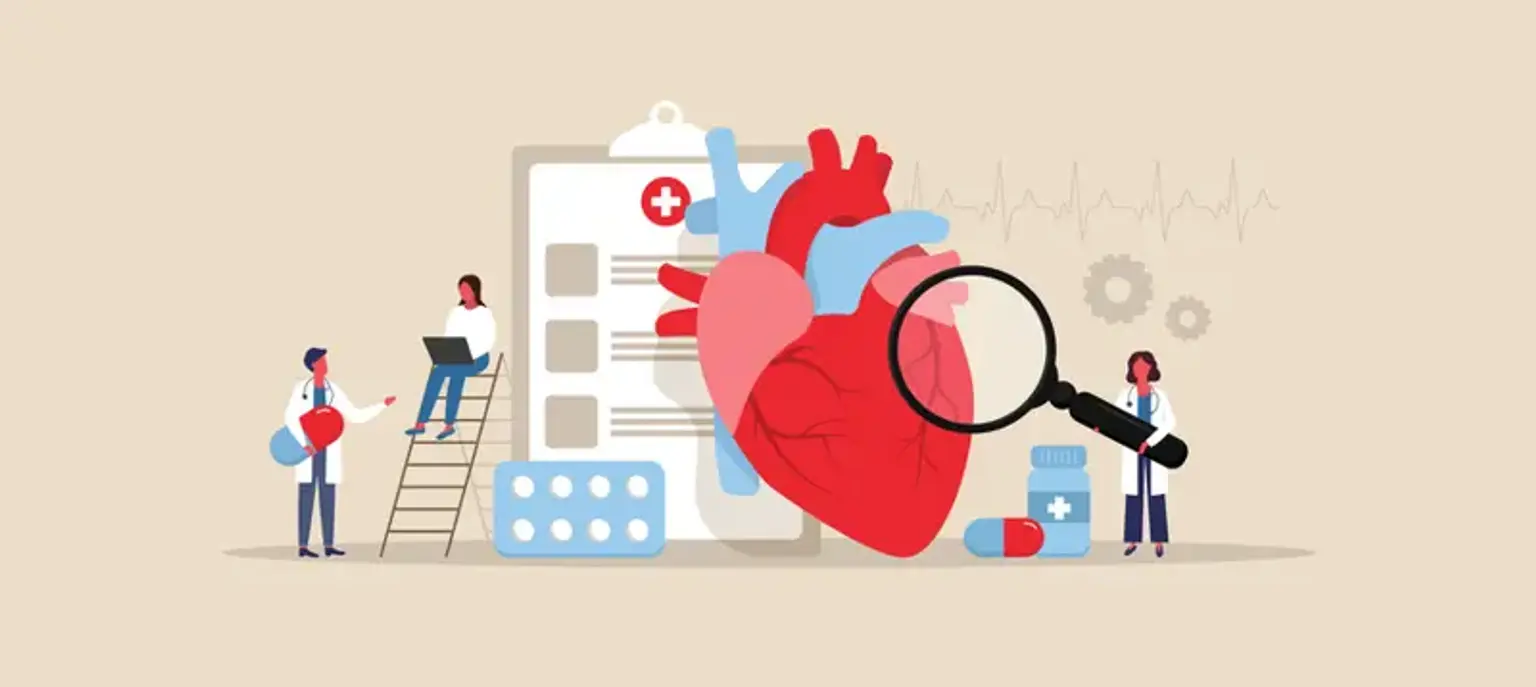
Heart failure is a major public health problem. Left ventricular dysfunction, also known as LV dysfunction of the heart, is frequently followed by congestive heart failure, which can lead to a variety of cardiac problems.
What is Left ventricular systolic dysfunction (LVSD)?
The left ventricle is the heart's thickest chamber, and it is largely responsible for pumping oxygenated blood to the body's important organs. LV dysfunction occurs when the left ventricle is either faulty or injured, interfering with normal function.
LV function can be disrupted for a variety of reasons. Certain heart abnormalities, such as valve malformations or illnesses, prevent blood from entering the body. To compensate for the shortage of oxygen-rich blood in the bodily tissues, the left ventricle is forced to work harder and pump blood quicker. The heart muscles thicken and weaken throughout time, and the walls extend to accept more blood. This results in cardiac dysfunction, particularly left ventricular dysfunction.
Systolic dysfunction is characterized by inadequate ventricular contraction (loss of inotropy). This is most likely due to alterations in the signal transduction systems that regulate cardiac excitation-contraction coupling in chronic heart failure. The loss of cardiac inotropy (i.e., decreased contractility) leads the Frank-Starling curve to move downward (red curve in figure). Because of incomplete ventricular emptying, this causes a drop in stroke volume and a compensatory rise in preload (typically measured as ventricular end-diastolic pressure or pulmonary capillary wedge pressure), resulting in an increase in ventricular end-diastolic volume and pressure.
End-diastolic pressure increase is a significant and negative consequence of systolic dysfunction. Left atrial and pulmonary venous pressures rise when the left ventricle is implicated. As a result, lung congestion and edema may develop. The rise in end-diastolic pressure will be reflected back into the right atrium and systemic venous vasculature if the right ventricle is in systolic failure. Ascites and peripheral edema might result from this.
- Ejection fraction
Heart failure with low ejection fraction is another name for systolic heart failure. The ejection fraction assesses how successfully the left side of the heart pumps blood. The portion of blood inside the heart that is ejected by the heart. A healthy, normal heart pumps blood with an ejection fraction of 55% to 70%. A smaller fraction suggests left ventricular failure.
Systolic heart failure Causes
According to the National Heart, Lung, and Blood Institute, systolic heart failure can have the following causes including:
- Faulty heart valves
- An irregular heartbeat
- A heart attack or blockages in the blood vessels
- A blood clot in the lung
- Coronary heart disease
- Hereditary causes
Systolic heart failure is also heritable, which means that people are more likely to develop the illness if the same health issues have happened in older members of their family.
Diabetes and hypertension, two of the most prevalent causes of heart failure, are also genetic. If certain problems are inherited genetically, numerous generations may be predisposed to heart failure.
Symptoms
Those suffering from systolic heart failure may not detect symptoms until the problem has progressed. The first indication a person may notice is that they get particularly out of breath while performing routine duties.
Other symptoms associated with left-sided heart failure include:
- Cough
- Fatigue, even after rest
- General weakness
- Bluish fingers
- Bluish lips
- Sleepiness
- Difficulty concentrating
- Difficulty sleeping when lying flat
- Weight gain or swelling
Classification of heart failure
The first, according to the American Heart Association, is the New York Heart Association (NYHA) Functional Classification, which considers a person's physical ability:
|
Stage |
Symptoms |
|
I |
Their physical activity has not changed. The individual's physical exertion does not result in fatigue, palpitations, or shortness of breath. |
|
II |
Physical activity is slightly restricted for the individual. When the person is at rest, he or she feels at ease. Their regular physical exertion causes fatigue, palpitations, and shortness of breath. |
|
III |
Physical activity is severely restricted for the individual. The individual is at ease while resting. Physical activities that should be easier on their bodies than their regular jobs produce weariness, palpitations, or shortness of breath. |
|
IV |
The individual is unable to perform any task without discomfort. At rest, the person has signs of heart failure. Their discomfort rises when they undertake any type of physical exercise. |
Tests to Diagnose LV Dysfunction
To examine the extent of damage due to left ventricular dysfunction, cardiologists prescribe the following tests:
- Blood tests: To test abnormal levels of certain blood components
- Echocardiogram: Sound waves are used to obtain video data of your heart’s condition
- Electrocardiogram: Measures electrical signals
- Chest X-ray: Checks heart size to test for enlargement and congestion
- Exercise tests: To judge how the heart reacts during stress
- Cardiac catheterization: A long slender tube is inserted into the vein or artery at the neck, arm, or groin. It is then slowly moved through the blood vessels to the heart. This catheter can then be used to diagnose the damage to the heart valves.
- Coronary angiography: For patients who show signs of angina or ischemic chest pain
The Management of Left Ventricular Systolic Dysfunction (LVSD)
If no established diagnosis of heart failure, please see referral for suspected diagnosis of heart failure page
For patients with an established diagnosis of heart failure:
Classify severity according to NYHA score
- Class I - No symptoms and no limitation in ordinary physical activity, e.g. shortness of breath when walking, climbing stairs etc.
- Class II - Mild symptoms (mild shortness of breath and/or angina) and slight limitation during ordinary activity.
- Class III - Marked limitation in activity due to symptoms, even during less-than-ordinary activity, e.g. walking short distances. Comfortable only at rest.
- Class IV - Severe limitations. Experiences symptoms even while at rest. Mostly bedbound patients.
Optimise patient management as per pathway attached - pharmacological, management of comorbidities, self-management and lifestyle
- Stop contraindicated medicines including: NSAIDs and calcium-channel blocker
- Consider a loop diuretic as symptom management. Titrate dose to control symptoms
- Monitor urea and electrolytes at baseline and 1-2 weeks after medication initiation (or dose increase)
- Beta blockers should only be considered when the patient is stable
- Consider lifestyle management advice
- Monitor weight and hydration status
- See relevant pathways elsewhere on website for management of co-morbidities
Indications for referral:
- Severe heart failure - NYHA Class IV (symptomatic at rest) – refer to specialist multidisciplinary heart failure team
- Heart failure not responding to treatment – refer to specialist multidisciplinary heart failure team
- Heart failure that can no longer be effectively managed in the community – refer to specialist multidisciplinary heart failure team
- If the patient is pregnant, or planning a pregnancy – refer to cardiology for specialist advice
- If the patient has certain co-morbidities (listed below) – refer to cardiology if the co-morbidity is: severe, contributing significantly to HF in the patient or complicating the management of HF
- Angina
- Renal impairment (e.g. Creatinine >200)
- Anaemia
- Thyroid disease
- Asthma or COPD
- Gout
- Peripheral arterial disease
- Cardiac valve disease
- LV ejection fraction <35% and previous MI – refer to cardiology for consideration of complex cardiac conditions CRD-D or ICD
- NYHA Class III or IV symptoms and known left bundle branch block (LBBB) on ECG – refer for consideration of CRT-D or ICD
Systolic heart failure Surgery
In extreme situations of LV dysfunction, physicians may recommend surgical therapy. There are three such procedures, depending on the symptoms and degree of left ventricular dysfunction:
- Coronary artery bypass: A surgical operation used to increase blood flow into the arteries by locating and eliminating blood vessel blockages.
- Valve Repair or replacement surgery: When LV dysfunction arises as a result of malfunctioning cardiac valves, this type of surgery is performed in which the valve is replaced with a donor or prosthetic valve. If the valve is not entirely damaged, it can be repaired to restore normal or near-normal blood flow. In the event of valve repair, individuals may be required to take blood-thinning medicine for the remainder of their lives.
- Use of a defibrillator: To restore the heart's normal rhythm, a defibrillator is employed. A defibrillator operates by giving an electrical signal or shock to the heart muscle, which restores and revives the regular heartbeat. Implantable defibrillators may also be utilized; these devices often include a pacemaker to prevent the heart from beating too quickly or too slowly.
- Heart transplant: For extremely severe LV dysfunction, doctors advise heart transplant procedures.
Congestive heart failure
Congestive heart failure (CHF) is a kind of heart failure that need prompt medical intervention, while the two names are occasionally used interchangeably.
As blood flow from the heart decreases, blood returning to the heart through the veins backs up, creating tissue congestion. Swelling (edema) is frequently the outcome. Swelling is most common in the legs and ankles, although it can occur in other regions of the body as well.
Fluid can build up in the lungs and interfere with breathing, causing shortness of breath, especially while lying down. This is known as pulmonary edema, and it can induce respiratory difficulty if left untreated.
Heart failure impairs the kidneys' capacity to excrete salt and water. This trapped water causes swelling in the tissues of the body (edema).
Hypertension and left ventricular failure
Patients with hypertension who have normal conventional resting echocardiogram may develop diastolic and systolic left ventricular function anomalies after exercise. Early research suggested that people with mild-to-moderate hypertension and neither LVH or CAD may have a decline in left ventricular ejection fraction (LVEF) after exercise.
Recent research has also found reduced long-axis performance, as well as left ventricular twist and suction. All of these aberrant ventricular functional alterations, which are only seen during exercise, might be the first signs of hypertensive heart disease. However, the therapeutic consequences of these discoveries have yet to be defined.
FAQs
- What is the life expectancy of patients diagnosed with left ventricular systolic dysfunction?
Life expectancy is determined by the stage of LV dysfunction. Patients who receive early diagnosis and treatment have a brighter outlook and live longer lives than those who are diagnosed later in life. In general, 50% of individuals with left ventricular dysfunction live for more than 5 years after being diagnosed. - What are some of the treatment guidelines for left ventricular diastolic dysfunction?
Although LV diastolic dysfunction is not completely curable, individuals can alleviate their symptoms and improve their heart health by taking the prescribed drugs, making healthy lifestyle changes, and undergoing surgery if necessary. - Can severe LV dysfunction be cured?
There is no treatment for severe LV dysfunction, which results in heart failure. Personalized treatment programs provided by skilled cardiologists can assist improve health and quality of life. - How can LV function be restored?
Defibrillators are devices that are used to restore normal LV function. To restore a normal heartbeat, it sends an electrical pulse or shock through the heart. In the event of a sudden cardiac arrest caused by left ventricular malfunction, defibrillators can restore heart activity.
Conclusion
Heart failure is a prevalent disabling ailment that causes considerable morbidity and death, as well as rehospitalization and societal expenditures. Left ventricular systolic dysfunction (LVSD) is a common and critical consequence of myocardial infarction (MI) that increases the risk of sudden death and heart failure significantly. For such individuals, efficient and cost-effective therapy is available, which can minimize morbidity and death. Family doctors face a difficult task in identifying individuals with LVSD.
Though guidelines recommend a thorough history and examination prior to further diagnostic testing, quantification of the predictive value of elements from history and examination and their impact on the likelihood of LVSD has only been done in hospital-based settings, not in community-based settings. Patients can improve their quality of life and prognosis by discussing an appropriate treatment plan with a medical practitioner.