Introduction
Vitreoretinal disorders are conditions that affect the retina (the light-sensitive tissue at the back of the eye) and the vitreous (the gel-like substance inside the eye). These disorders can lead to significant vision problems and, if left untreated, may result in permanent blindness.
Common vitreoretinal conditions include macular degeneration, diabetic retinopathy, retinal detachment, and vitreous hemorrhage. As populations age and diabetes rates increase globally, these conditions have become more prevalent, making early diagnosis and intervention critical.
What are Vitreoretinal Disorders?
Vitreoretinal disorders are diseases that affect both the retina and the vitreous humor. The retina is responsible for converting light into signals that the brain interprets as images, while the vitreous humor helps maintain the shape of the eye. When either of these structures is damaged, vision can be impaired.
Some of the most common vitreoretinal conditions include:
Macular Degeneration: A leading cause of vision loss in older adults, where the macula (the central part of the retina) deteriorates.
Diabetic Retinopathy: A complication of diabetes that damages blood vessels in the retina.
Retinal Detachment: Occurs when the retina separates from the back of the eye, often causing sudden vision loss.
Vitreous Hemorrhage: When blood leaks into the vitreous humor, obscuring vision.
Understanding these conditions is essential for proper treatment and to prevent long-term vision impairment.
Diagnosis of Vitreoretinal Disorders
Diagnosing vitreoretinal disorders requires a thorough examination by a retinal specialist. Several diagnostic tools are used to assess the condition of the retina and vitreous humor:
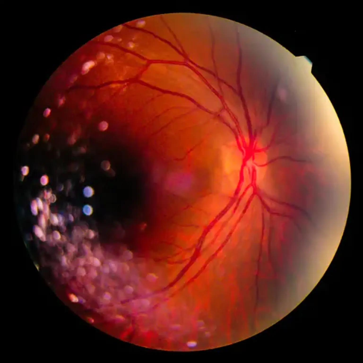
Fundus Imaging: This technique allows doctors to take detailed pictures of the retina to look for abnormalities.
Optical Coherence Tomography (OCT): A non-invasive imaging test that creates cross-sectional images of the retina, helping to detect early signs of conditions like macular degeneration.
Fluorescein Angiography: A dye is injected into the bloodstream, and special cameras capture images of blood flow in the retina. This is particularly useful for detecting diabetic retinopathy and retinal vascular diseases.
Ocular Ultrasound: Often used in cases of vitreous hemorrhage or retinal detachment to assess the internal structure of the eye.
Routine eye exams are crucial, especially for individuals with diabetes or those over 50, as these groups are more prone to retinal conditions.
Signs and Symptoms of Vitreoretinal Disorders
Recognizing the signs and symptoms of vitreoretinal disorders early can significantly improve the chances of successful treatment. Common symptoms include:
Blurry or Distorted Vision: This may occur in conditions like macular degeneration or diabetic retinopathy.
Floaters: Small spots or lines that drift across your field of vision. These can be a sign of retinal tears or vitreous hemorrhage.
Flashes of Light: Often reported in cases of retinal detachment.
Sudden Vision Loss: Especially in retinal detachment or advanced diabetic retinopathy, sudden vision loss is an emergency.
If you experience any of these symptoms, it’s important to see an eye care professional immediately to prevent further damage.
Macular Degeneration: Diagnosis and Care
Macular degeneration is one of the leading causes of vision loss in older adults, affecting the central part of the retina (the macula). This condition causes the macula to deteriorate over time, resulting in blurry or distorted central vision, though peripheral vision may remain unaffected.
Diagnosis:
Macular degeneration is typically diagnosed through fundus imaging, OCT, and fluorescein angiography. These tests help to assess the damage to the macula and monitor any changes.
Care and Treatment:
While there is no cure for macular degeneration, several treatment options can help slow its progression and improve quality of life:
Anti-VEGF Injections: These injections help prevent the growth of abnormal blood vessels that can damage the retina.
Laser Therapy: Used to destroy abnormal blood vessels or reduce leakage in wet macular degeneration.
Nutritional Supplements: High-dose antioxidants and vitamins may be recommended to slow progression in early stages.
Regular monitoring and early intervention can help manage the condition and reduce the risk of severe vision loss.
Diabetic Retinopathy: Early Detection and Treatment
Diabetic retinopathy is a complication of diabetes that affects the blood vessels in the retina. High blood sugar levels damage these vessels, leading to leakage, bleeding, and in severe cases, retinal detachment.
Diagnosis:
Diabetic retinopathy is diagnosed using a combination of retinal exams, fundus imaging, and OCT to assess the extent of damage to the retinal blood vessels.
Care and Treatment:
Early detection is key to preventing vision loss. Treatment options for diabetic retinopathy include:
Laser Therapy (Laser Photocoagulation): Helps seal leaking blood vessels and prevent further damage.
Intraocular Injections: Anti-VEGF drugs and steroids are used to reduce swelling and inhibit the growth of abnormal blood vessels.
Vitrectomy Surgery: In cases of severe bleeding or retinal detachment, vitrectomy may be necessary to remove damaged tissue and improve vision.
Maintaining good blood sugar control is essential to prevent the progression of diabetic retinopathy.
Vitreous Hemorrhage: Causes and Treatment
Vitreous hemorrhage occurs when blood leaks into the vitreous humor, the gel-like substance in the eye. This can cause floaters, blurry vision, and in severe cases, complete vision loss.
Causes:
Vitreous hemorrhage is often caused by retinal tears, diabetic retinopathy, or trauma to the eye. It can also occur in conditions like age-related macular degeneration.
Diagnosis:
An ocular ultrasound or OCT is often used to confirm the presence of blood in the vitreous humor and to assess the underlying cause.
Care and Treatment:
Observation: In some cases, vitreous hemorrhage may resolve on its own as the body reabsorbs the blood.
Vitrectomy Surgery: If the hemorrhage is severe or persistent, surgery may be required to remove the blood and any damaged retinal tissue.
Laser Treatment: In cases where a retinal tear is causing the hemorrhage, laser therapy can be used to prevent further bleeding.
Most cases of vitreous hemorrhage improve with proper treatment, though recovery may take time.
Retinal Tears and Detachments: Immediate Care and Surgery
A retinal tear occurs when the retina is torn due to trauma or other conditions, and if left untreated, it can lead to retinal detachment, where the retina separates from the back of the eye. This is a medical emergency and can cause permanent vision loss if not treated promptly.
Diagnosis:
Retinal tears and detachment are often diagnosed through OCT, fundus imaging, and a comprehensive eye exam. Symptoms like sudden flashes of light, floaters, and vision loss can indicate retinal detachment.
Care and Treatment:
Laser Treatment: Laser therapy can be used to seal retinal tears and prevent detachment.
Vitrectomy Surgery: This procedure involves removing the vitreous gel from the eye to allow the retina to reattach. It may be used for advanced detachment cases.
Pneumatic Retinopexy: A gas bubble is injected into the eye to help the retina reattach to the back of the eye.
Prompt treatment of retinal tears and detachment is essential to prevent permanent damage.
Innovations in Vitreoretinal Surgery
Vitreoretinal surgery has advanced significantly in recent years, with new technologies improving outcomes and reducing recovery times. These innovations have made surgeries more precise, less invasive, and safer.
Minimally Invasive Techniques:
Advances like microincision vitreoretinal surgery allow surgeons to perform procedures with smaller incisions, leading to faster recovery and reduced risk of complications.
Robotic-Assisted Surgery:
Robotic technology is being increasingly used in retinal surgeries, allowing for more precise movements and better outcomes, particularly in delicate procedures like retinal detachment repair and vitrectomies.
Improved Laser Systems:
New laser systems are more effective and safer, enabling targeted treatments for conditions like diabetic retinopathy and macular degeneration with minimal damage to surrounding tissues.
These innovations are not only improving the precision of surgery but also enhancing recovery times, offering patients better outcomes.
Laser Therapy for Vitreoretinal Disorders
Laser therapy is a cornerstone in the treatment of various vitreoretinal disorders. It is used to treat retinal tears, diabetic retinopathy, and macular degeneration, among others.
How It Works:
Laser therapy works by using focused light to either destroy abnormal retinal tissue (in cases of macular degeneration or diabetic retinopathy) or to seal retinal tears and holes (in retinal detachment). The goal is to prevent further damage and preserve vision.
Types of Laser Therapy:
Argon Laser: Often used to treat diabetic retinopathy and retinal tears.
YAG Laser: Commonly used for posterior capsulotomy and vitreous floaters.
Laser Photocoagulation: This technique is used in diabetic retinopathy to reduce leakage and prevent further damage to retinal blood vessels.
Recovery and Effectiveness:
Laser therapy is usually an outpatient procedure with minimal recovery time. While it can help slow the progression of retinal conditions, it may not fully restore lost vision, especially in advanced stages.
Role of Intraocular Injections in Retinal Treatment
Intraocular injections are increasingly used in the treatment of retinal conditions like macular degeneration, diabetic macular edema, and retinal vein occlusion. These injections deliver medication directly into the eye to reduce swelling, prevent abnormal blood vessel growth, and manage inflammation.
Common Injections:
Anti-VEGF (Vascular Endothelial Growth Factor): These injections block the growth of abnormal blood vessels in the retina, which can cause vision problems in diseases like macular degeneration.
Steroids: These may be injected to reduce inflammation and control swelling in conditions like diabetic macular edema.
Gene Therapy: An emerging treatment that aims to treat retinal diseases at the genetic level by delivering healthy genes into retinal cells.
Benefits and Considerations:
Injections can improve vision or slow disease progression, especially in wet macular degeneration. However, multiple injections may be needed over time, and potential risks include infection, increased eye pressure, or cataracts.
Vitreoretinal Surgery Recovery
Recovery from vitreoretinal surgery depends on the type of procedure performed, but it generally involves a period of healing and follow-up care to ensure proper healing and to monitor for complications.
Types of Surgery and Recovery:
Vitrectomy Surgery: Involves the removal of the vitreous gel to repair retinal tears, detachments, or hemorrhages. Post-surgery, patients may need to maintain a face-down position for a period to help the retina reattach.
Retinal Detachment Surgery: This procedure often requires several weeks of recovery, with close monitoring to ensure the retina stays in place.
Laser Surgery: Laser procedures often involve minimal downtime, with patients typically returning to normal activities within a few days.
Post-Surgery Care:
Patients should follow post-surgical instructions closely, including using prescribed eye drops to prevent infection, limiting physical activity, and attending follow-up appointments. It's also essential to avoid rubbing the eye or exposing it to excessive light.
Managing Complications:
While complications are rare, patients should be aware of potential risks like infections, retinal re-detachment, or increased eye pressure. Regular check-ups will help catch any issues early and ensure proper healing.
Managing Complications of Vitreoretinal Disorders
Vitreoretinal disorders can sometimes lead to complications, even with timely treatment. Managing these complications is essential for preserving vision and preventing long-term damage.
Common Complications:
Retinal Re-Detachment: This can occur after surgery if the retina does not stay in place.
Infection: Post-surgical infections can be serious and require immediate treatment.
Increased Eye Pressure: Conditions like glaucoma can develop following surgery or due to untreated retinal disease.
Management:
Regular follow-up appointments are crucial for detecting and addressing any complications early. In some cases, additional treatments or surgeries may be needed to correct issues like retinal re-detachment or elevated eye pressure.
Preventive Care and Risk Factors
Preventing vitreoretinal disorders involves addressing risk factors and adopting a proactive approach to eye health.
Risk Factors:
Age: Older adults are more likely to develop conditions like macular degeneration.
Diabetes: Poor blood sugar control increases the risk of diabetic retinopathy.
Family History: Genetic factors can increase the likelihood of certain retinal diseases.
Eye Trauma: Injuries to the eye can lead to retinal tears or detachment.
Prevention:
Routine eye exams, controlling diabetes, wearing protective eyewear, and maintaining a healthy lifestyle (e.g., not smoking, eating a balanced diet) can help reduce the risk of vitreoretinal disorders.
Global Prevalence and Awareness of Vitreoretinal Disorders
Vitreoretinal disorders affect millions worldwide, with rates increasing due to aging populations and the rise of diabetes.
Prevalence:
Macular Degeneration: Affects over 170 million people globally, particularly those over 50.
Diabetic Retinopathy: One of the leading causes of blindness in working-age adults, with millions of cases worldwide.
Retinal Detachment: Occurs in about 1 in 10,000 people annually.
Raising Awareness:
Increasing global awareness of these disorders is crucial, especially in high-risk populations. Early detection and proper management can significantly reduce the incidence of blindness caused by these conditions.
The Future of Vitreoretinal Disorder Treatment
The future of vitreoretinal disorder treatment is promising, with continuous advancements in research and technology.
Innovative Treatments:
Gene Therapy: Early-stage gene therapies aim to treat hereditary retinal diseases by directly altering the genetic material in retinal cells.
Stem Cell Therapy: Research into using stem cells to regenerate damaged retinal tissue holds potential for treating conditions like macular degeneration.
Artificial Retinas: Experimental treatments, such as bionic retinas, may offer solutions for patients with severe retinal degeneration.
Personalized Medicine:
As understanding of the genetic factors behind retinal diseases grows, treatments may become more personalized, offering targeted therapies tailored to an individual’s specific needs.
Psychological Impact of Vitreoretinal Disorders
The impact of vitreoretinal disorders extends beyond physical health, affecting mental and emotional well-being. Vision loss, even partial, can lead to anxiety, depression, and a sense of isolation.
Emotional Effects:
Depression and Anxiety: Many patients experience depression or anxiety due to the fear of permanent vision loss, especially with progressive conditions like macular degeneration or diabetic retinopathy.
Reduced Quality of Life: Vision impairment can affect daily activities, from reading to driving, leading to a loss of independence.
Coping Strategies:
Support from family, friends, and counselors is crucial for mental health. Joining support groups can help patients connect with others facing similar challenges. Early intervention for psychological well-being, in addition to medical treatment, can improve overall quality of life.
Cost of Vitreoretinal Disorder Treatment
The treatment of vitreoretinal disorders can be costly, depending on the type and stage of the condition. Surgery, injections, and long-term monitoring contribute to the financial burden.
Treatment Costs:
Intraocular Injections: These can cost several thousand dollars per injection, and patients may need multiple injections over time.
Vitreoretinal Surgery: Surgical procedures like vitrectomies or retinal detachment repairs can cost tens of thousands of dollars.
Follow-up Care: Regular eye exams, imaging, and monitoring can also add to the long-term financial burden.
Insurance and Financial Aid:
Most insurance plans cover many treatments, but costs may vary based on location and type of procedure. Patients may also seek financial aid programs or government assistance for costly treatments.
Patient Education and Empowerment
Educating patients about vitreoretinal disorders is crucial for encouraging early diagnosis, treatment, and proper care. Empowering patients with knowledge helps them take an active role in their health management.
Educational Resources:
Retinal Specialist Consultations: Patients should ask questions during appointments about their condition, treatment options, and what to expect.
Online Resources: Trusted websites like the American Academy of Ophthalmology provide reliable information on symptoms, treatment options, and prevention.
Support Groups: Patients can connect with others facing similar conditions for advice and emotional support.
An informed patient is better equipped to manage their condition and make decisions in partnership with their healthcare provider.
Conclusion
Vitreoretinal disorders, though diverse and potentially debilitating, have seen tremendous advancements in both diagnosis and treatment. Through innovative techniques, including minimally invasive surgeries, cutting-edge laser therapies, and intraocular injections, patients now have better options to manage conditions like macular degeneration, diabetic retinopathy, and retinal detachment.
However, effective treatment doesn't stop at surgery or injections. Preventive care, regular monitoring, and patient education play key roles in maintaining eye health and preventing progression. The psychological impact of vision loss is significant, highlighting the importance of emotional and mental support as part of the overall care plan.
As research continues to evolve, therapies such as gene therapy and stem cell treatments offer exciting possibilities for restoring vision and improving patient outcomes. With ongoing advancements, empowered patients, and improved access to care, the future of vitreoretinal disorders looks brighter, offering hope for those affected to maintain their vision and quality of life.