Introduction
Vitreous detachment is a natural age-related process that affects the vitreous humor—the clear gel that fills the inside of your eye. The vitreous gel helps maintain the shape of the eye and plays a role in focusing light onto the retina. As we age, the vitreous begins to shrink and pull away from the retina, which can lead to a condition called posterior vitreous detachment (PVD). Though it may sound concerning, vitreous detachment is a common occurrence, often affecting people over the age of 50.
In most cases, this condition doesn’t cause permanent damage, but it can be alarming when symptoms like eye floaters or flashes of light appear. Vitreous detachment is not usually dangerous, but if left unchecked, it can sometimes lead to more serious complications like retinal tears or detachment, which is why it’s important to seek medical advice.
What is Vitreous Detachment?
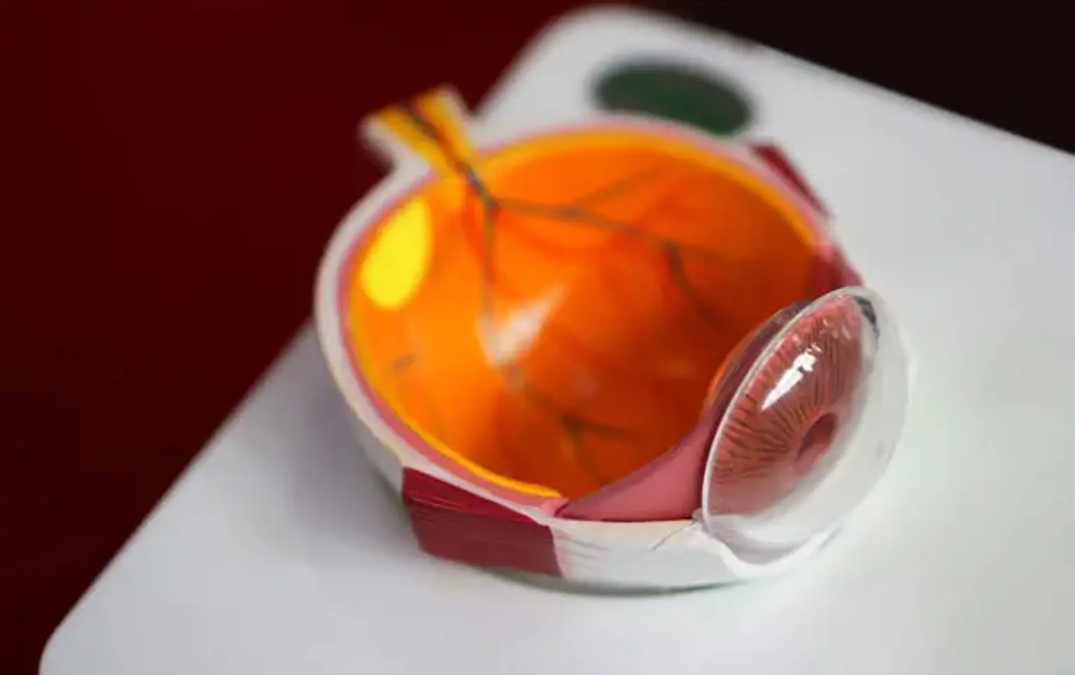
Vitreous detachment refers to the separation of the vitreous gel from the retina. The vitreous is a jelly-like substance that fills the eye, and it’s attached to the retina at certain points. Over time, typically after the age of 50, the vitreous begins to shrink and lose its gel-like consistency. It starts pulling away from the retina, which is the light-sensitive tissue at the back of the eye responsible for vision.
Posterior vitreous detachment (PVD) is the most common form, and it happens when the vitreous separates from the back of the eye. Most people experience no symptoms, but some may notice changes in their vision, such as flashes of light or the appearance of floaters—small specks or threads that drift across their vision. Although this is a common, typically non-threatening condition, it’s important to monitor for complications that could lead to retinal tears or detachment.
Causes of Vitreous Detachment
The primary cause of vitreous detachment is aging. As people get older, the vitreous becomes more liquid, which causes it to shrink and detach from the retina. However, other factors can increase the risk of developing vitreous detachment:
Age: Most people over the age of 50 experience some form of vitreous detachment.
High Myopia (Nearsightedness): People who are highly nearsighted may experience vitreous detachment earlier than those with normal vision.
Eye Trauma: An injury to the eye can trigger premature vitreous detachment.
Diabetes: Diabetic patients, particularly those with diabetic retinopathy, are at higher risk of complications like vitreous detachment.
Previous Eye Surgery: Surgeries like cataract removal can increase the risk of vitreous detachment.
Vitreous detachment is often considered a normal part of the aging process, but people with any of the risk factors mentioned above should be vigilant about their eye health and get regular eye exams.
Symptoms of Vitreous Detachment
Vitreous detachment doesn’t always come with clear symptoms, but when it does, the signs can be alarming. The most common symptoms include:
Eye Floaters: These are small, shadowy shapes that appear to float across your vision. They can look like dots, lines, or cobwebs and are caused by tiny clumps of gel or debris inside the vitreous. Floaters can be distracting, especially when looking at bright backgrounds like a clear sky or white paper.
Flashes of Light: Patients may experience brief flashes of light, often in the peripheral vision. These flashes can occur when the vitreous pulls on the retina or when the gel shifts inside the eye.
Blurred or Distorted Vision: In some cases, the detachment can cause temporary blurred vision or visual distortion. This might be due to the changes in the vitreous shape or any minor retinal changes that happen as the detachment progresses.
While these symptoms can be alarming, they are not always dangerous. However, if a sudden increase in floaters or flashes occurs, or if you notice a shadow or curtain-like effect over your vision, it’s essential to seek immediate medical attention, as these could be signs of a retinal tear or detachment.
Diagnosis of Vitreous Detachment
Diagnosing vitreous detachment involves a detailed eye exam by an ophthalmologist. Key diagnostic tools include:
Retinal Examination: Using a special light to check for retinal tears or detachment.
OCT (Optical Coherence Tomography): Provides cross-sectional images of the retina and vitreous.
Ultrasound: Helps detect detachment if the retina is not clearly visible due to opacity.
Early detection ensures timely monitoring and intervention if complications arise, such as retinal tears.
Treatment Options for Vitreous Detachment
Most cases of vitreous detachment do not require treatment, as the condition often stabilizes on its own. However, in certain situations, intervention may be necessary:
Observation: Regular monitoring for any changes or progression of the condition.
Eye Drops: Sometimes used to manage symptoms like inflammation or discomfort.
Surgical Intervention: Required if complications such as retinal tears occur.
In most cases, treatment focuses on managing symptoms, particularly floaters, which may improve over time or with medical intervention.
Managing Eye Floaters: Treatment and Prevention
Eye floaters are a common symptom of vitreous detachment. They often appear as small, dark shapes that drift across your vision. While floaters may diminish over time, treatments are available if they interfere with daily activities:
Laser Therapy: A laser can break up the floaters, making them less noticeable.
Vitrectomy: In rare cases, vitrectomy may be performed to remove floaters if they are severe and persistent.
Preventing floaters is challenging, as they are often part of the natural aging process. However, maintaining good eye health through regular check-ups and managing underlying conditions like diabetes can reduce the risk of complications.
Risk of Retinal Tear and Retinal Detachment
While vitreous detachment is usually harmless, it can sometimes lead to more serious conditions like retinal tears or retinal detachment. As the vitreous shrinks and pulls away from the retina, it can cause a tear in the retinal tissue. If untreated, this can lead to retinal detachment, which can result in permanent vision loss.
Signs that retinal complications may be occurring include a sudden increase in floaters, a shadow or curtain in your peripheral vision, or a sudden loss of vision. If you experience any of these symptoms, it’s important to seek immediate medical attention. Treatment, such as laser therapy or surgery, can repair retinal tears and prevent detachment.
Vitrectomy: Surgical Treatment for Severe Cases
For severe cases, particularly when there is a retinal tear or detachment, vitrectomy surgery may be recommended. This procedure involves removing the vitreous gel and replacing it with a saline solution or gas bubble.
Vitrectomy can prevent further retinal damage and restore vision, though it carries risks like infection, cataract formation, and increased intraocular pressure. The decision for surgery depends on the severity and progression of the detachment, as well as the patient’s overall eye health.
Recovery After Vitreous Detachment Treatment
In most cases, vitreous detachment resolves on its own, and no active treatment is needed. If surgery or other interventions are required, recovery is generally straightforward:
Post-Treatment Care: Patients may need to avoid heavy lifting or vigorous activity for a few weeks after surgery.
Follow-Up Visits: Regular eye exams ensure that the eye heals properly and that no further complications arise.
Managing Discomfort: Mild discomfort, like redness or light sensitivity, is common but typically temporary.
Most patients experience a full recovery, with symptoms like floaters decreasing over time, although some may remain. Your ophthalmologist will guide you through the recovery process and monitor your progress.
The Role of Ophthalmologists and Retinal Specialists
Ophthalmologists and retinal specialists are key to diagnosing and treating vitreous detachment. These professionals are trained to manage the condition, assess risks, and provide treatments to prevent further damage to the retina.
If you experience symptoms like floaters or flashes, an ophthalmologist should be your first point of contact. For more complex cases, particularly those involving retinal tears or detachment, a retinal specialist may be necessary to provide specialized care. They can offer advanced treatments, such as laser therapy or vitrectomy, depending on the severity of the condition.
Global Awareness and Advancements in Vitreous Detachment Treatment
As awareness of vitreous detachment grows, more people are seeking treatment and understanding the condition better. Advancements in diagnostic technology, such as OCT scans and laser treatments, have significantly improved the accuracy of detection and the effectiveness of treatments. These innovations help prevent complications and improve recovery outcomes.
Healthcare providers globally are working toward greater education about the risks associated with vitreous detachment, particularly as the aging population increases. Regular eye exams and early intervention remain the best strategies for managing the condition and preserving vision.
Costs of Vitreous Detachment Treatment
The costs of treating vitreous detachment vary depending on the severity of the condition and the type of treatment required. For most people, observation and regular check-ups are enough, which are typically covered by insurance. However, for patients requiring more advanced treatments such as laser therapy or vitrectomy, costs can rise significantly.
Laser Therapy: Typically less expensive than surgery, but can still be costly depending on the number of sessions.
Vitrectomy: This surgical procedure tends to be the most expensive, particularly if performed in a specialized clinic or hospital.
Insurance: Many insurance plans cover the costs of treatment for retinal issues, but coverage may vary based on the provider and the patient’s plan.
For those without insurance or with high out-of-pocket expenses, there may be financial assistance programs or payment plans available through clinics or organizations.
Long-Term Outlook and Prognosis for Vitreous Detachment
The long-term outlook for vitreous detachment is generally positive. In most cases, the condition resolves on its own without the need for surgery. However, some patients may continue to experience floaters or mild visual disturbances, which typically improve over time.
For patients who require surgical intervention, such as vitrectomy, the prognosis is usually good, with most recovering full vision. However, the recovery time may vary depending on the individual’s age, overall health, and the complexity of the surgery.
If vitreous detachment leads to more serious complications like retinal tears, timely treatment is crucial. With prompt care, the risk of permanent vision loss can be minimized, and vision can often be restored.
Preventive Measures and Maintaining Eye Health
Although vitreous detachment is often a natural part of aging, certain steps can help maintain eye health and potentially reduce the risk of complications:
Regular Eye Exams: Annual check-ups allow early detection of vitreous changes and other potential eye issues.
Healthy Lifestyle: Eating a balanced diet rich in antioxidants and omega-3 fatty acids may support retinal health.
Protecting Your Eyes: Wearing protective eyewear during sports or activities that could cause eye injury helps reduce the risk of trauma.
While you cannot prevent vitreous detachment entirely, a healthy lifestyle and proactive eye care can help manage the condition and promote long-term eye health.
Understanding the Emotional Impact of Vitreous Detachment
While vitreous detachment is often a mild condition, it can cause emotional distress due to changes in vision, especially with symptoms like floaters. Many patients report feeling anxious or worried about their vision, particularly if they experience frequent flashes or see shadows in their peripheral vision.
Support from healthcare professionals and a clear understanding of the condition can help alleviate fears. Talking to an ophthalmologist about your concerns and learning more about the process can provide reassurance. Additionally, joining support groups or talking to others who have experienced vitreous detachment may help patients cope emotionally.
It’s essential to remember that most cases of vitreous detachment are non-threatening and manageable, and with the right treatment, patients can maintain a good quality of life.
Frequently Asked Questions About Vitreous Detachment
Is vitreous detachment a serious condition? Vitreous detachment is generally not considered a serious condition. It often occurs naturally with aging and can be managed with regular monitoring. However, in rare cases, it can lead to complications like retinal tears or detachment, which require prompt medical attention.
Can vitreous detachment cause permanent vision loss? Vitreous detachment itself doesn’t usually cause permanent vision loss. However, if complications such as retinal tears or detachment occur, there is a risk of vision loss, which is why early detection and treatment are important.
How long do floaters last after vitreous detachment? Floaters can persist for several weeks or even months after vitreous detachment. Over time, they tend to become less noticeable, but some individuals may continue to experience them long-term. Treatment options like laser therapy or vitrectomy may help reduce their severity.
Is there anything I can do to prevent vitreous detachment? Since vitreous detachment is largely due to aging, it’s not entirely preventable. However, maintaining healthy eyes through regular eye exams, proper nutrition, and protecting your eyes from injury can help minimize complications.
Global Popularity of Vitreous Detachment Treatment
Vitreous detachment is a global concern, as it predominantly affects individuals over the age of 50, and the aging population worldwide continues to grow. Treatment for this condition varies by region, depending on access to healthcare and the availability of advanced diagnostic tools. In developed countries, OCT and laser therapies are widely used for early detection and symptom management.
In regions with limited healthcare resources, more basic forms of monitoring may be employed, but surgical treatments like vitrectomy are often available for advanced cases. As awareness increases, especially with the rise in eye health education campaigns, more people are seeking medical advice and treatment options for vitreous detachment, ensuring better outcomes globally.
Advancements in Vitreous Detachment Research
Research into vitreous detachment continues to evolve, with ongoing studies aimed at improving treatment options and understanding the condition. New technologies in imaging, such as adaptive optics and optical coherence tomography (OCT), allow for more detailed and precise evaluations of the vitreous and retina.
Additionally, scientists are exploring pharmacological treatments to slow down the process of vitreous liquefaction or prevent complications like retinal tears. While many of these innovations are still in the research phase, they hold promise for improving the management and treatment of vitreous detachment in the future.
Conclusion
Vitreous detachment is a natural part of the aging process that many people experience. While it can be unsettling, it is typically manageable with proper care and monitoring. In most cases, the condition improves on its own, and only severe cases require surgical intervention.
By maintaining regular eye exams and being aware of the symptoms, you can manage the condition effectively. If complications arise, advancements in diagnostic tools and treatments can help preserve vision and ensure a good quality of life. Understanding the condition, seeking prompt medical advice, and staying informed about new research and treatments can help you navigate vitreous detachment with confidence.