What is Rib Cartilage Grafting Surgery?
Definition and Explanation Rib cartilage grafting surgery is a specialized procedure where cartilage is harvested from the ribs and used to reconstruct or augment other parts of the body, most commonly the nose. This technique is especially favored in reconstructive surgery due to the durability, flexibility, and structural integrity of rib cartilage. In many cases, rib cartilage is the best option when nasal structures require significant support or when other grafting options, like ear cartilage, may not be sufficient.
In simple terms, rib cartilage grafting allows surgeons to create a durable framework within the nose or face that can restore both function and appearance. This procedure is most commonly used in rhinoplasty (cosmetic nose surgery) and nasal reconstruction after trauma or congenital defects, such as cleft lip and palate.
Unlike synthetic materials or cadaveric cartilage, rib cartilage is taken from the patient’s own body (autologous graft), meaning it is less likely to be rejected by the body, reducing the risk of complications.
Common Applications Rib cartilage grafting is versatile and can be applied in both aesthetic and reconstructive surgeries. Some of the most common applications include:
Nasal Reconstruction: After trauma or congenital defects, rib cartilage can help restore the structure of the nose, improving both appearance and function.
Rhinoplasty: In cosmetic surgery, rib cartilage is used to reshape the nose, especially in complex cases where other sources of cartilage may not provide enough support.
Cleft Lip and Palate Surgery: Rib cartilage grafts can assist in correcting deformities caused by congenital conditions, offering more durable and aesthetically pleasing results than traditional methods.
The Rib Cartilage Harvesting Process
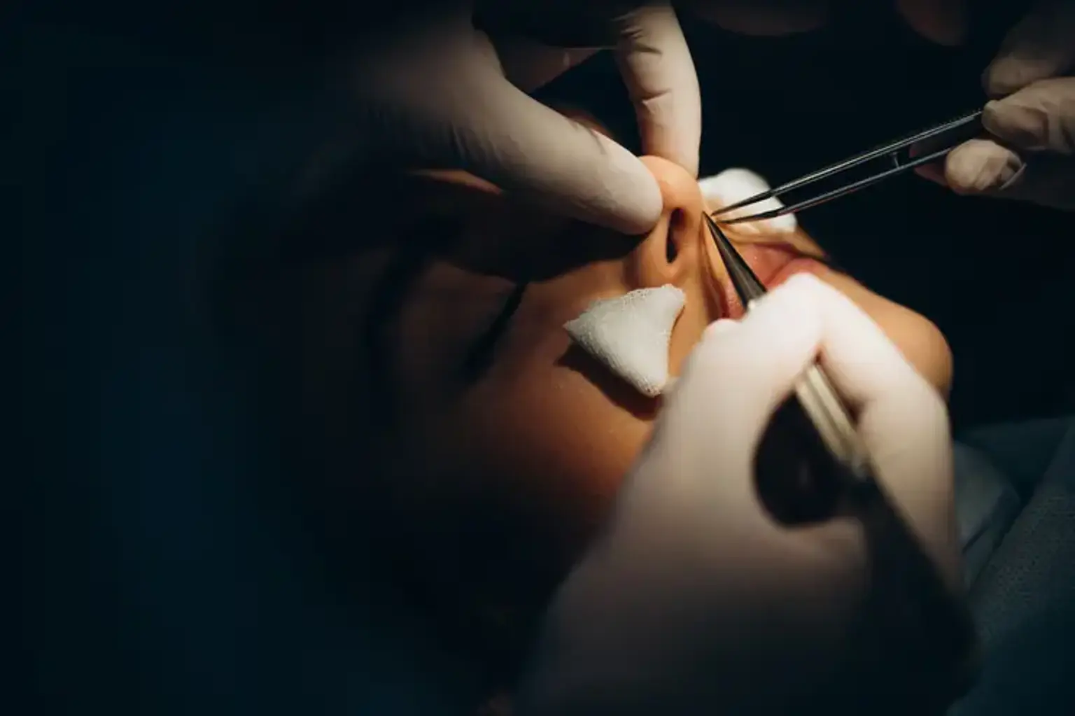
How Rib Cartilage is Harvested The process of harvesting rib cartilage is a delicate procedure that involves making an incision along the chest wall to access the ribs. The surgeon carefully removes a small section of cartilage, usually from the lower ribs, as this area offers sufficient cartilage without interfering with critical structures like the lungs or blood vessels. This cartilage is then shaped and used to support or reconstruct the desired area, most often in the nose.
Types of Rib Cartilage There are primarily two types of rib cartilage that may be harvested: costal cartilage (from the ribs) and sternal cartilage (from the breastbone). Costal cartilage is the more commonly used graft in nasal surgeries because it is flexible, strong, and less likely to cause complications when used in delicate areas like the nose. It is also relatively easy to access and provides a natural contour when reshaped.
Safety Protocols The procedure is performed under general anesthesia, ensuring that patients are comfortable throughout the process. To minimize risks and complications, surgeons take several precautions, including:
Using advanced imaging techniques to identify the correct rib for harvesting.
Ensuring a sterile environment to prevent infection.
Monitoring the patient’s vital signs carefully during and after the procedure.
While rib cartilage harvesting is generally considered safe, as with any surgery, there are inherent risks, such as infection, bleeding, or scarring. However, these risks are minimized through careful surgical technique and post-operative care.
Indications for Rib Cartilage Grafting Surgery
Reconstructive Surgery One of the most significant applications of rib cartilage grafting is in reconstructive surgery, where the goal is to restore the appearance and function of damaged or malformed nasal structures. Some of the most common conditions that benefit from rib cartilage grafting include:
Trauma to the Nose: Accidents or injuries that result in severe nasal deformities may require rib cartilage grafts to restore both form and function. Rib cartilage offers the strength needed to rebuild a structurally sound nasal framework.
Congenital Defects: Children born with congenital conditions such as cleft lip and palate may need rib cartilage grafts to reconstruct the nasal passages and correct deformities. Rib cartilage is particularly useful in these cases because of its flexibility and ease of shaping.
Nasal Septal Defects: If a patient has a damaged or deviated septum, rib cartilage grafts can help straighten the septum and improve airflow, resulting in better breathing.
Cosmetic Surgery In aesthetic surgery, rib cartilage grafting is often used in rhinoplasty to achieve a more natural and symmetrical appearance. The procedure allows for more significant changes to the shape of the nose, especially in patients who have had prior surgeries or have very thin nasal cartilage. Rib cartilage is ideal for creating a strong, stable structure that can support various cosmetic enhancements, such as:
Raising the Nasal Bridge: For patients who have a flat or collapsed nasal bridge, rib cartilage provides a solid base to lift and support the nose.
Creating a More Defined Tip: Rib cartilage grafts can help refine the shape of the nose, especially in patients with underdeveloped nasal tips.
Correcting Irregularities: Rib cartilage can be used to correct asymmetries or irregularities caused by previous surgeries or traumatic injury.
Benefits of Rib Cartilage Grafting
Functional Benefits The functional benefits of rib cartilage grafting are significant, particularly when it comes to restoring breathing function. After trauma or congenital deformities, the nasal septum may be misaligned, obstructing airflow. Rib cartilage grafts can restore a more natural, open passage, improving both appearance and function. Some of the key functional benefits include:
Improved Breathing: Rib cartilage can help realign the nasal septum, which is often necessary for patients with severe nasal airway obstructions.
Strength and Durability: Rib cartilage is known for its strength and ability to withstand the stress of reshaping, ensuring that it stays in place over time and continues to support nasal structures.
Aesthetic Improvements Rib cartilage is also prized for its aesthetic qualities in rhinoplasty and other cosmetic surgeries. Some of the key benefits include:
Natural Shaping: Rib cartilage can be sculpted to create a more natural and customized appearance, allowing for more significant changes to the nasal structure than other types of grafts.
Long-lasting Results: Because rib cartilage is a durable material, it provides long-lasting results with minimal risk of resorption or deformation, ensuring that the changes to the nose remain stable over time.
Autologous Nature One of the standout advantages of rib cartilage grafting is that it is an autologous graft, meaning it comes from the patient’s own body. This significantly reduces the risk of rejection or allergic reaction, which can occur with synthetic or cadaveric materials. Additionally, using one’s own tissue promotes better integration and healing, leading to more predictable and successful outcomes.
Risks and Potential Complications
Surgical Risks As with any surgery, rib cartilage grafting carries some risks. These include:
Infection: Although rare, infection can occur at the incision site, requiring antibiotics or further intervention.
Bleeding: Excessive bleeding is possible during the harvesting of cartilage, though it is usually manageable.
Scarring: A visible scar may form along the chest wall where the cartilage is taken. However, modern surgical techniques minimize scarring.
Post-surgical Complications Some patients may experience complications related to graft failure or misalignment, including:
Cartilage Resorption: Over time, the body may reabsorb the graft, leading to reduced effectiveness or shape alteration.
Graft Shifting: Occasionally, the graft can shift, requiring corrective surgery to reposition it.
Managing Risks To reduce these risks, it’s crucial to:
Choose an experienced surgeon familiar with rib cartilage harvesting.
Follow post-operative care instructions carefully, including proper wound care and follow-up visits.
Recovery After Rib Cartilage Grafting Surgery
Initial Recovery Recovery time varies, but in general:
First Week: Most patients experience swelling and discomfort around the chest and nose. Pain management is typically done with prescribed medications.
Chest Recovery: The site where the rib cartilage is harvested may feel sore for a few weeks, but this typically resolves with time.
Long-term Recovery
Full Recovery: Most patients can return to normal activities within 4 to 6 weeks. However, heavy lifting or strenuous activity should be avoided for several months to ensure proper healing.
Post-operative Care: Following instructions for nasal care is crucial to prevent complications. Most swelling in the nose will subside within a few weeks, but complete results may take several months to be fully visible.
Pain Management Pain is usually mild to moderate and is managed with over-the-counter medications or prescribed painkillers. Ice packs and rest are also effective in reducing swelling and discomfort.
Rib Cartilage Grafting Surgery for Different Conditions
Nasal Deformities Rib cartilage grafting is often used to correct nasal defects caused by trauma, disease, or congenital conditions such as cleft lip and palate. It is particularly effective for restoring nasal function and shape, especially when other grafting options are not sufficient.
Trauma and Injury Following facial trauma, rib cartilage grafting can help rebuild the nasal structure, providing support and allowing the patient to regain normal breathing function. Rib cartilage is sturdy, making it an ideal material for reconstruction in such cases.
Cosmetic Enhancements In rhinoplasty, rib cartilage is used for complex reshaping of the nose. It’s especially beneficial for patients who require substantial changes, such as building a more defined nasal bridge or tip. The strength of rib cartilage ensures that the nose retains its new shape over time.
Comparison to Other Grafting Methods
Ear Cartilage vs. Rib Cartilage While ear cartilage is commonly used in some nasal surgeries, rib cartilage is often preferred when more volume or support is needed. Rib cartilage is stronger and more flexible, providing better structural support, especially for more complex procedures like rhinoplasty. However, ear cartilage has the advantage of being easier to harvest and results in a smaller incision.
Synthetic and Cadaveric Grafts Compared to synthetic and cadaveric grafts, rib cartilage is more natural and better integrated into the body. Synthetic grafts, while convenient, carry risks of rejection or movement. Rib cartilage, being autologous, offers superior durability and less risk of complications.
How to Prepare for Rib Cartilage Grafting Surgery
Pre-Surgical Assessment Before undergoing rib cartilage grafting surgery, patients will have a comprehensive evaluation, which may include:
Physical Examination: To assess overall health and ensure the patient is a good candidate for surgery.
Imaging Tests: X-rays or CT scans to evaluate the structure of the ribs and nasal area.
Blood Tests: To check for any underlying conditions, such as anemia or infections, which might affect healing.
Instructions for Preparation Patients are typically advised to:
Avoid Smoking: Smoking impairs healing and increases the risk of complications.
Stop Certain Medications: Blood thinners and certain supplements may need to be discontinued before surgery.
Arrange for Post-Op Care: It’s crucial to have someone available to help during the initial recovery phase.
Expected Results and Long-Term Outcomes
Short-term Results After rib cartilage grafting, most patients see immediate improvements in their nasal structure and function. The nose may appear more symmetrical, and breathing may improve if the procedure was aimed at correcting a deviated septum or structural deformities.
Long-term Results The final results can take several months to fully manifest as swelling continues to subside. In the long term:
The graft will integrate well with the surrounding tissue.
The nose will maintain its new shape and function, with minimal risk of the cartilage shifting or resorbing if proper care is followed.
Durability Rib cartilage provides a long-lasting, stable result. Unlike synthetic materials, it doesn’t risk rejection or degradation over time, offering patients a reliable and permanent solution.
Cost of Rib Cartilage Grafting Surgery
Factors Affecting the Cost The cost of rib cartilage grafting surgery can vary significantly based on several factors, including:
Geographic Location: Prices vary depending on the country or region. Major cities or areas with high living costs may have higher surgical fees.
Surgeon’s Expertise: A highly experienced and well-known surgeon may charge more for their services.
Complexity of the Surgery: If the procedure involves significant reconstruction, the cost will likely be higher due to the longer surgery time and need for specialized equipment.
Average Cost Range On average, the cost of rib cartilage grafting surgery can range from $5,000 to $15,000. This includes the surgeon's fee, anesthesia, and post-operative care. Additional costs such as hospital fees or consultations may increase the overall price.
Insurance Coverage Insurance may cover rib cartilage grafting if the surgery is deemed medically necessary, such as in cases of trauma or congenital defects. However, if the procedure is cosmetic, it is typically not covered by insurance.
Frequently Asked Questions (FAQs)
1. Is rib cartilage grafting surgery painful? While some discomfort is expected, especially during the initial recovery period, pain is usually manageable with medication. The chest area where the cartilage is harvested may be sore, but this typically resolves within a few weeks.
2. How long does recovery take? Most patients can return to normal activities within 4 to 6 weeks, though more strenuous activities should be avoided for up to 3 months. Full recovery may take several months, especially for the nose to fully heal and settle into its new shape.
3. Are there any alternatives to rib cartilage grafting? Yes, alternatives include ear cartilage, septal cartilage, synthetic grafts, or cadaveric cartilage. However, rib cartilage is often preferred in complex cases due to its strength and longevity.
4. Will I need another surgery in the future? In most cases, rib cartilage grafts offer permanent results. However, rare instances of graft resorption or shifting may require additional minor procedures.
5. How can I ensure the best results? To ensure optimal results, it’s important to follow your surgeon’s post-operative care instructions closely, avoid smoking, and attend follow-up appointments to monitor healing.
The Role of Rib Cartilage Grafting in Enhancing Quality of Life
Improving Physical Appearance One of the most significant benefits of rib cartilage grafting is its ability to improve physical appearance, particularly in cases of nasal deformities. For individuals born with congenital defects or those who have suffered trauma, rib cartilage grafting can restore balance and symmetry to the nose, boosting self-esteem and confidence. Many patients report feeling more comfortable in social situations and more positive about their overall appearance after the surgery.
Enhancing Functionality Beyond aesthetics, rib cartilage grafting can significantly improve nasal function. For those with breathing difficulties due to structural nasal issues, such as a deviated septum, the procedure can provide relief and improve airflow. The ability to breathe better can lead to better sleep, more energy, and overall improved well-being.
Psychosocial Benefits The psychological benefits of a successful surgery can be profound. Reconstructive nasal surgeries that use rib cartilage often lead to an increase in quality of life. By improving both appearance and function, patients can experience a reduction in social anxiety and an enhanced sense of normalcy. This can be especially life-changing for individuals who have long struggled with breathing problems or self-consciousness about their nasal appearance.
Conclusion
Final Considerations Rib cartilage grafting surgery is a highly effective and versatile procedure that offers both functional and aesthetic benefits. It is particularly well-suited for patients with complex nasal deformities, whether due to trauma, congenital conditions, or cosmetic desires. The strength and durability of rib cartilage make it an ideal material for reconstructing and reshaping the nose, ensuring long-lasting and natural results.
However, like all surgeries, it comes with potential risks and a recovery period that requires careful attention and patience. Understanding the procedure, the recovery process, and the potential benefits can help you make an informed decision about whether rib cartilage grafting is the right choice for your needs.
Consulting a Specialist If you’re considering rib cartilage grafting, the most important step is consulting a skilled and experienced surgeon. They will guide you through the process, provide personalized advice based on your unique situation, and help you set realistic expectations for both the surgery and recovery.
Whether for reconstructive purposes or cosmetic enhancement, rib cartilage grafting offers patients a chance to restore both form and function, enhancing their quality of life in profound ways.