Bone and Cartilage Transplantation

Last updated date: 23-Oct-2023

Originally Written in English

Bone and Cartilage Transplantation

Full-thickness cartilage abnormalities can be treated with osteochondral grafts. Patients who suffer from focal cartilage abnormalities have a wide range of therapeutic choices. Different methods for treating cartilage problems have been described. There is little prospect of self-repair when the defect is full thickness. For lesser problems, micro-fracture surgery has traditionally been used. Repeated weight-bearing and compressive stresses, which can be harmful to the joint over time and be connected to worse patient outcomes, boost healing through the production of fibrocartilage, which is not ideal. The use of autologous cells in other treatments has been described. The drawback of autologous chondrocyte implantation, which involves inserting chondrocytes into the defect and stitching them in place, has been noted as being the procedure's technical complexity; nonetheless, it may be a good option for larger lesions. A single plug or several plugs may be utilized in osteochondral graft transplantation to fill a bigger lesion.


What is Bone and Cartilage Transplantation

Bone and cartilage transplantation is a treatment for cartilage injuries that uncover underlying bone. A piece of tissue called an osteochondral graft is taken from the patient himself or a deceased donor and contains both bone and cartilage. It is used to replace damaged cartilage that lines the ends of bones in a joint. To repair the damage, the graft tissue is transplanted after being precisely sculpted to match the defect in the patient's damaged joint.
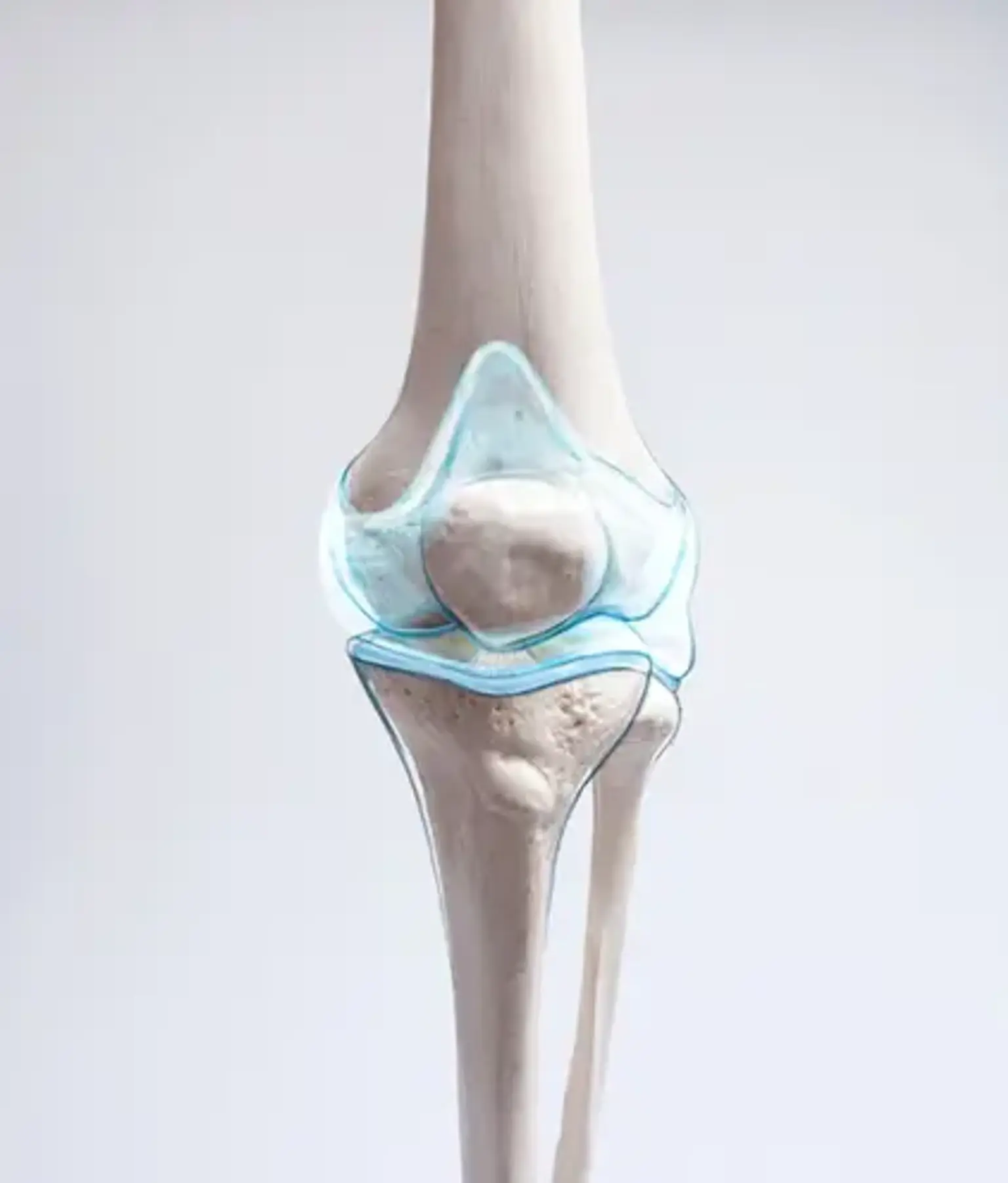
 

Anatomy and Physiology

Bone and Cartilage Transplantation Anatomy

Hyaline, fibroelastic, fibrocartilage, elastic, and physeal cartilage are the five forms of cartilage that can be found in the human body. Hyaline cartilage predominates among the types of cartilage that line the articular surfaces of native joints. Hyaline cartilage serves as a smooth, friction-free surface that can sustain the repeated loading and unloading of the joint. Hyaline cartilage mostly consists of water, type 2 collagen, and proteoglycans.

Hyaline cartilage normally contains between 70 and 80 percent water by weight. When osteoarthritis is present, water content rises, which causes strength to decline. The superficial zone, intermediate zone, deep layer, tidemark, and subchondral bone are the five separate layers that make up articular cartilage. Fibrocartilage develops when there is an injury to the articular cartilage that extends deep into the tidemark. Pluripotent mesenchymal cells have been released from the bone marrow and have created fibrocartilage.


Techniques for Bone and Cartilage Injury

Bone and Cartilage Injury

A special type of tissue is articular cartilage. Its matrix supports innumerable cycles during the person's lifetime and enables movement between the joint surfaces with the least amount of friction.  Harm to this structure, which does not compromise the subchondral bone's integrity, will not heal on its own and, because it is asymptomatic, can cause the joint to gradually deteriorate up to severe osteoarthritis, whose only treatment option would be to replace it with a mechanical prosthesis. Hilar cartilage injuries can be treated using one of three methods: palliative, reparative, or substitutive (often known as bone and cartilage graft or transplantation).

Different treatments have been designed to create a repair tissue with structure, histological composition, and functional behavior similar to that of native articular cartilage to stop the progression of this process. There are three different approaches to treating these injuries:

  • Palliative. This typically entails joint lavage, also known as a joint washout, and debridement.
  • Reparative. They include treatments focused on the subchondral bone's capacity for regeneration.
  • Substitutive. Osteochondral or cartilage and bone grafts serve as substitutes.


Bone and Cartilage Transplantation Benefits

Mosaicplasty is the process of filling a defect using several plugs. For the single plug technique to be effective, the recipient site's architecture and the donor surface architecture must be identical. Since many donor plugs are inserted into the defect during mosaicplasty, the donor site architecture may be less consistent. Fibrocartilage will fill the spaces between the several plugs. In the past, subchondral bone abrasions or drilling at the location of focal injury have been used to treat articular cartilage lesions. Bulk autografts and allografts have been applied to osteochondral lesions. However, these only apply to major lesions greater than 10 cm2. The following explanations explain why autogenous or allogenic osteochondral plugs have gained popularity:

  • They can all be done in one operation.
  • They give the possibility of real hyaline cartilage resurfacing.
  • There is no need for outside laboratory assistance.
  • It can be carried out using reusable tools.


Bone and Cartilage Transplantation Advantages

Bone and Cartilage Transplantation

There are various benefits to fresh bone and cartilage transplantation. The ability to perform it with any size and measurement given from a tissue bank is one of them. They can also be applied to big lesions without endangering the donor site. After many years of transplantation, chondrocyte viability is still seen, with new grafts having the highest viability.

Since frozen chondrocytes and even those cryopreserved at -196°C do not keep the chondrocytes alive, as They showed in many doctoral theses in 2006, most of these cells die after the transplant, rendering the tissue no longer viable. In some circumstances, fresh bone and cartilage transplantation can greatly improve the treatment of degenerative cartilage lesions.

Due to its avascular features (i.e., the absence of vascular structures inside), cartilage is regarded as an immunologically favored tissue since it reduces the host's immune response. As a result, there are no rejection reactions in these fresh cartilage transplant cases as there are with transplants of the heart, kidney, or liver.


Bone and Cartilage Transplantation Indications

Bone and Cartilage Transplantation Indications

There are various reasons for bone and cartilage transplantation. To increase the likelihood of success, Hangody offered recommendations for choosing patients. This includes restricting surgery to individuals under 45 with focal lesions and in good physical condition. A patient with a symptomatic little focal traumatic lesion is a better prospect than one with widespread arthrosis. Any age is eligible for a surgical indication as long as a bone healing response is anticipated. The knee joint is most easily accessed using this technique because of its size and diverse pathologies.

A femoral condyle can be reached either openly or arthroscopically, although an open approach is required for the retro-patellar region and trochlear groove. The tibia provides a special challenge since open or orthoscopic approaches cannot provide direct perpendicular access; however, an indirect retrograde approach may be used. Oblique grafts from the donor should be carefully selected to fit the recipient tunnel surface angle. Most experts agree that lesions with a diameter of between 1 cm and 2.5 cm are acceptable for full-thickness cartilage. Larger osteochondral grafts have shown positive results in studies, although these numbers are not generally recognized.


Bone and Cartilage Transplantation Contraindications

Bone and cartilage transplantation is not recommended in the following situations:

  • Global arthrosis
  • Opposing kissing lesions
  • Inflammatory arthropathy
  • Infection (septic arthritis)
  • Biomechanically altered joint line
  • Significant degeneration

Global arthrosis is the only absolute contraindication. However, this method might work for focal lesions in two or more different areas of the knee. However, the procedure's effectiveness is diminished where secondary changes, such as osteophytes or joint space constriction, are present.


How to Obtain Hyaline Cartilage

Hyaline Cartilage

The specialized cells of cartilage tissue known as chondrocytes work to repair damaged articular cartilage. They do this by producing more proteoglycans, the proteins in charge of creating and maintaining the structure of cells. Instead of stimulating type II collagen, which promotes the formation of hyaline cartilage, the repair that is thus achieved stimulates type I collagen, which produces fibrocartilage.

This needs to be mentioned since fibrocartilage has different biomechanical properties than hyaline cartilage and as a result, causes more wear and friction. When it has been impossible to obtain type II collagen, the key component of the hyaline cartilage of the joints, this has always been the mainstay of reparative treatments. The osteochondral cartilage can cross this barrier.


Equipment and Personnel

This operation will need to be performed in an operating room. The arthroscopic camera/tower and a regular operation room table with a leg holder will be used. Any of the surgical techniques designed to collect and deliver bone and cartilage grafts may be applied.

The operating room will need to be staffed with all personnel required to carry out this procedure safely. Orthopedic surgeons, surgical assistants, scrub nurses, circulating nurses, anesthesia providers, supplementary operating room employees, preoperative care team members, postoperative care team members, and medical device specialists are required personnel.


Bone and Cartilage Transplantation Preparation

Patients are assessed clinically and put via diagnostic imaging tests before the surgical operation. Full-thickness cartilage lesions are frequently found in conjunction with other ligamentous knee injuries. Patients frequently describe a traumatic event such as a falling, a pivot-shift trauma, a patellar dislocation, or a direct blow to the affected knee. Knee radiographs (AP and Lateral) are part of the radiographic workup.

Radiographs are neither sensitive nor specific enough to identify cartilage abnormalities, hence additional imaging techniques like MRI are frequently employed. The depth and extent of the chondral defect can be determined using MRI. When considering several treatment options, the extent of the defect becomes crucial. Lesions larger than 2.5 cm in diameter and less than 1 cm in diameter may both benefit from various restorative methods, as was previously indicated.


Bone and Cartilage Transplantation Procedure

Bone and Cartilage Transplantation Procedure

To achieve the best results, an osteochondral autograft must be performed in a step-by-step process. When pre-operatively planning, there are a few guidelines and ideas that may be helpful. It's crucial to keep in mind that maintaining perpendicularity to the joint surface being restored is crucial. Preserving the capacity to hyperflex the knee is essential for visualization because many of these injuries are situated posteriorly on the femoral condyles.

A first diagnostic arthroscopy, including examination of all joint surfaces, ligaments, menisci, and medial/lateral gutters, should be performed. It is best to remove any chondral fragments and loose bodies. The detection of the chondral flaws is essential, as was already indicated. Equally crucial is the ability to access the flaw through portal placement. Appropriate portal placement can be tried with a spinal needle to ensure perpendicular access while inserting the new cartilage plug.

The defect should be cleaned up, and its borders should be identified, once the location of the accessory portal has been determined. Chondral flaps that are loose should be cut off. Following the measurement of the deficiency, the choice of using one or more bone plugs (mosaicplasty) might be made. A drill bit that matches the size of the graft being put in is then used to drill the defect. It is necessary to drill the defect parallel to the articular surface. Numerous drill bits contain measurements for the depth of the flaw on their sides. The drill bit is used to determine the depth, which is then compared to the height of the adjacent cartilage. Graft harvesting becomes the primary concern after the defect has been prepared and its depth determined. The donor site is chosen depending on contact pressure; the medial and lateral trochlea are frequent sites for grafts.

Since a simultaneous ACL injury is common, many surgeons opt to harvest from the lateral intercondylar notch instead of the notchplasty, which destroys this region. A collecting tool is hit into the donor area and driven into the proper depth, which corresponds to the depth to which the recipient site was drilled, depending on the technique being employed. To avoid graft prominence, the harvesting tool must be inserted perpendicular to the joint surface. The graft is subsequently placed into the delivery system and withdrawn from the donor site.

The graft is inserted into the defect using the delivery system. Numerous studies concentrate on graft insertion procedures and how to prevent graft damage during insertion. The majority of researchers agreed that the insertion force should not exceed 400N and that the graft should not protrude or recede more than 1 mm. Graft failure might result from leaving the graft visible.


Bone and Cartilage Transplantation Complications

Osteochondral autografting has a variety of well-known complications:

  • During insertion, the cartilage may sustain an injury, which could cause degeneration and graft failure.
  • The graft can sit proudly and not conform to the recipient joint's surface topography, which puts too much pressure on the graft's edges and increases the risk of infection and graft loosening.
  • For osteochondral transplantation, donor site morbidity, which includes significant postoperative bleeding and donor site pain, remains a problem.


Transplantation Bone Marrow

Transplantation Bone Marrow

Patients with specific malignancies or other disorders may benefit from the particular therapy known as bone marrow transplantation (BMT). A bone marrow transplant includes extracting stem cells, which are often present in the bone marrow, purifying them, and then returning the cells to the donor (patient) or someone else. After a person's diseased bone marrow has been treated to remove the defective cells, normal bone marrow cells are then transplanted into that person. Since 1968, leukemias, lymphomas, aplastic anemia, immune deficiency syndromes, and various solid cancers have all been successfully treated with bone marrow transplants.


Conclusion

Articular cartilage can be repaired with the use of bone and cartilage transplantation. When done properly, 73% of patients can still have positive results after ten years. This course of treatment has advantages over microfracture surgery because it slows the growth of fibrocartilage, a factor in the development of arthritis.