Cholecystolithiasis
Overview
Gallstones, also known as cholecystolithiasis, are hardened deposits of digestive fluid that can form in the gallbladder. The gallbladder is a tiny organ underneath the liver. The gallbladder stores bile, a digestive fluid that is discharged into the small intestine. Gallstones affect 6% of men and 9% of women in the United States, with the majority of cases being asymptomatic.
The chance of developing symptoms or consequences in persons with asymptomatic gallstones identified inadvertently is 1% to 2% every year. Asymptomatic gallbladder stones discovered in a healthy gallbladder and biliary tree do not require treatment unless they cause symptoms.
However, roughly 20% of these asymptomatic gallstones will develop symptoms during the course of a 15-year follow-up. Complications from gallstones include cholecystitis, cholangitis, choledocholithiasis, gallstone pancreatitis, and, in rare cases, cholangiocarcinoma.
What is Cholecystolithiasis?
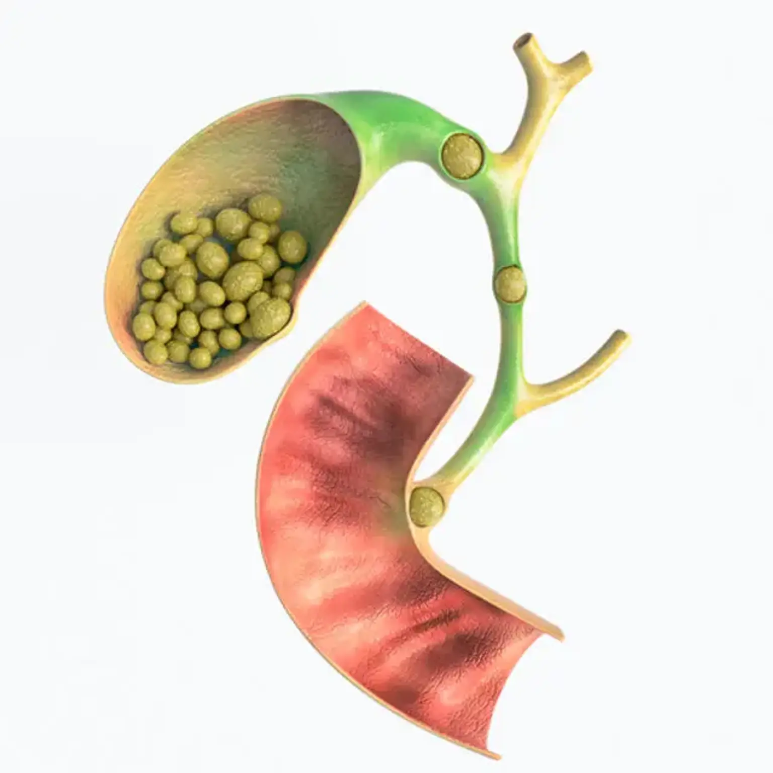
Cholelithiasis is characterized by the presence of gallstones, which are concretions formed in the biliary system, most often in the gallbladder. The existence of gallbladder stones is not considered a disease until they induce symptoms. Gallstones are tiny stones that occur in the gallbladder and are generally formed of cholesterol. Most of the time, they do not create any symptoms and do not require treatment.
The gallbladder
The gallbladder is a tiny pouch-like organ that is located underneath the liver. Its primary function is to store and concentrate bile. Bile is a liquid generated by the liver to aid in fat digestion. It travels from the liver to the gallbladder via a network of channels known as bile ducts.
Bile is stored in the gallbladder and grows more concentrated over time, making it better at digesting fats. When bile is required, the gallbladder discharges it into the digestive system.
Gallstone disease — The term gallstone disease refers to gallstones that cause symptoms.
Uncomplicated gallstone disease — Biliary colic in the absence of gallstone-related consequences is referred to as simple gallstone disease.
Epidemiology
Cholelithiasis is rather prevalent, affecting roughly 6% of men and 9% of women. Native American populations had the greatest rate of cholelithiasis. Gallstones are uncommon in Africa and Asia. Obesity is thought to have exacerbated the rise in gallstones.
Regardless matter how common gallstones are, more than 80% of patients are asymptomatic. Biliary discomfort, on the other hand, will develop in 1% to 2% of previously asymptomatic people each year. Major problems (cholecystitis, choledocholithiasis, gallstone pancreatitis, cholangitis) occur at a rate of 0.3 % per year in those who began to develop symptoms.
Cholecystolithiasis causes
There are three main pathways in the formation of gallstones:
- Supersaturation of cholesterol: Normally, bile can dissolve the quantity of cholesterol discharged by the liver. However, if the liver generates more cholesterol than bile can breakdown, the extra cholesterol may crystallize. Gallbladder sludge is formed when crystals become caught in gallbladder mucus. With time, the crystals may expand to form stones and obstruct the ducts, resulting in gallstone disease.
- Bilirubin excess: Bilirubin, a yellow pigment generated from red blood cell disintegration, is released into bile by liver cells. Certain hematologic disorders lead the liver to produce an excessive amount of bilirubin during the breakdown of hemoglobin. Excess bilirubin may also result in gallstone development.
- Gallbladder hypomotility or reduced contractility: Bile can get concentrated and produce gallstones if the gallbladder does not drain properly.
Gallstones are composed differently depending on their cause. Cholesterol gallstones, black pigment gallstones, and brown pigment gallstones are the three most prevalent forms. Gallstones made up of cholesterol account for 90% of all cases.
Each stone has its own set of dangers. Obesity, age, female gender, pregnancy, heredity, complete parenteral feeding, fast weight loss, and certain drugs are all risk factors for the formation of cholesterol gallstones (oral contraceptives, clofibrate, and somatostatin analogs).
Black and brown pigment stones account for around 2% of all gallstones. These can be detected in people who have a high hemoglobin turnover. The pigment is largely made up of bilirubin. Black pigment stones are common in patients with cirrhosis, ileal disorders, sickle cell anemia, and cystic fibrosis. Brown pigments are found mostly in Southeast Asian populations and are uncommon in the United States. Intraductal stasis and persistent bile bacterial colonization are risk factors for brown pigment stones.
Patients with Crohn's disease and ileum illness (or resection) are unable to reabsorb bile salts, increasing their risk of gallstones.
Pathophysiology
Cholesterol gallstones arise primarily as a result of excessive cholesterol release by liver cells and hypomotility or delayed gallbladder emptying. Bilirubin concentrations in bile may be greater than usual in pigmented gallstones due to excessive heme turnover. Bilirubin can then crystallize and create stones.
Cholelithiasis symptoms and problems occur when stones restrict the cystic duct, bile ducts, or both. Temporary blockage of the cystic duct (as when a stone lodges in the duct before it dilates and the stone returns to the gallbladder) causes biliary discomfort but is typically temporary. This is referred to as cholelithiasis. More prolonged cystic duct blockage (as when a big stone becomes permanently lodged in the neck of the gallbladder) might result in acute cholecystitis. A gallstone can pass through the cystic duct and get stuck and impacted in the common bile duct, causing blockage and jaundice. Choledocholithiasis is the medical term for this problem.
If gallstones pass through the cystic duct, common bile duct, and become dislodged at the ampulla of the distal portion of the bile duct, acute gallstone pancreatitis can occur as a result of fluid backing up and increased pressure in pancreatic ducts, as well as in situ activation of pancreatic enzymes. Large gallstones can perforate the gallbladder wall and form a fistula between the gallbladder and the small or large bowel, resulting in intestinal blockage or ileus.
Gallbladder stones symptoms
Patients with gallstone disease typically present with symptoms of biliary colic (intermittent episodes of constant, sharp, right upper quadrant (RUQ) abdominal pain often associated with nausea and vomiting), normal physical examination findings, and normal laboratory test results. It may be accompanied by diaphoresis, nausea, and vomiting.
- Biliary colic is typically caused by the gallbladder contracting in response to some form of stimulation, allowing a stone to pass through the gallbladder and into the cystic duct opening, resulting in increased gallbladder wall tension and pressure, which frequently results in pain known as biliary colic. The stones frequently slide back into the gallbladder when the gallbladder relaxes, and the discomfort usually goes away within 30 to 90 minutes.
- Fatty foods are a frequent cause of gallbladder constriction. The discomfort generally begins an hour after a fatty meal and is reported as strong and dull, lasting 1 to 5 hours. However, the relationship with meals is not ubiquitous, and a considerable number of individuals have discomfort at night. The frequency of recurring episodes varies, although most individuals do not have symptoms on a daily basis.
- A comprehensive physical exam can help differentiate between biliary discomfort caused by acute cholecystitis, simple cholelithiasis, and other problems.
- The patient is afebrile and has a basically benign abdominal examination with no rebound or guarding in simple biliary colic.
Acute cholecystitis occurs when a persistent stone lodges in the cystic duct, causing the gallbladder to swell and become inflamed. Fever, discomfort in the right upper quadrant, and soreness above the gallbladder may also be present.
When fever, prolonged tachycardia, hypotension, or jaundice are present, it is necessary to look for cholelithiasis complications such as cholecystitis, cholangitis, pancreatitis, or other systemic causes. Choledocholithiasis is a complication of gallstones that occurs when stones plug the common bile duct, preventing bile from flowing from the liver to the gut. Pressure increases, causing an increase in liver enzymes and jaundice.
Cholangitis is caused by bacterial colonization and overgrowth in stagnant bile above an obstructing common duct stone. Purulent inflammation of the liver and biliary tree results. Charcot's triad is characterized by significant RUQ discomfort, fever, and jaundice, and is typical with cholangitis. To treat this disease, surgical removal of the stone blockage along with intravenous antibiotics is necessary.
Gallbladder stones diagnosis
CBC, CMP, PT/PTT, lipase, amylase, Alk Phos, total bilirubin, and urine analysis are common first labs used to examine gallstones.
Ultrasound is still the best imaging technique for diagnosing gallstones. According to a systematic study, the sensitivity was 84 percent and the specificity was 99 percent, which was higher than that of other modalities. Biliary disease can be detected using either a radiology ultrasound scan or a point-of-care ultrasound. Several studies in the literature have indicated that doctors' use of point-of-care ultrasonography to diagnose or rule out biliary illness is accurate and trustworthy.
Gallstones show as hyperechoic masses inside the gallbladder with distal acoustic shadowing on ultrasonography. Sludge in the gallbladder may be observed, as well as hyperechoic layers within the gallbladder. Unlike stones, sludge does not cast acoustic shadows. If the following additional symptoms are observed, acute cholecystitis should be suspected:
- Thickened anterior gallbladder wall (greater than 3 mm),
- The presence of pericholecystic fluid or
- Positive sonographic Murphy's sign.
Furthermore, ultrasonography can be used to get common bile duct (CBD) measures, which, if elevated, can indicate choledocholithiasis. In patients under the age of 40, the usual CBD range is four millimeters, with a further one millimeter permitted for each consecutive decade of life. Because the common duct becomes the bile reservoir after the gallbladder is removed, post-Cholecystectomy patients are permitted up to 10 mm diameter.
If an ultrasound study is inconclusive in excluding acute cholecystitis, a nuclear medicine cholescintigraphy scan, commonly known as a HIDA scan, can be conducted. A radioactive tracer injected into a peripheral vein is transported to the liver, where it enters the biliary network and is taken up by the gallbladder within 4 hours in a typical healthy gallbladder. A sick gallbladder with a blocked cystic duct prevents the tracer from accessing the gallbladder. HIDA scan has a sensitivity of up to 97 percent and specificity of 94 percent for the diagnosis of acute cholecystitis.
Abdominal CT imaging does not improve sensitivity or specificity in identifying gallstones or cholecystitis. It can identify pancreatic inflammation and problems, as well as determine whether CBD dilatation is present (masses, pseudo-cysts, necrotizing features). If RUQ ultrasound excludes biliary illness and alternative reasons of stomach discomfort are being explored, CT imaging might be helpful.
Furthermore, endoscopic or magnetic retrograde cholangiopancreatography (ERCP/MRCP) is occasionally beneficial in working up individuals with jaundice and dilated CBD or suspected cholangitis, although it is generally acquired after an ultrasound. ERCP is an invasive test that uses contrast dye, but it also has the advantage of allowing intervention if pathology is discovered (e.g., stenting, stone extraction, biopsy). MRCP, on the other hand, is non-invasive and does not necessitate the use of contrast dye.
Treatment for cholecystolithiasis
Gallstone management is separated into two categories: asymptomatic gallstones and symptomatic gallstones.
Asymptomatic gallstones necessitate patient counseling on biliary colic symptoms and when to seek medical assistance. Once the diagnosis has been established and alternate diseases have been ruled out, cholelithiasis can be treated promptly in the emergency room or urgent care center with oral or parenteral analgesics. Patients should also be given nutritional guidance to lessen the likelihood of recurring episodes, and they should be sent to a general surgeon for elective laparoscopic cholecystectomy. Laparoscopic cholecystectomy is now the standard of treatment, and the majority of patients are handled as outpatients.
Patients presenting with symptoms and a workup consistent with acute cholecystitis will require hospitalization, a surgical consult, and intravenous antibiotics. Patients with choledocholithiasis or gallstone pancreatitis will additionally require hospitalization, GI consultation, and an ERCP or MRCP. Patients with acute ascending cholangitis are often sickly and septic. They frequently require extensive resuscitation and ICU-level care, as well as surgical surgery to empty a biliary tract infection.
Ursodeoxycholic acid medical therapy is a possibility, however it is impractical. The patient must have stones that are smaller than 1 cm in diameter and have a high cholesterol level. However, only 50% of the time does the therapy take 9-12 months to remove the stone. Another treatment option for non-calcified gallstones is extracorporeal shockwave lithotripsy.
Differential Diagnosis
- Bile Duct Strictures
- Bile Duct Tumors
- Diabetic ketoacidosis
- Emergent Treatment of Gastroenteritis
- Esophageal spasm
- Hepatitis
- Irritable bowel syndrome
- Pancreatitis (acute or chronic)
- Peptic Ulcer Disease
Prognosis
Only around half of people with gallstones develop symptoms. An elective cholecystectomy has a 0.5 percent death rate and a morbidity rate of less than 10%. The mortality rate for an emergency cholecystectomy is 3% to 5%, with morbidity ranging from 30% to 50%.
Stones in the bile duct may return after cholecystectomy. Separately, single-incisional laparoscopic cholecystectomy appeared to be related with an 8% incisional hernia rate, with age (50 years) and body mass index (BMI) (30 kg/m2) acting as independent predictors.
An related choledocholithiasis affects 10% to 15% of individuals. The prognosis of choledocholithiasis patients is determined by the existence and severity of sequelae. 45 percent of patients who refuse or are unable to undergo surgery remain asymptomatic from choledocholithiasis, while 55 percent experience varying degrees of complications.
Complications
- Pancreatitis
- Bile duct stones
- Acute cholecystitis
- Gallbladder empyema, necrosis
- Gallbladder cancer
- Cholecystoenteric fistula
Seek care right away for a gallbladder attack
See a doctor right away if you are having these symptoms during or after a gallbladder attack:
- Pain in your abdomen lasting several hours
- Nausea and vomiting
- Fever—even a low-grade fever—or chills
- Yellowish color of your skin or whites of your eyes, called jaundice
- Tea-colored urine and light-colored stools
These symptoms might indicate a severe infection or inflammation of the gallbladder, liver, or pancreas. Gallstone symptoms may be similar to those of other disorders such as appendicitis, ulcers, pancreatitis, and gastroesophageal reflux disease, all of which should be treated as soon as possible by a specialist. If your bile ducts remain clogged, you may develop gallstone issues. Blockages in the bile ducts or pancreatic ducts can be lethal if left untreated.
Prevention of Gallstones
The use of ursodeoxycholic acid can help to prevent the development of gallstones. This has been shown in the context of fast weight reduction produced by very low-calorie diets or bariatric surgery, both of which are linked with an increased incidence of developing cholesterol gallstones (20 -30 % within 4 mo). In this situation, using 600 mg of ursodeoxycholic acid daily for 16 weeks reduced the risk of gallstones by 80%.
It is wise to recommend dietary adjustments that reduce fat consumption; this may reduce the frequency of biliary colic events. However, it has not been demonstrated that it causes stone disintegration.
- Diet and Activity
There is no evidence that food composition influences the natural course of gallstone disease in humans. Obese individuals who embark on severe weight-loss programs or have bariatric surgery are at risk of developing gallstones. Consumption of coffee appears to be linked to a lower incidence of gallstone disease. Exercise on a regular basis may help to lessen the frequency of cholecystectomy.
Long-Term Monitoring
About 5% To 10% of individuals have persistent diarrhea after cholecystectomy. This is mainly related to bile salts. When the gallbladder is removed, the frequency of enterohepatic circulation of bile salts rises, resulting in more bile salt reaching the colon. Bile salts increase the mucosal production of salt and water in the colon.
Postcholecystectomy diarrhea is generally moderate and can be controlled with over-the-counter antidiarrheal medications such as loperamide. More frequent diarrhea can be controlled by taking a bile acid-binding resin on a regular basis (eg, colestipol, cholestyramine, colesevelam).
Following cholecystectomy, a few people report recurring discomfort similar to biliary colic. This illness is sometimes referred to as postcholecystectomy syndrome.
Many patients with postcholecystectomy syndrome experience long-term functional discomfort that was initially misdiagnosed as biliary in nature. It is not uncommon if symptoms persist after cholecystectomy. Diagnostic and treatment efforts should be focused on determining the root cause.
Some people with postcholecystectomy syndrome have an underlying sphincter of Oddi motility condition called biliary dyskinesia, in which the sphincter fails to relax correctly after eating. Endoscopic biliary manometry can be used to make the diagnosis at specialist centers. Endoscopic retrograde sphincterotomy is typically helpful in treating established instances of biliary dyskinesia.
Conclusion
Gallstone disease is defined as the presence of stones in the gallbladder or common bile duct, as well as the symptoms and consequences that these stones produce. Gallstone disease symptoms can range from moderate, non-specific symptoms that can be difficult to identify to severe pain and/or problems that are frequently identified as gallstone disease by health experts. The sickness is discovered by chance as a consequence of tests for other disorders.
There is disagreement over the best technique to treat gallstone disease. There are several endoscopic, surgical, and medicinal therapies available, however it is unknown which ones are best for certain people. There is also debate over the timing of cholecystectomy, and whether it should be performed during the acute presentation of the illness or after the acute symptoms have passed.