Coronary Angioplasty

    Last updated date: 11-Feb-2023

    Originally Written in English

    Coronary Angioplasty

     Coronary Angioplasty

    Overview

    Coronary heart disease (CHD) is common among the elderly globally. 15.5 million individuals in the United States have CHD According to the American Heart Association. It is a substantial cause of mortality and morbidity in industrialized nations, accounting for almost one-third of all deaths in persons over the age of 35.

    Because to prompt percutaneous coronary intervention with stenting, CHD mortality has gradually decreased over the previous few decades. As a result, angioplasty is a game changer in terms of lowering morbidity and mortality. For acute myocardial infarction, angioplasty is the preferred therapy. There are two primary catheterization approaches: traditional and transfemoral.
     

    What is Coronary Angioplasty?

    Coronary Angioplasty definition

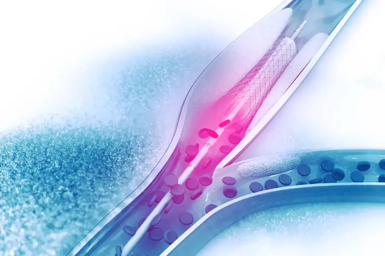
    Angioplasty, with or without stenting, is a nonsurgical surgery for opening blocked or narrowed coronary arteries caused by underlying atherosclerosis. The treatment entails inserting an inflated balloon-tipped catheter through the skin in the extremities and inflating the balloon once it has passed through the stenosed artery site. It forces atherosclerotic intraluminal plaque against the artery wall, restoring luminal diameter. As a result, it restores normal blood flow to the myocardium. Percutaneous coronary intervention (PCI), formerly known as angioplasty with stent, is the preferred therapy for unstable angina, myocardial infarction, and spontaneous coronary artery perforation.

    With the debut of "plain old balloon angioplasty" (POBA) without stenting 40 years ago, the PCI idea was launched. POBA usage was limited in the mid-1980s due to an early problem of vascular recoil property and restenosis after balloon deflation, which led to the development of bare metal stents (BMS).

    Professionals employ a tube-like metallic meshwork during the surgery, and its scaffolding features resist vascular recoil, preventing early POBA restenosis owing to vascular recoil. Long-term, in situ BMS, on the other hand, can cause wall stress, endothelial discontinuity, and permanent presence of the metallic foreign body in arteries, causing inflammation with fibrin deposition and promoting myofibroblast migration, resulting in in-stent restenosis (IRS) due to a neointimal hyperplasia mechanism.

    This problem prompted the invention of drug-eluting stents (DES). When compared to BMS, DES technology employs an antiproliferative drug coating on top of the metallic framework of stents, resulting in reduced neointimal hyperplasia and stent restenosis. Late stent thrombosis is also related with DES as a result of delayed arterial repair with a lack of re-endothelialization and fibrin deposition owing to underlying chronic inflammation, which occurs more frequently in first-generation DES. Second-generation DES contains an additional biocompatible polymer layer for improved endothelial repair.

    cobalt-chromium everolimus-eluting stents (second-generation DES) are safer than paclitaxel-eluting stents (first-generation DES) and BMS due to improved vascular healing and re-endothelialization of stent struts. Recent research indicates that second-generation DES with biodegradable polymer coatings are more effective than BMS in lowering target-vessel revascularization (TVR), target-lesion revascularization (TLR), in-stent late loss (ISLL), and late-stent thrombosis. DES was also found to be more effective in complicated lesions than BMS in studies.

    Due to the absence of metallic meshwork in stent platforms, which serves as a triggering agent for late-onset problems such as IRS and stent thrombosis, the newest revolutionary agent bioresorbable scaffolds system (BRS) preserves cyclic pulsatility with lower possibilities of vascular remodeling and IRS. However, optimum implantation procedures and strut size are required for BRS. The constraint of BRS is strut thickness because restenosis in the early post-procedural period is due to vascular rebound, which is counteracted by a metallic scaffold of BMS and DES. If the size of the BRS struts is lowered, vascular recoil cannot be appropriately mitigated.

    This attribute has been attained in some way by second-generation BRS. BRS eventually vanishes owing to resorption, which may be monitored using intravascular ultrasonography (IVUS). IVUS and optical coherence tomography (OCT) can be utilized to properly implant BRS. Although there is little evidence on the safety of BRS, the notion of the metal-free stent that helped design BRS has been challenged since scaffold thrombosis has been documented. Brown et al. recently proposed that following BRS implantation, both pre- and post-dilatation with pressures greater than 20 ATM are required for minimizing acute vascular recoil, improved scaffold expansion, and reduced rates of scaffold thrombosis, which is best predicted by minimum luminal area on IVUS.


    Who Needs Coronary Angioplasty?

     coronary artery disease

    Coronary angioplasty is performed to restore blood flow to the heart when the coronary arteries have constricted or become obstructed as a result of coronary artery disease (CAD). When medications and lifestyle modifications, like as eating a balanced diet, stopping smoking, and increasing physical exercise, do not help your CAD symptoms, your doctor will discuss alternative treatment options with you. Angioplasty and coronary artery bypass grafting (CABG), a form of open-heart surgery, are two of these alternatives.

    When selecting the optimal surgery for you, your doctor will consider a variety of criteria. These considerations include the severity of your blockages, their location, and any other ailments you may have.

    When there is less severe narrowing or blockage in your arteries and the obstruction can be accessed during the treatment, angioplasty is frequently employed. CABG may be recommended if you have significant heart disease, numerous blocked arteries, diabetes, or heart failure.

    Compared with CABG, some advantages of angioplasty are that it:

    • Has fewer risks than CABG
    • Isn't surgery, so it won't require a large cut
    • Is done with medicines that numb you and help you relax. Unlike CABG, you won't be put to sleep for a short time
    • Has a shorter recovery time

    Angioplasty is also done as an emergency operation in the event of a heart attack. Plaque can rupture in the coronary arteries, causing a blood clot to develop on its surface. If the clot becomes large enough, it can fully or partially restrict blood flow to a portion of the heart muscle.

    Opening a blockage as soon as possible reduces the damage to the heart during a heart attack and restores blood flow to the heart muscle. Angioplasty can open the artery fast and is the best option during a heart attack.

    An benefit of angioplasty over CABG is that the artery may re-narrow over time. When stents, particularly medicine-coated stents, are utilized, the likelihood of this occurring is reduced. These stents, however, are not without danger. Blood clots can develop in the medicine-coated stents and trigger a heart attack in some situations. Your doctor will go through your treatment choices with you and determine which surgery is best for you.


    How do I prepare for a coronary angioplasty?

    coronary angioplasty preparation

    It is commonly acknowledged that the metallic surface of a stent is thrombogenic; as a result, a prominent feared consequence is acute vascular closure due to atheroma rupture, platelet activation, and tissue factor release during and after angioplasty. To avoid acute stent thrombosis, PCI should be performed under anticoagulation (AC), with a balanced risk of thrombosis and access site hemorrhage complication.

    Anticoagulants include heparin (low molecular weight heparin (LMWH) or unfractionated heparin), bivalirudin, P2Y12 blockers, direct thrombin inhibitors, and glycoprotein IIb/IIIa inhibitors. Bivalirudin, on the other hand, is related with a lower risk of access site bleeding complications, thrombocytopenia, and mortality than heparin, but studies reveal that it is associated with a slightly higher risk of acute stent thrombosis than heparin. Despite this, heparin can occasionally produce Heparin-induced thrombocytopenia (HIT).

    Bivalirudin should be used for AC in patients who have previously experienced HIT. To control periprocedural heparin usage, activated clotting time is employed. The thigh or wrist region is shaved with informed permission. To sedate the patient before to surgery, propofol is administered intravenously. Because midazolam has been linked to respiratory depression, propofol is preferable. The artery is perforated using the Saldinger procedure, and the 5F sheath is inserted. Coronaries are catheterized using dye under fluoroscopic supervision.

    A more complicated lesion may be identified in 5% to 10% of instances. These lesions may be very lengthy, chronic with complete blockage and calcification, non-dilatable, or have anatomical changes such as being placed at a bifurcation or an ostium. These need lesion pretreatment prior to stent insertion. The purpose of lesion preparation is to improve stent delivery and expansion while lowering the danger of distant plaque embolization. To achieve this purpose, several techniques are available, including directional or rotational coronary atherectomy, cutting balloon, FX miniRAIL catheter, and arthroplasty for highly calcified plaques. In these circumstances, OCT guidance is required to precisely place a stent.


    How is coronary angioplasty done?

    Coronary Angioplasty Procedure

    In a catheterization room, you recline on a couch. Above the couch is an X-ray machine. A thin, flexible 'guide' tube (catheter) is placed into a blood artery in the groin or arm by a broad needle or tiny puncture in the skin. A local anaesthetic is injected into the skin above the blood vessel to provide pain relief. As a result, inserting the catheter into the blood vessel should be painless. The catheter is carefully pushed up the blood artery towards the heart by the doctor. Low-dose X-rays are utilized to monitor the catheter tip's progress while it is gently pushed into the proper position. The catheter's progress may be seen on the X-ray display.

    The catheter tip is inserted into a heart (coronary) artery and advanced to a constricted segment caused by fatty patches, or 'plaques' (atheroma). The 'guide' catheter is then passed through with a second, thinner 'balloon catheter.' At the tip of the balloon catheter, there is a balloon and a tiny tube (a stent). For 30-60 seconds, the balloon is inflated. This compresses the atheroma and expands the constricted artery. When the balloon is inflated, the blood flow is stopped. As a result, you may have angina-like pain for a brief period of time. However, this quickly fades as the balloon is deflated.

    In most cases, a stent is left in the enlarged area. The stent is a wire mesh tube that provides support to the artery and aids in keeping the artery open. The 'collapsed' stent covers the balloon and opens when it is inflated. Some stents include a chemical coating that helps to keep the artery from being clogged again. People who have a nickel allergy may require a nickel-free stent. The treatment might be repeated for one or more more constricted portions of the coronary arteries.

    The catheter cannot be felt inside the blood vessels. During the operation, you may notice an odd missing or additional pulse. This is natural and unimportant. During the process, electrodes put on your chest detect your heartbeat and generate a trace on an electrocardiograph (ECG) equipment. If you are nervous, you may be given a sedative before the test.


    Transradial Approach

    Because the radial artery is fairly superficial, it is readily punctured, and bleeding is controlled by manual compression. There are no important nerves or veins in the vicinity. As a result, there is a low risk of neurovascular injury. However, because the radial artery has a relatively tiny diameter, small size catheters are necessary. Transradial approaches are less expensive than transfemoral approaches and are linked with earlier hospital discharge. With the progress of interventional cardiology hardware, the transradial technique has emerged as a viable alternative to the traditional transfemoral route.

    Because of the strong collateral blood supply of the hand by ulnar artery via palmer arch, the transradial route is linked with a low risk of access site hemorrhage or hematoma development, pseudoaneurysm formation, morbidity and mortality, and a decreased risk of hand ischemia. Allen's test or a pulse oximetry examination can be used to evaluate palmar arches. Transradial approaches are linked with increased radiation exposure, longer operation length, anatomical variances leading to catheterization failure, and radial artery spasm, which can be treated with local injection of vasodilatory medicines such as nitrates and calcium channel blockers.


    Transfemoral Approach

    Transfemoral Approach

    The more traditional technique is the transfemoral approach, which is linked with easier access, shorter radiation duration, and less contrast usage. Access site issues, on the other hand, are more prevalent, particularly in obese individuals. Access site hemorrhage, hematoma, significant retroperitoneal bleeding necessitating a blood transfusion, arteriovenous fistula development and pseudoaneurysm, and neurovascular injury are among the complications. Because the femoral artery is the primary route of blood to the leg, there is a higher risk of ischemia than with the transradial technique.


    Leaving hospital after an angioplasty

    after Coronary Angioplasty

    Most individuals can go home the same day or the following day, but if you had an emergency angioplasty, you may need to stay in the hospital for a little longer.

    Check the region where the catheter was placed when you return home. Expect some bruising and pain, but call your doctor if you see any redness or swelling, or if the bruising increases.

    Someone will talk to you about your recuperation and what you can and cannot do before you leave the hospital. It's common to feel fatigued afterward, but most individuals return to normal after a few days. However, if you've also suffered a heart attack, your recovery will be slower.

    • It’s best to avoid doing any demanding activities, such as heavy lifting, for a week or so.
    • You shouldn’t drive for at least a week after having angioplasty – longer if you also had a heart attack.
    • If you’ve had a planned angioplasty with no complications you may be able to return to work within a few days, depending on the type of work you do.
    • If you’ve had an emergency angioplasty or a heart attack you may need to take a few weeks off.

    You should also be encouraged to participate in a cardiac rehabilitation program, which includes exercise and educational sessions designed to help you recover as rapidly as possible. To assist lower the danger of blood clots developing in and around the stent, you'll need to take anti-platelet medications such as aspirin (unless you're allergic to it) or other drugs such as clopidogrel, ticagrelor, or prasugrel if you have a stent. Airport security systems and MRI scans have no effect on stents.


    Outcome

    Coronary Angioplasty Outcome

    The treatment normally takes around 30 minutes if only one portion of the artery is enlarged. The technique takes longer if numerous parts have to be expanded. Following the surgery, you may be required to stay in the hospital overnight for observation.

    More than 9 out of 10 operations are effective in treating angina. Coronary angioplasty, on the other hand, cannot be utilized for everyone who has angina. This is due to an excess of constricted portions in the heart (coronary) arteries in many situations. Alternatively, the constricted parts are too lengthy, too thin, or too far down a coronary artery or branch artery for this treatment.


    Cardiac Rehabilitation

    Your doctor may recommend that you participate in a cardiac rehabilitation (rehab) program. Cardiac rehabilitation enables persons with heart disease to heal more quickly and return to work or regular activities. Cardiac rehab consists of supervised physical exercise, instruction on heart healthy living, and counseling to reduce stress and assist you in returning to an active lifestyle. Your doctor can direct you to a cardiac rehab program near you.


    Complications

    Coronary Angioplasty Complications

    Iatrogenic coronary artery perforation (CAP) owing to an underlying complicated lesion is an uncommon but significant complication after angioplasty, occurring in 0.1% to 0.8% of all patients undergoing angioplasty. CAP can occur as a result of an angioplasty guide wire perforation, balloon oversizing, or the use of atherectomy devices. The severity of the lesion, hemodynamic condition, and Ellis class type of CAP all influence CAP management. Class 1 is often benign, however class 3 is linked with an increased risk of cardiac tamponade and the need for emergency heart surgery.

    Mild CAP can be treated with anticoagulation reversal (protamine sulfate if heparin is being used), extended balloon inflation, polytetrafluoroethylene-covered stents (CS), and trans-catheter embolization using autologous fat particles. Stent thrombosis is a risk with CS usage, and a few cases of coronary arteriovenous fistula have been recorded as a result of CS failure. ST-segment elevation myocardial infarction and early or delayed cardiac tamponade with or without hemodynamic instability are CAP complications that may necessitate emergency pericardiocentesis.

    In-stent restenosis (ISR) is described as a decrease in vascular luminal diameter following percutaneous intervention (PCI). The pathophysiology of ISR is determined by the kind of stent utilized during PCI. POBA has an abrupt start due to elastic recoil and vascular remodeling. Neointimal Hyperplasia is a distinctive feature of BMS. Late stent thrombosis in DES is caused by a number of underlying pathogenic factors, including reduced vascular re-endothelialization, polymer coating hypersensitivity, and increased fibrin deposition as a result of metal generating chronic inflammation.

    Patients with unstable angina or acute coronary syndrome who had PCI are more likely to develop ISR owing to chronic inflammation, as indicated by greater C-reactive protein levels (CRP) in these individuals compared to patients with stable angina who had PCI. Stent fracture (SF) is a seldom documented adverse effect of DES administration during PCI that can occur either periprocedurally or after drug elution has been completed. The development of ISR and stent thrombosis has also been associated to stent breakage. ISR can be caused by neoatherosclerosis regardless of the kind of intervention used during PCI.

    All of the above-mentioned causes of ISR produce angina symptoms or acute coronary syndrome owing to decreased blood supply to the myocardium and may necessitate reintervention such as coronary artery bypass graft surgery or re-PCI. This type of reintervention is known as target lesion revascularization.


    Conclusion 

    Coronary angioplasty is a procedure used to expand constricted parts of the coronary arteries in the heart. Percutaneous coronary intervention is another name for it (PCI). It does not need extensive heart surgery, but rather the insertion of a thin, flexible tube (a catheter) into coronary arteries via massive blood channels