Double valve replacement

Last updated date: 04-Mar-2023

Originally Written in English

Double Valve Replacement

Overview

Heart valve disease can be severe, resulting in double (mitral-aortic, mitral-tricuspid, or mitral-aortic-tricuspid) or triple (mitral, aortic, and tricuspid) valvular regurgitation. The surgical treatment of severe valvular regurgitation often consists of repairing or replacing all valves damaged by a pathologic condition.
 

What is Heart Valve Surgery?

Heart Valve Surgery

Heart valve surgeries and treatments are conducted to repair or replace a heart valve that is not functioning correctly due to valvular heart disease (also called heart valve disease). Heart valve surgery is an open-heart procedure that is performed via the breastbone into the chest. It is a big procedure that can take two hours or more to complete, and recuperation can take several weeks. There are newer, less invasive techniques available for specific forms of valvular heart disease, but these are only available at a few facilities.


Why is it done?

In a healthy heart, valves guide the flow of blood in one direction through the heart and the body. If a valve fails to function properly, blood flow and the delicate network of blood arteries that transport oxygen throughout the body suffer.

If your valve problem is modest, your doctor may keep an eye on your symptoms or prescribe medication. If your problem is more severe, surgery to repair or replace the valve is typically necessary to prevent long-term damage to your heart valve and heart.


What is done?

Depending on the problem, there are a several different procedures for repairing or replacing valves.

  1. Surgical valve repair

Surgical techniques are often utilized to treat mitral or tricuspid valve abnormalities.

  • Commissurotomy is a procedure used to treat a clogged valve. The valve flaps (leaflets) are removed to slightly relax the valve, enabling blood to flow freely.
  • Annuloplasty is performed to repair a leaking valve. The annulus is a ring of fibrous tissue at the base of the heart valve. Sutures are sewed around the ring to close the opening in an expanded annulus. Alternatively, a ring-like device is placed to the exterior of the valve opening to support the valve and allow it to close more firmly.
  • Valvulotomy is a surgical operation used to widen constricted heart valves. It is also possible to use a balloon to do this.


     2. Non-surgical valve repair

Percutaneous or catheter-based operations are performed without the need for any incisions in the chest or the halting of the heart. Instead, a catheter is put into a blood vessel in your groin or arm and then threaded through the blood vessels into your heart.

  • Percutaneous or balloon valvuloplasty/valvotomy is performed to treat stiffened or narrowed (stenosed) pulmonary, mitral, or aortic valves. The catheter's balloon tip is inserted into the constricted valve and inflated to expand the aperture.
  • Percutaneous mitral valve repair procedures, such as edge-to-edge repair, can repair a leaking mitral valve in a patient who is deemed high risk for surgery. A catheter with a clip is placed into the groin and up into the left side of the heart. The open clip is positioned beyond the leaking valve and then pushed back to catch the mitral valve flaps (leaflets). When closed, the clip binds the leaflets together and prevents the valve from leaking.


    3. Heart valve replacement

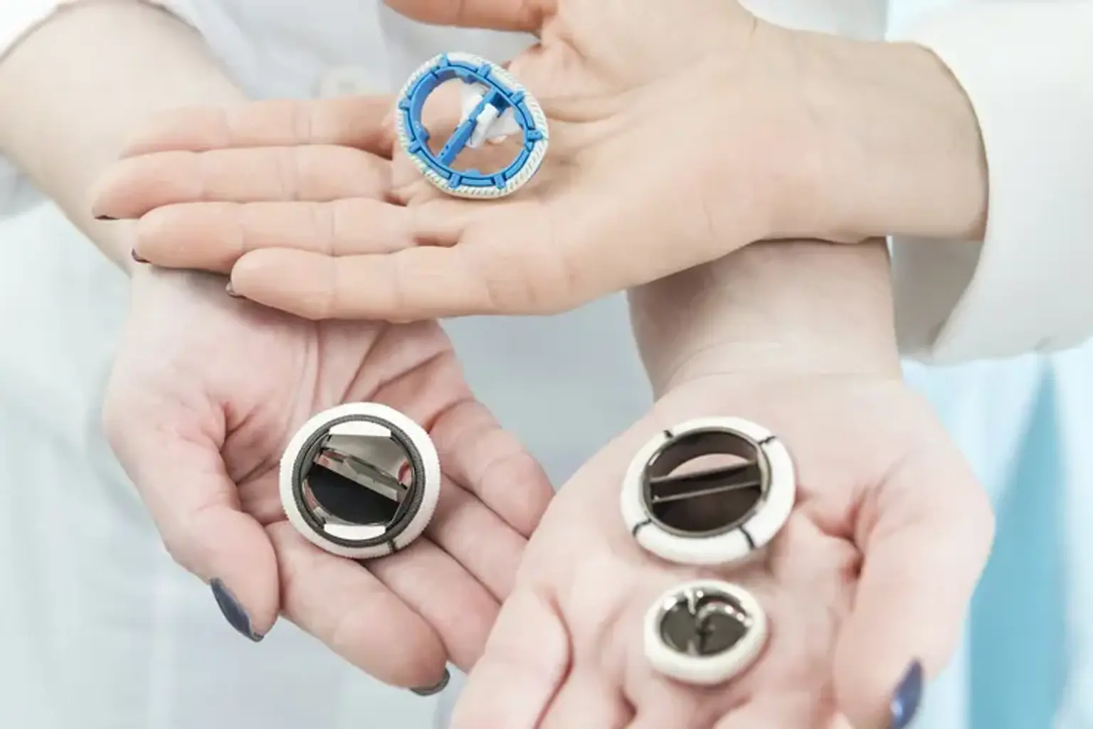
If your heart valve is too seriously damaged to heal, surgery to replace it with a new mechanical or biological valve may be required. In most cases, age plays a role in determining which kind to utilize; biological valves are often chosen for elderly persons. You and your doctor will talk about your options and determine which is best for you and your situation.

  • Mechanical valves composed of long-lasting metals, carbon, ceramics, and plastic were the first to be employed in valve replacement surgery. They have been modified and enhanced since their introduction in the 1960s.
    • The major advantage of a mechanical valve is durability – they can last a long time.
    • A fabric ring is used to sew the valve to heart tissue. 
    • Mechanical valves can produce blood clots, which can lead to a heart attack or stroke. People with mechanical valves must take blood-thinning drugs (anticoagulants) every day for the remainder of their lives to avoid clots. This has ramifications for women of reproductive age and anyone with a history of heavy bleeding. Depending on the type of blood thinners you are taking, you may need to have routine blood tests to check your INR (international normalized ratio), which measures the likelihood of your blood to clot.
    • A mechanical valve makes a soft clicking sound when it closes. This can bother some people.


  • Biological (also called bioprosthetic) or tissue valves are specially prepared natural valves that come from human donors or animals.
  • Animal source valves (a xenograph) derived from cows or pigs are similar to human heart valves. They are well tolerated and less prone to cause blood clots than mechanical valves.
  • Human heart valves derived from a donated heart (allografts or homograft) are well tolerated and endure longer than animal valves. Human valves are seldom used.
  • Valves can be produced from your own tissue (an autograft). Your working pulmonary valve is utilized to replace your damaged aortic valve in a Ross (or switch) surgery. After that, your pulmonary valve is replaced with a donor one.
  • People with biological valves need to take blood thinners in the short term.
  • Biological valves are not as durable as mechanical valves. They are more durable in the aortic position and in older patients.


     4. Minimally invasive valve repair and replacement

Minimally invasive surgery, as opposed to traditional surgery, does not include cutting through the breastbone and opening the chest. It does not necessitate the stopping of your heart or the use of a heart-lung machine. The surgeon operates with long-handled surgical equipment placed through small incisions while watching your heart on a television screen. Robotic arms are employed in various circumstances. Although minimally invasive valve repair and replacement are appropriate for some kinds of valvular heart disease, they are only offered in a few institutions. It is also known as endoscopic or robotic cardiac surgery.

  • Transcatheter aortic valve implantation (TAVI) is also known as transcatheter aortic valve replacement (TAVR). TAVI is a minimally invasive surgical valve replacement treatment used to treat symptomatic aortic valve stenosis. It differs from typical valve replacement surgery in two important ways. TAVI is performed through minor incisions in the groin or chest rather than opening up the chest. Instead than mending or replacing the damaged aortic valve, a new one is implanted immediately on top of the old one.
  • Through tiny incisions in the groin or chest, the surgeon inserts a catheter holding a replacement, collapsible aortic valve.
  • The catheter is directed to the right spot in the heart using ultrasound and chest x-rays, and the new valve is inserted and inflated.
  • When the replacement valve is installed, it instantly begins to regulate blood flow.
  • TAVI patients recover faster and have shorter hospital stays (on average three to five days) than open-heart valve surgery patients.


Types of Valve Replacement Surgery

Types of Valve Replacement Surgery

Aortic Valve Replacement

Aortic Valve Replacement

The aortic valve is an outflow valve on the left side of the heart. Its function is to allow blood to exit the heart's primary pumping chamber, the left ventricle. Its role is also to keep blood from leaking back into the left ventricle. If you have a congenital defect or condition that causes stenosis or regurgitation, you may require aortic valve surgery.

A bicuspid valve is the most prevalent form of congenital defect. The aortic valve normally includes three portions of tissue known as leaflets. This is known as a tricuspid valve. Because a bicuspid valve has just two leaflets, it is faulty. According to a recent research, aortic valve replacement surgery has a 94 percent five-year survival rate. Survival rates are determined by:

  • Your age
  • Your overall health
  • Other medical conditions you have
  • Your heart function


Mitral Valve Replacement

Mitral Valve Replacement

The mitral valve is found on the heart's left side. It functions as an inflow valve. Its function is to enable blood to flow from the left atrium into the left ventricle. If the valve does not fully open or close, surgery may be necessary. When the valve is excessively thin, blood may have difficulty entering. This can cause it to back up, creating pulmonary hypertension. Blood might seep back into the lungs if the valve fails to shut correctly. This might be caused by a congenital abnormality, an infection, or a degenerative condition.

The faulty valve will be replaced with a metal artificial valve or a biological valve. The metal valve will last a lifetime, but it will necessitate the use of blood thinners. The biological valve will last between 15 and 20 years, and you will not be needed to take blood-thinning medication. The five-year survival rate is around 91%. The following factors also influence survival rates:

  • Your age
  • Your overall health
  • Other medical conditions you have
  • Your heart function

Ask your doctor to help assess your personal risks.


Double Valve Replacement

A double valve replacement involves replacing both the mitral and aortic valves, as well as the complete left side of the heart. This form of surgery is less prevalent than others and has a somewhat greater fatality rate.


Pulmonary Valve Replacement

The pulmonary valve is a valve that connects the pulmonary artery, which transports blood to the lungs for oxidation, with the right ventricle, one of the heart's chambers. Its primary function is to allow blood to pass from the heart to the lungs through the pulmonary artery. The most common reason for pulmonary valve replacement is stenosis, which inhibits blood flow. A congenital abnormality, illness, or carcinoid disease can all induce stenosis.


What is Double Valve Replacement?

Double Valve Replacement

A double valve replacement involves the replacement of both the mitral and aortic valves, or the entire left side of the heart. This type of operation is not as common as others, yet it is recommended in difficult cases.

The four coronary valves serve a critical part in the organ's functionality by controlling blood supply via the heart and blood avoidance. The heart has four chambers: two upper atria and two lower ventricles. There are two valves in each ventricle (lower heart chamber), a one-way entrance valve and a one-way exit valve. 

The tricuspid valve is the right ventricle's entrance valve that transports blood from the pulmonary arteries to the lungs. The mitral valve is the valve that connects the left ventricle to the left atrium; the aortic valve is the conduit that connects the aorta to the right atrium and collects oxygenated blood from the lung.

Heart valves are responsible for allowing new blood to enter the heart chambers. Following the introduction of blood flow, each valve should be completely closed. Heart valves that are damaged cannot always do their function effectively.


Preoperative preparation

Preoperative preparation

Anesthesia is administered in accordance with established protocol, and the procedure is carried out under general intravenous anesthesia. For patient monitoring, peripheral arterial and venous accesses are acquired. If an endoaortic balloon occlusion is planned, both right and left radial artery lines must be obtained to manage any potential balloon migration.

Two percutaneous sheath introducers are placed in the jugular vein: 

  • One (standard 4-lumen-7.0 or 8.5 Fr) is used for drug administration and central venous pressure monitoring, and 
  • The other one (8.5 Fr) for eventual placement of endocavitary pacemaker leads. 

In most cases, a single lumen tube is utilized for intubation. A double-lumen endotracheal tube may be useful when a lengthy surgical preparation (difficult cannulation, pleural adhesions) is anticipated, as well as in redo-thoracotomy operations. A transesophageal echocardiography (TEE) probe and two defibrillator pads are inserted across the chest wall. The patient is positioned supine with an air sac beneath the right scapula to allow for minor elevation of the right chest in order to obtain optimal exposure of the working field.

To allow for functional port placement, the patient's right arm should be somewhat deviated from the body. Following that, antiseptic solutions are applied to the patient's skin, and the patient is draped, exposing the anterior and right lateral chest walls, as well as both groin regions.


Surgical technique

Surgical technique

The double valve operation is quite similar to the previously described solitary aortic or mitral valve repair/replacement with a single-access right anterolateral minithoracotomy. TEE is used intraoperatively to check the function of the valves and the cause of regurgitation.

During the surgery, your chest is opened to provide the surgeon access to your heart and the damaged valve. The technique varies from patient to patient and might take up to two hours or more. You are under general anesthesia at this period.

The surgeon will remove any tissue or calcium deposits that are interfering with the valve's proper function. Your faulty valve might be totally removed. The replacement valve will then be sewed into the space where your old valve was. After the surgeon confirms that your valve is functioning properly, blood flow to your heart will be restored and the incisions will be closed. Your operation is carried out while your heart's function is taken over by a heart-lung machine known as cardiopulmonary bypass (CPB).

cardiopulmonary bypass (CPB)

Before systemic heparinization, the venous introducer sheath is inserted into the right femoral vein. This helps to reduce bleeding from femoral artery punctures performed prior to heparin administration.

The procedure begins with a 6-8 cm incision in the third intercostal region. The length of the incision varies amongst surgeons and is determined by individual 'feasibility' concerns. For the double valve (mitral and tricuspid) treatment, the incision begins 5-6 cm from the sternal edge and from the sternal edge projection if the aortic valve is involved.

In most circumstances, heart valve replacement is performed as an open-heart surgery. The surgeon will open your chest and heart to remove the damaged valve. In rare circumstances, the valve can be replaced through a tiny incision near the sternum or beneath the right chest muscle. This is referred to as minimally invasive surgery.


Recovery from Surgical Heart Valve Replacement Management

Surgery recovery

The ICU team will monitor your heart rate, temperature, blood pressure, and other crucial physiological indicators. Because of all the monitoring equipment linked to you, you may feel a bit uneasy. The nurses will make every effort to keep you as comfortable as possible. Friends and relatives are normally welcome to pay you a visit.

When you no longer require intensive care monitoring, you will be transferred to a standard hospital room. Depending on how quickly you heal, you might be in the hospital for three to ten days.


Cardiac Rehabilitation

Cardiac Rehabilitation

Cardiac rehabilitation is a tailored regimen of exercise, education, and counseling to assist you in recovering from heart valve problems. Rehab will assist you in regaining your strength and lowering your risk of future cardiac issues. Consult your doctor for information on finding a program in your region, or contact your local public health agency or hospital. The Canadian Association of Cardiac Rehabilitation also maintains a list of cardiac rehabilitation programs to assist you discover one in your area.

  • Lifestyle changes can help

Healthy lifestyle choices can aid in the management of heart disease. Become practical suggestions and advice on how to get healthy from Heart & Stroke professionals. Learn how to:

  • Eat well
  • Get moving
  • Maintain a healthy weight
  • Stop smoking
  • Drink less alcohol
  • Manage your stress 


Conclusion 

Multiple valve surgery accounts for 8-12% of valve surgeries and carries significant operating risk. Combined mitral and tricuspid valve surgery has a reported operative mortality of 10%, simultaneous Mitro-aortic surgery has a reported operative mortality of 11%, and aortic and tricuspid valve surgery has a reported operative mortality of 13.2%.