Heart valve disease
Overview
Valvular heart disease (VHD) refers to a group of prevalent cardiovascular disorders that account for 10% to 20% of all cardiac surgical operations performed in the United States. A greater understanding of the natural history, along with significant breakthroughs in diagnostic imaging, interventional cardiology, and surgical methods, has resulted in more accurate diagnosis and patient selection for therapeutic procedures.
A detailed understanding of the various valvular abnormalities is required to assist in the care of VHD patients. A thorough history for evaluation of causes and symptoms, accurate assessment of the severity of the valvular abnormality by examination, appropriate diagnostic testing, and accurate quantification of the severity of valve dysfunction, as well as therapeutic interventions, are all part of an appropriate work-up for patients with VHD.
In assessing outcomes, it is also critical to determine the role of treatment interventions against the natural course of the illness. Infective endocarditis prophylaxis is no longer suggested unless the patient has a history of endocarditis or a prosthetic valve.
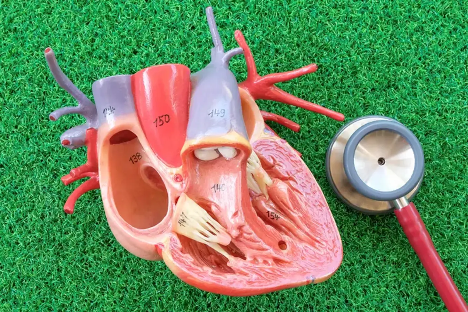
Heart anatomy
The heart is made up of four chambers. The top chambers are known as the left and right atrium, while the lower chambers are known as the left and right ventricle. The four valves at the exit of each chamber keep blood flowing one way through the heart to the lungs and the rest of the body.
The tricuspid valve, pulmonary valve, mitral valve, and aortic valve are the four valves:
- The right atrium receives oxygen-depleted blood from your body when it enters your heart. The tricuspid valve connects the right atrium to the right ventricle. It opens to allow blood to flow to the right ventricle.
- The pulmonary valve regulates the flow of blood between the right ventricle and the lungs. It opens to let the heart to flow blood from the ventricles into the pulmonary artery and into the lungs to pick up oxygen. The oxygenated blood returns from the lungs to the left atrium.
- The mitral valve connects the left atrium to the left ventricle. It opens to allow oxygen-rich blood from the left atrium to enter the left ventricle.
- The aortic valve regulates the flow of blood from the left ventricle into the aorta (the main artery in your body). When this valve opens, oxygen-rich blood is pushed to the aorta before being expelled to nourish the rest of your body.
The valve closes between each stride to prevent blood from flowing backwards and mixing oxygen-poor blood with oxygen-rich blood. The continual one-way flow of blood provides oxygen throughout your body.
When one or more of the heart valves fails to open or close correctly, this is referred to as heart valve disease. Multiple valvular heart disease occurs when more than one heart valve is affected.
- Stenosis occurs when the aperture of the valve narrows and inhibits blood flow.
- When a valve slides out of position or the valve flaps (leaflets) do not close correctly, this is referred to as prolapse.
- Regurgitation occurs when blood seeps backward through a valve, which can occur as a result of prolapse.
What is Heart Valve disease?
Valvular heart disease (VHD) is defined by damage to or a congenital defect in one or more heart valves, including the mitral, aortic, tricuspid, and pulmonary valves. Heart valves have just one purpose: to allow unhindered forward blood flow through the heart. Damaged or malfunctioning valves can create two sorts of problems: they either fail to open correctly (a condition known as stenosis), restricting blood flow, or they leak, allowing reverse flow.
Regurgitation happens when a valve fails to seal securely, allowing blood to flow back into the prior chamber from which it originated. Prolapse, a disease in which the valve leaflets expand into the left atrium during a heartbeat, is the most common cause of mitral regurgitation. Stenosis occurs when the heart valve cannot fully open because the valve flaps or ring have thickened, stiffened, or fused together, restricting proper blood flow through the valve.
Valve Diseases might be congenital, the result of inflammation, or the result of infection complications. Mild to moderate illness is typically asymptomatic at first, but due to the disease's progressive and degenerative nature, it can develop to serious heart failure and death if left untreated. VHD is generally associated with aging. Prevalence is rising as the United States' population ages and lives longer. The most frequent valve disease in the United States is aortic stenosis, which is followed by mitral regurgitation, aortic regurgitation, and mitral stenosis.
In the United States, degenerative valve disease is the most frequent kind of valvular heart disease, but rheumatic heart disease accounts for the majority of valve pathology in developing countries. Physicians are anticipated to see more patients with degenerative valve problems as the US population ages. Because individuals from underdeveloped countries continue to move to the United States, rheumatic valve disease may become increasingly common. As a result, knowing the complete spectrum of valvular illnesses is critical to providing appropriate patient care.
Types of valvular heart disease
1. Valvular stenosis (narrowing)
Heart valve stiffening can limit the size of the valve opening and hinder blood flow. The narrowing is referred to as valve stenosis. It prevents the valve from fully opening, limiting the volume of blood that may flow through. In extreme situations, the valve opening can become so small that blood flow to the rest of the body is compromised.
- Tricuspid valve Stenosis. Blood cannot entirely flow from the right atrium to the right ventricle if your tricuspid valve narrows. The atrium may grow as a result, influencing pressure and blood flow in the surrounding chambers and veins. It can also cause the right ventricle to shrink, resulting in less blood flowing to your lungs to pick up oxygen.
- Pulmonary valve Stenosis: When your pulmonary valve narrows, the movement of oxygen-depleted blood from the right ventricle to the lungs is reduced. This impairs your blood's capacity to pick up oxygen and transmit it to the rest of your body. With pulmonary valve stenosis, the right ventricle needs to work harder to pump blood through the constricted pulmonary valve, and heart pressure is frequently elevated.
- Mitral valve Stenosis: Blood flow from the left atrium to the left ventricle is diminished when the mitral valve narrows. Because the amount of blood delivering oxygen from the lungs is diminished, this can produce weariness and shortness of breath. Blood pressure in the left atrium can cause the atrium to expand and fluid to accumulate in the lungs.
- Aortic valve stenosis: When the aortic valve narrows, blood flow from your heart to your aorta (your body's major artery) and then to the rest of your body is reduced. As a result, the left ventricle must contract more forcefully in order to push blood past the aortic valve. This frequently results in thickening of the left ventricle (left ventricular hypertrophy), making the heart less effective.
2. Valvular prolapse (slipping out of place)
Prolapse occurs when the valve flaps (leaflets) move out of position or produce a bulge. This might result in an uneven or incorrect closing of the heart valve. Blood may leak backwards through the valve as a result of the prolapsed valve, disrupting one-way blood flow.
- Mitral valve prolapses: Mitral valve prolapse occurs when the valve fails to shut evenly. When the two ventricles contract, a portion of the mitral valve bulges upward into the atrium. A little quantity of blood may escape backward through the valve as a result of this (regurgitation).
- Tricuspid, pulmonary and aortic valve prolapse: These prolapses occur less frequently than mitral valve prolapses. The valve leaflets do not shut entirely and fail to produce a tight seal, similar to mitral valve prolapse.
3. Regurgitation (leaking)
Regurgitation occurs when the valve fails to seal correctly, allowing blood to flow backwards. This disturbance of one-way blood flow in the heart strains your heart, diminishes its pumping efficiency, and inhibits its capacity to give oxygen-rich blood to your body.
- Tricuspid valve regurgitation: When the tricuspid valve fails to shut correctly, blood pushed forward from the right ventricle to the lungs might flow backward into the right atrium, causing it to expand.
- Pulmonary valve regurgitation: This happens when the pulmonary valve fails to shut correctly. The lower right chamber of the heart (right ventricle) sends blood through the pulmonary artery into the lungs to pick up oxygen. When the pulmonary valve fails to seal entirely, blood can seep back into the heart from the lungs. This reverse blood flow combines oxygen-poor and oxygen-rich blood, reducing the availability of oxygen-rich blood to feed the rest of your body.
- Mitral valve regurgitation: Some blood spills backward into the left atrium from the lower chamber as the mitral valve closes in mitral valve regurgitation. This decreases the amount of blood flowing to the rest of the body. Regurgitation causes an increase in blood volume and pressure in the left atrium. In extreme situations, the increased volume and pressure may cause atrial enlargement and fluid buildup (congestion) in the lungs.
- Aortic valve regurgitation: With each pulse, oxygen-rich blood seeps backward from the aorta into the left ventricle. Your body does not receive enough blood, and your heart needs to work harder to compensate. The walls of the ventricle may thicken with time (hypertrophy). This might raise your chances of developing heart failure.
Valvular heart disease Causes
Valvular heart disease can occur before or during birth (congenital causes), or normal valves might be damaged over time (acquired causes). It is not always possible to determine the etiology of valvular heart disease. More funding for research into the causes of valvular heart disease is required.
Congenital causes
- Congenital valvular heart disease: This is a congenital abnormality in which a heart valve is either the wrong size or form, or its valve flaps (leaflets) are not correctly linked to the heart.
- Bicuspid aortic valve disease: A congenital condition affecting the aortic valve. The bicuspid aortic valve contains just two leaflets instead of the usual three. Without the third leaflet, the valve cannot adequately open or close, is more prone to aortic valve stenosis, and may result in regurgitation.
- Marfan syndrome: This is a hereditary condition that affects the connective tissue of the body. All of the cells, organs, and tissues in the body, including the heart, are held together by connective tissue. Marfan syndrome patients may experience mitral valve prolapse and aortic valve regurgitation.
Acquired causes
- Rheumatic fever: This is an inflammatory illness that, if not treated appropriately, can damage the heart valves. Rheumatic fever generally begins as strep throat or a strep infection (streptococcal bacteria). As the body battles the strep infection, heart valves may be damaged or scarred.
- Infective (bacterial) endocarditis: Germs from the circulation can enter the heart and infect the surface of the heart, including the heart valves. People who have valvular heart disease are more likely to develop infective endocarditis.
- Radiation therapy: Cancer patients who had chest radiation therapy are more prone to develop valvular heart disease.
- Age: Heart valve abnormalities can be caused by degenerative changes or by the natural "wear and tear" of aging.
Other causes
- Coronary artery disease
- Damage to the heart muscle from a heart attack
- Other diseases of the heart muscle (cardiomyopathy)
- Metabolic disorders such as high blood cholesterol
- Tumor in the heart
- Certain medications.
Symptoms of Heart Valve Diseases
Many people do not notice any symptoms until their blood flow has been significantly reduced by valvular heart disease. Symptoms can include:
- Chest discomfort, pressure or tightness (angina) along the front of your body between your neck and upper abdomen.
- Palpitations (irregular or fast heartbeats produced by electrical system issues in the heart) can be an indication of valvular heart disease. Your heart may be working harder than usual. This can cause your heart to expand and interfere with regular cardiac rhythm, resulting in arrhythmia.
- Shortness of breath, particularly while exercising. Valvular heart disease limits the quantity of oxygen available to power your body, resulting in shortness of breath.
- Weakness or fatigue: Routine tasks such as walking or cleaning may become more difficult.
- Aortic stenosis is most commonly associated with light-headedness, dizziness, or fainting.
- Swelling can develop when valve issues cause blood to back up in other regions of the body, causing fluid buildup and swelling in the belly, feet, and ankles.
If you don't have many symptoms, or if they are moderate and don't bother you too much, your doctor may decide to keep a close eye on your situation and wait until it is required to treat your symptoms. It is critical to note that the symptoms of valvular heart disease do not always represent the severity of the illness. Maintain frequent check-ups with your doctor and explain any changes in your health that you detect.
Women are more likely than males to develop valvular heart disease as a result of rheumatic fever. Women should be extra cautious if they have a strep infection (streptococcal bacteria).
If a woman has a history of heart illness, she should visit her doctor before getting pregnant. Pregnancy frequently causes considerable changes in blood flow and blood pressure, which can worsen valvular heart disease and raise the risk of a cardiac catastrophe.
Valvular heart disease Diagnosis
The symptoms you mention to your healthcare provider and the findings of a physical exam are typically used to make a diagnosis of valvular heart disease. During the exam, your doctor will use a stethoscope to listen to your heart. The stethoscope can detect an odd sound known as a cardiac murmur. A cardiac murmur may not always indicate a heart disease because persons with normal hearts might have murmurs as well.
Your doctor will listen to your lungs to check for fluid buildup. They will also look for swelling in your abdomen, feet and ankles.
Test for valvular heart disease also include:
- Echocardiogram
- Angiogram
- Chest X-ray
- Electrocardiogram (ECG)
- Stress test
- Heart MRI
Management
The severity of your valvular heart disease determines your treatment. If your heart valve condition is minimal, you may not require any treatment. You will be examined on a regular basis to evaluate whether your condition worsens. If your heart valve condition is generating symptoms, medication may be suggested.
If your disease is severe, you may require more intense therapy. Valve repair or replacement in conjunction with medicine is an option. The strategy adopted will be determined by your age, general health, the impacted valve, and the kind and severity of your illness.
You and your doctor will talk about your treatment choices and determine which is best for you and your situation.
Medication
Medication cannot cure valvular heart disease, but it can alleviate symptoms such as edema, irregular heart rhythm, high blood pressure, and others.
Your doctor may prescribe:
- Diuretics (water pills) to reduce swelling and fluid buildup in the body.
- Blood thinners to prevent blood clots and reduce the risk of other cardiac problems.
- Antiarrythmics to prevent irregular or rapid heartbeats (arrhythmias).
If you have a heart ailment in addition to valvular heart disease (such as coronary heart disease or heart failure), drugs to lower the burden on your heart and improve your symptoms may be administered. There are suggestions and resources available to assist you in managing your drugs, such as keeping track of prescriptions, travel, storage, interactions, and so on.
Surgeries and other procedures
Heart valve surgery to repair or replace your heart valves may be required to avoid long-term heart damage.
Valve repair
- Patching holes or rips, reshaping the valve, or splitting valve leaflets to allow it to open and close properly are all methods for repairing heart valves.
- The constricted valve can be opened by introducing a thin catheter with a balloon at the tip through a blood vessel to the narrowed valve. The valve aperture is then widened by inflating the balloon. This is known as balloon valvulopasty.
- Annuloplasty is a surgical procedure used to correct an enlarged annulus (a ring of fibrous issue at the base of the heart valve). Sutures are sewed around the ring to close the gap. Alternatively, a ring-like device is connected to the exterior of the valve opening to allow it to seal more firmly.
Valve replacement
A defective heart valve is removed and replaced with a mechanical or biological valve if it cannot be mended. You and your doctor will talk about your options and determine which is best for you and your condition.
- Mechanical valves are constructed from long-lasting metals, carbon, ceramics, and polymers.
- Animal tissue, donated human tissue, or a patient's own tissues are used to create biological valves. Biological valves do not last as long as mechanical valves.
Transcatheter aortic valve implantation is a less invasive treatment that can be used instead of open-heart surgery to replace a failing aortic valve. A new valve is implanted using an ultrasound and chest x-rays to guide a catheter to your heart.
Lifestyle
Knowing and controlling your blood pressure, diabetes, and cholesterol levels can reduce your chance of developing additional heart illnesses including stroke. It is also critical to maintain a healthy lifestyle.
- Be smoke-free.
- Be more active.
- Aim for a healthy weight.
- Eat a healthy balanced diet – there are some specific diets you can follow that have been proven to reduce the risk of heart disease.
- Drink less alcohol.
- Manage stress.
Living with Valvular heart disease
Many people who have valvular heart disease lead full lives. Here are some things to be aware of as you learn to live with your heart condition.
- Valvular heart disease increases your chances of getting heart failure, which occurs when your heart muscle is unable to pump enough blood through your body. That is why it is critical to be aware of your symptoms and to notify your healthcare provider of any changes in your health.
- You may be more susceptible to infective endocarditis. If you require dental work, have your teeth cleaned, or are having any medical procedures involving your respiratory system, consult with your dentist or doctor (for example bronchoscopy, tonsillectomy or adenoidectomy). Antibiotics may be required both before and after these surgeries.
- Another proactive strategy to remain healthy is to get flu and pneumonia vaccines. Many persons with chronic heart disease have a reduced ability to fight infections. You may be more prone to respiratory tract infections, which can cause serious consequences and even death. Consult your doctor about getting a flu shot.
- Because men and women have distinct hearts, they require different therapy and care. This is particularly true for women who intend to establish a family. Make sure to talk to your doctor about your alternatives.
Conclusion
The heart valves function by ensuring that blood flows forward and does not back up or leak. When you have a heart valve issue, the valve is unable to perform this function adequately. This can be caused by a leaking of blood, known as regurgitation, or a narrowing of the valve opening, known as stenosis, or a combination of the two. Heart valve problems can damage any of your heart's valves. The severity of the condition and the symptoms determine the treatment for heart valve abnormalities.