Endoscopic Choledochoduodenostomy

    Last updated date: 08-Jan-2023

    Originally Written in English

    Endoscopic Choledochoduodenostomy

    Endoscopic Choledochoduodenostomy

    Overview

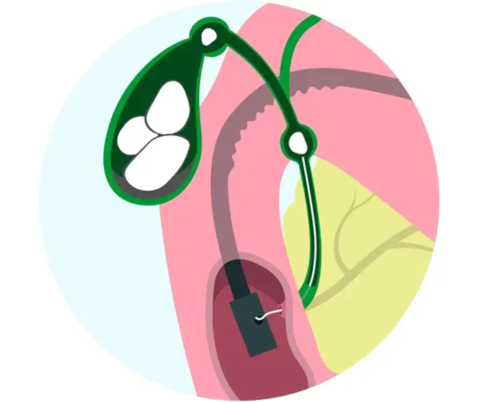
    Endoscopic ultrasonography (EUS) is a therapeutically important technique not only for diagnostic purposes during EUS-guided fine needle aspiration, but also for interventional purposes. Endoscopists with experience devised and conducted EUS-guided biliary drainage. In patients with biliary blockage, EUS-guided choledocoduodenostomy (EUS-CDS) is a rather well-established alternative biliary drainage procedure for biliary decompression.


    What is EUS-guided choledocoduodenostomy

    EUS-guided choledocoduodenostomy

    Endoscopic ultrasonography (EUS)-guided biliary drainage was used to treat patients with obstructive jaundice after endoscopic retrograde cholangiopancreatography (ERCP (failed. 

    Obstructive jaundice is a serious side effect of pancreatic or biliary cancer. This adverse effect necessitates treatment, particularly in individuals who are unable to have surgery owing to concomitant chemotherapy. Endoscopic biliary stenting (EBS) is considered the gold standard therapy for obstructive jaundice. This procedure, however, is associated with a number of complications, including post-endoscopic retrograde cholangiopancreatography (ERCP) pancreatitis.

    Furthermore, EBS cannot be done in individuals who have selective cannulation failure of the main papilla or a papilla that is inaccessible owing to duodenal invasion. Percutaneous transhepatic biliary drainage is an alternate treatment option in these cases (PTBD). However, PTBD can cause a variety of complications, including cholangitis, bile leakage, and pneumothorax. Furthermore, the prevalence of significant problems, such as prolonged hospital stay and lasting unfavorable sequelae, ranges from 4.6% to 25%, while procedure-related fatalities range from 0% to 5.6%. Cosmetic concerns caused by external drainage reduce the patient's quality of life.

    Endoscopic ultrasonography (EUS) is therapeutically valuable not only as a diagnostic tool during EUS-guided FNA, but also during interventional EUS. Endoscopic ultrasound-guided biliary drainage (EUS-BD) is a type of interventional EUS developed and done by skilled endoscopists. The approach path determines the EUS-BD method. Transgastric EUS-BD is accomplished using EUS-guided hepaticogastrostomy (EUS-HGS), in which the intrahepatic bile duct is perforated through the stomach and a stent is placed from the intrahepatic bile duct to the stomach.

    Transduodenal EUS-BD is accomplished using EUS-guided choledochoduodenostomy (EUS-CDS), in which the common bile duct (CBD) is perforated and a stent is placed from the CBD to the duodenum. EUS-guided gallbladder drainage (EUS-GBD) is conducted for transgastric or transduodenal EUS-BD, in which the gallbladder is pierced and a stent is inserted from the gallbladder to the stomach or duodenum. Furthermore, like EUS-BD, the EUS-guided rendezvous technique (EUS-RV) is incorporated.

    While EUS-HGS and EUS-GBD show therapeutic advantages in certain patients, they are linked with a number of side effects, including stent migration. If stent migration occurs, it can be deadly. To avoid the problems associated with these operations, innovative approaches or new equipment are necessary. EUS-RV is only appropriate in patients whose ampulla of Vater can be reached by duodenoscopy. This operation might be challenging at times and takes a long time.

    EUS-CDS has a claimed technical success rate of 50% to 100%, and a clinical success rate of 92% to 100%. Additionally, the overall technical success rate was 93%, while the clinical success rate was 98%. According to the current research, the total adverse event rate for EUS-CDS is 16%. The total technical and clinical success rate for EUS-CDS is encouraging. However, EUS-CDS can still cause a number of issues, thus more viable and safe approaches or devices must be developed. Although standardizing the method in multicenter clinical trials and randomized clinical studies comparing it to endoscopic biliary drainage are still required, EUS-CDS has the potential to become a first-line biliary drainage therapy.


    Indications

    Indications

    EUS-CDS is mostly used for individuals who have failed an EBS excluded prospective clinical study. This operation is used to clear blockages in the middle and lower bile ducts.

    Pancreatic cancer, ampullary carcinoma, and cholangiocarcinoma were the major illnesses in patients who received EUS-CDS. This suggests that pancreato-biliary cancer is the most common reason for EUS-CDS. However, as previously noted, EUS-CDS for benign biliary stricture was only conducted in two individuals.

    EUS-CDS is not recommended in individuals who have surgically changed anatomy, such as a Roux-en-Y anastomosis or a duodenal blockage caused by tumor invasion that cannot be passed through with an endoscope. An EUS-guided hepaticogastrostomy may be suggested in such circumstances. However, if the duodenum is not implicated, EUS-CDS can be used with duodenal stenting.

    The indications for EUS-CDS versus ERCP for benign illness are yet unknown. Versus establish the procedure's clinical effectiveness, prospective randomized controlled studies comparing EUS-CDS to ERCP are required. As a result, the following are the EUS-CDS indications:

    1. Failed EBS; 
    2. Inaccessibility of the ampulla of Vater, such as due to duodenal invasion by the tumor; 
    3. Contraindications for percutaneous transhepatic biliary drainage (PTBD); and 
    4. Middle or lower bile duct obstruction


    Device selection and technical tips

    Device selection and technical tips

    Puncture of the common bile duct

    The EUS scope is pushed into the duodenum, tilted slightly to the left, and oriented downwards to see the CBD. To prevent any intervening vessels, the CBD is pierced with a 19-G needle under Doppler vision. A tiny quantity of contrast medium is administered after aspirating bile juice. It is critical to prevent puncturing the duodenal mucosa and cystic duct during this procedure. To prevent puncture and stenting through double duodenal mucosa, the CBD should not be pierced when a double duodenal mucosal line is observed on EUS.


    Guidewire insertion into the bile duct

    The guidewire is inserted deep into the intrahepatic bile duct after the contrast is introduced into the CBD. When the CBD is parallel to the FNA needle on EUS imaging, the guidewire can be readily moved toward the hepatic hilum.

    A 19-gauge needle should be used to allow a firm guidewire to pass through the needle. It is advisable to use a 0.025 inch guidewire with a very flexible tip, enough stiffness, and easy seeking ability. To eliminate wire sharing, a new guidewire with a coil top shape was developed.

    When inserting the guidewire with other devices, it is critical to be able to see the devices under both EUS and fluoroscopic guidance to confirm that they match the axis.


    Devices used to dilate the fistula

    Several devices have already been reported for dilatation of the fistula following CBD puncture. The dilator, balloon catheter, or needle knife are the most often used devices for transmural tract dilatation. Park reported a 27% total adverse event rate for EUS-BD, which included EUS-CDS and EUS-HGS.

    They also identified the main risk factor for unfavorable procedural events after EUS-BD as the use of a needle knife for fistula dilatation. Because of the acute angulation of the EUS scope, when the catheter is placed at the duodenal bulb, the needle knife points tangentially, which might result in an incidental incision with a risk of pneumoperitoneum or hemorrhage. As a result, they came to the conclusion that fistula dilation should be avoided wherever feasible.

    However, it is true that using a needle knife makes fistula dilation simpler. A diathermic dilator has recently become accessible. The guidewire is always coaxial with this device. As a result, its usage for fistula dilation may minimize the incidence of EUS-BD-related adverse events when compared to needle knife dilation. Comparative studies of these technologies are thus necessary.


    Stent selection

    During EUS-CDS, both plastic and metallic stents were utilized. Plastic stents with sizes ranging from 5 to 10 Fr were previously utilized. Because the working channel of the EUS is 3.7 mm in diameter, a 7 or 8.5 Fr plastic stent is often employed. However, bile leakage can occur after the implantation of a plastic stent. This patient experienced gastrointestinal discomfort and fever for up to three days following EUS-CDS, and computed tomography and duodenoscopy revealed bile leakage. Because of the fine gauge of the plastic stent, bile leakage from the space between the stent and the fistula is possible if a big fistula is established during stent implantation.


    Success Rate

    Success Rate

    EUS-CDS has a claimed technical success rate of 50% to 100%, and a clinical success rate of 92% to 100%. Additionally, the overall technical success rate was 93%, while the clinical success rate was 98%. When compared to ERCP, this is a reasonably high success rate. As a result, if EUS-CDS is linked with a low rate of adverse events and lengthy stent patency, it has the potential to replace EBS as the preferred bile drainage approach. To compare endoscopic biliary drainage with EUS-CDS, a prospective randomized clinical study is required.


    Adverse Events

    Adverse events

    According to the current research, the total adverse event rate for EUS-CDS is 16%. EUS-CDS has the potential to induce a number of side effects, including:

    1. Infection (peritonitis, cholangitis, cholecystitis); 
    2. Pneumoperitoneum; 
    3. Bile leakage, biloma; 
    4. Bleeding; 
    5. Abdominal pain;
    6. Perforation; and 
    7. Stent migration.

    According to earlier research, the most common adverse event was pneumoperitoneum (28%). If adverse effects do occur in most EUS-CDS instances, they may be managed conservatively. The bile leak in both of these individuals was caused by the use of an uncovered SEMS. Even if a completely covered SEMS is chosen, the danger of biliary peritonitis persists.

    This adverse event might occur as a result of a bile or air leak during fistula dilatation when installing the stent delivery device. To prevent these complications, more advanced technologies that allow for one-step stent implantation (without dilation of the fistula) are urgently needed. The use of color Doppler ultrasonography to detect vascular structures can help to reduce the risk of bleeding.


    Conclusion 

    Biliary drainage

    The most frequent technique for biliary drainage in patients with obstructive jaundice is endoscopic transpapillary biliary stenting. However, some patients were unable to gain bile duct access due to unsuccessful biliary cannulation or an inaccessible papilla as a result of severe duodenal stenosis induced by tumor invasion. Endoscopic ultrasonography (EUS)-guided choledochoduodenostomy has recently been reported as another biliary drainage procedure.