Endoscopic Epidural Neuroplasty

Last updated date: 22-Aug-2023

Originally Written in English

Endoscopic Epidural Neuroplasty

Along with surgical procedures and other modalities, interventional techniques in the management of low back pain, such as spinal endoscopy and epidural neuroplasty, remain to be some of the more controversial ones. Low back pain is the most prevalent chronic painful condition, affecting 20 to 40% of the population and having significant economic and social consequences. Apparently, among musculoskeletal conditions, low back pain comes in first. Numerous studies have shown proof to the contrary of the generally held belief that most episodes of low back pain are transient and that 90% of patients recover in around 6 weeks. According to studies, up to 80% of individuals still experience chronic or recurrent low back pain after a year.

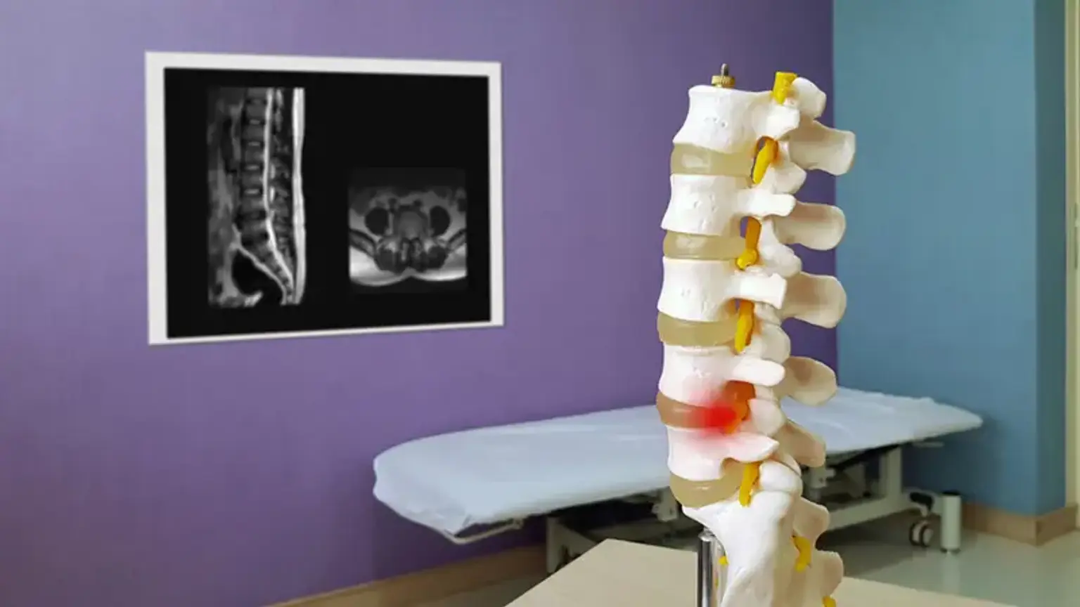
Low back pain is primarily caused by disc and joint disorders, but failed low back surgery syndrome, also known as failed management syndrome or postlumbar laminectomy syndrome, is a growing medical condition. It is estimated that 10 to 40 percent of lumbar surgical interventions result in failed back surgery syndrome, with failure rates as high as 67 percent, according to surprising statistics. Although these frequently follow surgery that was inadequate, inappropriate, or unnecessary, this condition can also occur after a surgical operation that was properly indicated and carried out.

The idea behind endoscopic epidural neuroplasty is that flexible fiberoptic catheters inserted through the sacral hiatus can securely access the epidural area. It enables a three-dimensional view of the epidural space's contents and gives the operator the power to direct the catheter toward certain structures. Through this method, it is possible to examine a particular nerve root's pathology, cure it by injecting a medication straight into the root, and widen the epidural space with normal saline.


What is Endoscopic Epidural Neuroplasty?

Endoscopic Epidural Neuroplasty

One-third to one-half of cases with medically symptomatic fibrous adhesions, which are typically a result of repeated back surgery, have exhibited improvement in chronic back pain after undergoing endoscopic epidural neuroplasty, also known as epiduroscopy. During the technique, a flexible spinal endoscope is used to deliver drugs, commonly ozone and ciprofloxacin, and mechanically dissolve adhesions while the target sites are visible. Patients with chronic low back pain that is resistant to non-invasive treatments like physiotherapy, oral and topical painkillers, and epidural injections are candidates for therapy. Epiduroscopy provides clinicians with a direct vision of the epidural space and pathologic adhesions in comparison to percutaneous epidural neuroplasty. There is less research to support the use of endoscopic epidural neuroplasty than there is for the percutaneous method.


Endoscopic Epidural Neuroplasty Benefits

Endoscopic Epidural Neuroplasty Benefits

According to many doctors, less invasive techniques provide the patient several benefits over traditional operations:

  • Less chance of nerve injury.
  • Smaller epidural fibrosis.
  • Speedier recovery and return to work.
  • Fewer infections occurring.
  • Less secondary surgical trauma and small surgical cut.

The major benefits of endoscopic and minimally invasive procedures are a new surgical strategy. Contrary to surgery, no long surgical cut is made, preventing damage to the back muscles, whose long-term care is otherwise necessary. One surgical entry with a length of 8 to 10 mm is adequate for minimally invasive and endoscopic procedures, where working instruments and an endoscopic camera are placed into the spinal space. The fact that there is no hospitalization required and that you can start your recovery right away in the comfort of your own home after the treatment is an advantage.


Endoscopic Epidural Neuroplasty Indications

Endoscopic Epidural Neuroplasty Indications

With epiduroscopy, structures in the epidural space can be detected, including hyperemia, vascularity alterations, fibrosis and adhesions, lateral recess stenosis, disk protrusion, and ligamentum flavum hypertrophy. Epiduroscopy has relative clinical indications for diagnosing and treating pain disorders.

Epiduroscopy used to be primarily utilized to diagnose the causes of pain. Due to the capacity to precisely treat these areas, treatment has recently been the primary indication of epiduroscopy. Utilizing the method improves the ability to diagnose pathological and anatomical components and conditions. The underlying condition affects how well a treatment works. As a result, the effectiveness of a given treatment plan can be used to evaluate the validity of the diagnostic data obtained during epiduroscopy.

Epiduroscopy is used to perform treatments such as direct administration of medication, epidural neuroplasty, lysis of scar tissue, catheter insertion, implantation of stimulation electrodes (for radio frequency treatment and spinal cord stimulation), and discectomy. To respond to stimulated pain-generating regions, conscious sedation is also given to patients. Additionally, epiduroscopy has the potential to alleviate post-lumbar surgical syndrome in addition to reducing the incidence of surgery.

Epiduroscopy is also recommended if radiculopathy is present together with any of the following signs or symptoms: epidural steroid injection failure, filling adhesion-caused abnormalities, post-laminectomy failed back syndrome, or ineffective conservative back treatment.

After spinal surgery, spinal surgery syndrome can happen when scar tissue puts pressure on spinal nerves, resulting in persistent discomfort in the lumbar region of the spine (lower back). Steroid injections are typically the first course of treatment. Epidural neuroplasty, however, frequently offers more powerful and long-lasting pain relief. Injections of steroids may lessen inflammation, but they do not remove scar tissue.

A narrowing of the spinal canal known as spinal stenosis compresses the nerves, resulting in discomfort, muscular weakness, and motor issues. Both spinal nerve inflammation and scar tissue may contribute to these symptoms. Individuals normally experience decreased back discomfort after epidural neuroplasty, while some patients experience no pain at all.


Endoscopic Epidural Neuroplasty Preparation

Endoscopic Epidural Neuroplasty Preparation

  • The patient should be informed of the procedure's potential advantages and dangers, and informed consent documents should be obtained. The patient should also be well informed about the target.
  • The patient's use of anticoagulant medications needs to be checked. According to the recommendations, these anticoagulants should be stopped if the patient's comorbid conditions are suitable.
  • It is important to inquire about and make note of any drug allergies, problems with the previous anesthetic, and interventional treatments.
  • It is important to analyze the results of the prothrombin time, partial thromboplastin time, bleeding time, white blood cell count, and platelet function tests.
  • For the patient's comfort, premedication is advised (midazolam, fentanyl, and ceftriaxone by intravenous catheter)
  • The patient should be watched closely for safety (Automated blood pressure cuff, electrocardiogram, and pulse oximeter).


Endoscopic Epidural Neuroplasty Procedure

Endoscopic Epidural Neuroplasty Procedure

The patient is positioned on his or her back. It is best to follow standard American Society of Anesthesiology guidelines for moderate sedation. Antibiotics should be given as a preventative measure before the procedure begins.

  1. A skin wheal is established over the sacral hiatus using 1-2 cc of 1% lidocaine and a 25-gauge needle after appropriate sterile preparation and draping. The hiatus is next penetrated with a 16-gauge RK needle while being viewed under lateral and AP fluoroscopy.
  2. Next, 10 ml of nonionic contrast dye is injected, and an epidurogram is done.
  3. The needle is used to introduce a 0.9-mm guide wire, which is subsequently advanced with the aid of a fluoroscope to the level of the suspected lesion and contrast filling defect. A 9-French dilator with a catheter (sheath) is then advanced over the guide wire after making a little cut with a blade.
  4. The guide wire is withdrawn when the catheter has been introduced up to its tip. Then, using video and fluoroscopic images as guides, a fiber-optic spinal endoscopic video-guided system is inserted into the catheter through the valve and pushed until the tip is situated at the distal end of the catheter. The suspicious side of the lesion should be toward which the endoscope is positioned ventrolaterally.
  5.  The catheter and fiber-optic endoscope are moved around and turned in different directions while being gently irrigated with normal saline to view the nerve roots at different levels. Slow, steady infusion is used to carry out gentle irrigation. Saline irrigation should be administered at a pace of no more than 30 ml per minute, with a maximum volume of no more than 100 ml. (Retrograde flow will occur; this flow should not be included in the overall volume.) A continuous subarachnoid needle may be implanted for continuous CSF pressure monitoring in extended events with higher irrigation volumes. Epidural neuroplasty and decompression are performed mechanically with the fiber-optic endoscope and by dilatation of the epidural space with normal saline. Only if the epidural space is constantly inflated by frequent saline injections will visualization be possible. The dura mater is one example of a structure that is simple to visualize. 
  6. An epidurogram is conducted at least twice, and confirmation is achieved by injecting nonionic contrast material. Following the surgery, lidocaine 1%, preservative-free, combined with 6-12 mg of betamethasone acetate or 40–80 mg of methylprednisolone is often given after checking for any signs of subarachnoid contrast leakage.
  7. If it is established that there are many levels of pathology, the procedure can be performed at each level, and the injectate should be administered in a series of divided dosages.


Endoscopic Epidural Neuroplasty Outcomes

In cases where other forms of pain management have failed, epidural neuroplasty combined with epidural steroids is highly beneficial for both short- and long-term pain control, according to the American Society of Interventional Pain Physicians' guidelines. Studies indicate that patients' overall health has improved. Following epidural neuroplasty, patients also claimed that they used their painkillers less frequently and that both their physical and mental wellbeing had improved.

There is a chance of complications, as there are with all surgical treatments. Many persons who experience back discomfort are thought to benefit from epidural neuroplasty as a form of treatment. To determine whether the advantages outweigh the hazards in your particular circumstance, you should consult a pain specialist.


Endoscopic Epidural Neuroplasty Complications

Endoscopic Epidural Neuroplasty Complications

An invasive epidural surgery carries the usual complications. Dural puncture, hemorrhage, infection, injury to blood vessels and nerves, unintended dural puncture, medicine injection errors, and catheter shearing are the most often reported problems in percutaneous and endoscopic epidural neuroplasty.

Frequent adjusting of the catheter against the needle might lead to catheter shearing. The back edge of a Tuohy needle is a cutting surface and should not be utilized for this procedure since it will shear the catheter. The initial needle tip should be positioned toward the probable lesion as a way to reduce this. By doing this, the catheter won't need to be directed and adjusted as frequently. Retained hardware can be an issue after catheter shearing. Epidural endoscopy or surgical intervention may be required for catheter removal. Manchikanti gave an example of a situation where endoscopy by itself was insufficient to remove the catheter, and arthroscopy forceps had to be used instead.

Other cases where pieces of the Racz catheter were sheared off and lodged in an L5-S1 foramen have been documented. In a case study, Karaman and colleagues describe a situation in which a catheter piece was left in the epidural area. After a year of meticulous monthly monitoring showed that the patient was responding favorably to the neurolysis, it was decided not to proceed with an aggressive surgical resection to retrieve the tiny fragment.

Serious side effects such as arachnoiditis, paralysis, or bowel or bladder problems were not previously reported in the literature. The example of a 73-year-old woman who chose endoscopic epidural neuroplasty for extensive scarring of the epidural space is discussed by Justiz and colleagues. The patient thereafter experienced urine retention and a neurogenic bladder. She recovered from the neurogenic bladder symptoms three years after beginning to use the antibiotic nitrofurantoin. The patient noticed that she was unable to urinate on her own after stopping the antibiotic. The patient, who had been off nitrofurantoin for more than three years, was able to successfully void once again after having it reintroduced.

Any neuraxial method has the potential risk of infection. Even though using a strict aseptic technique should be a routine procedure, infections still happen. An uncommon but documented consequence of this specific technique is meningitis. After performing epidural neuroplasty to treat non-specific low back pain, Wagner and colleagues observed an incidence of severe meningitis with serious neurological consequences. They advised that a thorough aseptic approach should be used during this treatment. Before beginning this intervention, it is advised to proceed with thorough patient selection to lower overall risks.


Percutaneous vs Endoscopic Epidural Neuroplasty

Endoscopic Epidural Neuroplasty

More than one randomized controlled trial has shown the effectiveness of percutaneous epidural neuroplasty as a treatment for refractory low back and lower extremities pain, although endoscopic epidural neuroplasty has less support.

When carried out by skilled professionals, percutaneous epidural neuroplasty is a safe and reliable operation with few risks. In the future, epiduroscopy will be used more frequently for treating complicated spinal pain syndromes, but it should only be carried out by qualified professionals. Inexperienced users prolong the operation and require more infusion volume, which raises the risk of complications. To prevent complications, the overall amount, the rate, and the length of time should be limited. To choose technique preferences and indications, randomized controlled trials with high-quality data are necessary. The recommendation is to select the type that you have greater experience in challenging situations.


Conclusion

Chronic low back pain is a significant social and medical issue. endoscopic epidural neuroplasty for the treatment of refractory low back pain is sometimes confusing because it places too much focus on biopsychosocial issues and chooses the wrong patients for it. According to the preliminary cumulative data found in the literature, endoscopic epidural neuroplasty is just as effective as other existing treatment modalities for chronic low back pain, if not more so, than surgical intervention.

Even though this method is quite helpful for treating persistent low back pain, care must be taken because spinal cord trauma has a serious risk of complications. Doctors advise endoscopic epidural neuroplasty because it is a useful, safe, and cost-effective technique for treating chronic, intractable pain that is unresponsive to all other conservative modalities of treatment when carried out in an outpatient setting, with reasonable and customary charges for the facility and physician services. However, a pain practitioner must tailor the treatment options to each patient and personal experience.