Endoscopic Submucosal Dissection (ESD)
Overview
Niwa and Tsuneoka et al. developed gastric polypectomy in the 1960s, and Wolff and Shinya described colon polypectomy for the first time in 1969. Endoscopic mucosal resection (EMR) was first used to treat early esophageal cancer in 1971 by Karita et al, Dyehle et al, and Inoue et al. Lesions under 2 cm in diameter can be removed in one piece during EMR; for lesions larger than 2 cm, only surgery or piecemeal resection were available. A minimally invasive and efficient method for en bloc resecting early-stage tumors or precancerous lesions in the gastrointestinal system that are larger than 2 cm is endoscopic submucosal dissection (ESD).
Endoscopic resection with a local injection of the hyperosmotic saline-epinephrine solution was a method created by Hirao et al in the 1980s. In this operation, the submucosa is dissected after a circumferential incision is created with a needle knife. Ono and colleagues at the National Cancer Center Hospital eventually formed an insulated tipped knife and began stomach ESD in the 1990s. Yahagi et al. employed the snare tip (later the flex knife and eventually the dual knife) for stomach ESD at the same time as Oyama began utilizing the prototype of the hook knife, which they made by bending a needle knife.
After more than 20 years, professionals can now safely conduct esophageal and colorectal ESD. However, without the proper indications, tools, and a well-thought-out plan, ESD cannot be carried out consistently and safely. Additionally, the initial step is to correctly locate and diagnose lesions that are amenable to ESD utilizing image-enhanced endoscopy. A minimal criterion is also to get sufficient histological specimens for thorough pathologic assessment with high-quality ESD.
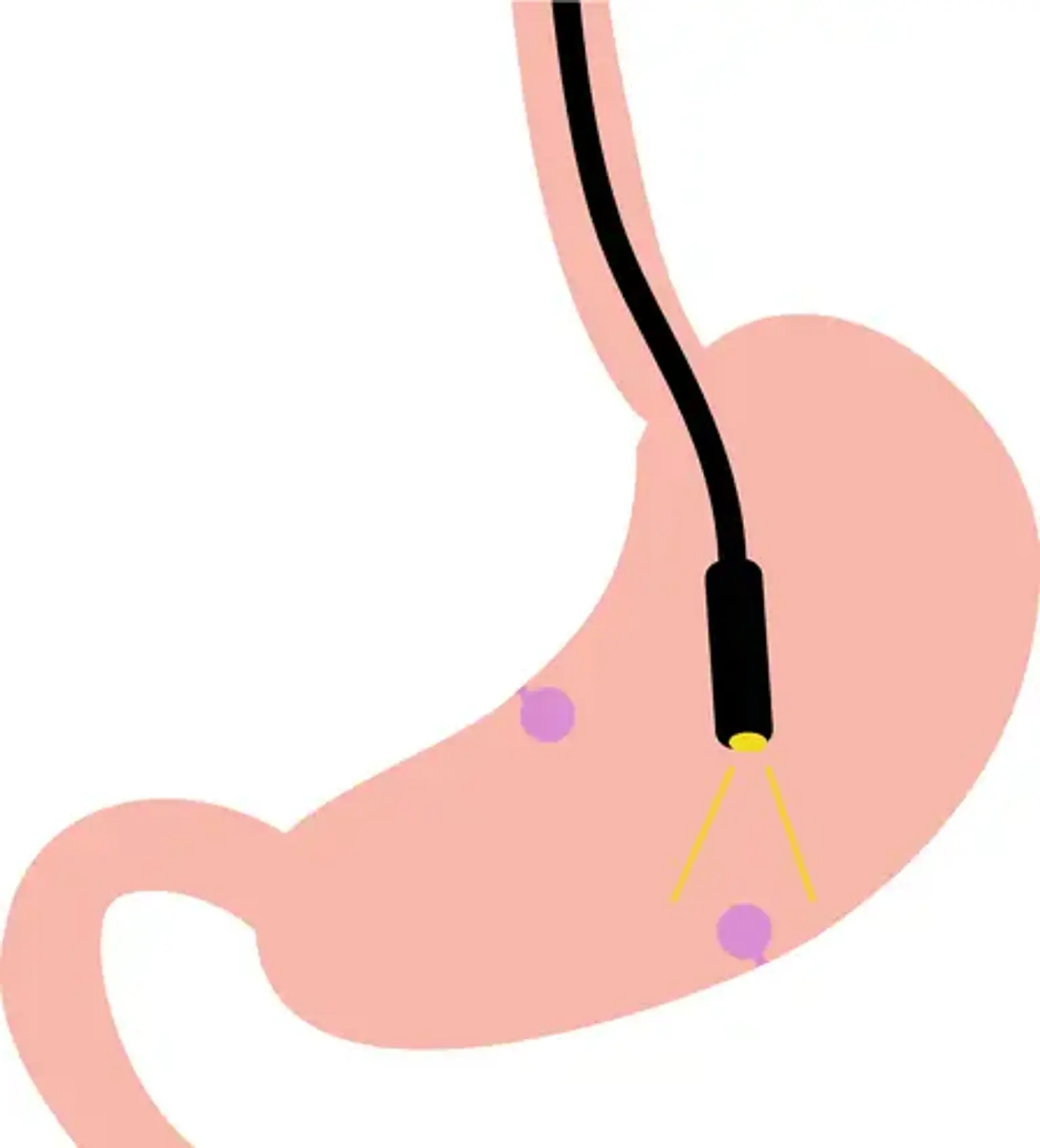
Endoscopic Submucosal Dissection
endoscopic submucosal dissection (ESD) is the use of the cutting-edge therapeutic endoscopy technique through which tumors and growths of the esophagus, stomach, small intestine, colon, and rectum can be removed without the need for open surgery. It makes use of an endoscope, a small, flexible instrument that is introduced through the mouth or anus. Once the growth has been located, it is carefully noted, and a specific solution is then injected between the thin layers of the digestive tract wall to create a cushion and a safe working environment. The growth is then meticulously dissected for removal using specialized instruments that are subsequently inserted through the endoscope. To speed up the healing process, tiny sutures and/or metallic clips may be applied. To ensure that the resection is curative, the growth is subsequently sent to the pathology lab for a microscopic examination.
Significance of Endoscopic Submucosal Dissection
ESD is important in part because many people undergo surgery for early malignancies that are confined to the GI tract's inner layer and haven't spread to deeper tissues. The patient typically avoids needing to have that portion of their GI system surgically removed, whether it be the esophagus, stomach, colon, or rectum, thanks to the removal of those lesions using ESD. Patients will benefit from ESD to eliminate lesions even if they are not malignant now since they could develop into cancer in the future. In the past, many doctors have referred people for surgical resection, but improved approaches might have avoided surgery. For some patients, ESD can be an organ-saving treatment, much like having a lumpectomy instead of a mastectomy for a breast tumor.
Endoscopic Submucosal Dissection Benefits
The main advantage of ESD is that it typically does not require surgery to provide curative care. Expert ESD is safe and efficient when done. When compared to after surgery, which would be a more invasive and riskier option, patients can typically return home the day after the operation and resume normal activities much sooner. After ESD, the majority of patients report minimal pain or discomfort.
Endoscopic Submucosal Dissection Indications
The following cancers and lesions that affect the submucosa, the region between the lining of internal organs and the muscle wall, can be treated with the ESD procedure:
- Barrett’s esophagus
- Early-stage colon polyps or malignant tumors, such as colorectal, stomach, or esophageal cancer.
- Colon, stomach, or esophageal cancers that have not yet spread to the deeper layer of the GI wall
For the removal of some growths, particularly those without distinct borders or those that are too large to be removed in one piece by conventional procedures, ESD may be a more effective choice than endoscopic mucosal resection (EMR). In these situations, ESD can reduce the danger of the tumor spreading. Your doctor can stage your GI cancer with the aid of ESD to further refine your treatment plan.
Endoscopic Submucosal Dissection Preparation
Your doctor will want to know your medical history and the medications you take before doing an endoscopic submucosal dissection. You will be required to provide the following details:
- The names and dosages of all prescription drugs, over-the-counter medications, and dietary supplements you take, including aspirin and diabetes prescription drugs, as well as any blood-thinning medications.
- Whether you currently have or have ever had a medication allergy, as well as the name of the medication,
- All of your medical issues, notably diabetes, heart disease, lung disease, and blood clotting issues
The day before the surgery, you'll get written instructions detailing what to do, including:
- Before the treatment, you might not be allowed to eat or drink anything for many hours. They refer to this as fasting. Usually, fasting starts at midnight on the night before your treatment, however, the exact timing can vary. You'll be told when to start. Additionally, you might not be permitted to smoke or chew gum at this time.
- Putting off taking specific medications. Some medications, such as those that affect blood coagulation or some diabetes treatments, may be temporarily stopped at your doctor's instruction.
- You will be instructed how to cleanse using a bowel preparation if you have a lower GI ESD, which involves the colon or rectum. It often entails taking laxatives and drinking a solution that cleans the colon.
- A special diet is followed both before and throughout the preparation period. To ensure that the treatment may be carried out safely and successfully, it is crucial to carefully read the instructions.
Endoscopic Submucosal Dissection Procedure
An endoscope, a lengthy, thin, flexible instrument with a diameter of about half an inch, is used to perform the ESD procedure. The patient's mouth is used to insert the endoscope. The tumor is accurately dissected (cut) using an endoscope, and then it is completely removed through the patient's mouth. The following esophageal cancer ESD method is carried out as follows:
- The treatment is performed when the patient is under general anesthesia.
- The patient's mouth is used to insert the endoscope.
- A stain is used to highlight the malignant lesion and make it stand out from the surrounding healthy tissue.
- To ensure that the entire tumor is removed, the doctor marks the margins of the tumor (the region surrounding the tumor).
- The lesion is lifted and made easier to remove by the doctor injecting a liquid into the submucosal layer beneath it. (The tissue layer immediately beneath the mucous membrane is known as the submucosa).
- The lesion is then dissected by the surgeon, who then takes it out of the patient's mouth.
Endoscopic Submucosal Dissection Recovery Time
You'll be in the post-anesthesia care unit (PACU) when you awaken. Your blood pressure, respiration, and heart rate will all continue to be observed by your nurse. You must spend the night in the hospital so that your nurse can keep an eye on you. When an inpatient bed becomes available, you will move to it. The next day, if nothing goes wrong overnight, you'll be free to leave the hospital. Within the first week following your procedure:
- Avoid consuming alcohol.
- Don't engage in any vigorous exercise (such as jogging and tennis).
- Lifting anything more than 10 pounds (4.5 kilograms) is discouraged.
- Stay close to home and avoid taking long trips, such as those abroad.
It is typical for your bowel movements to be irregular or out of the norm. Following your surgery, this could continue for up to a week. It is common to experience some minor rectal bleeding. Only a few drops of blood should be present, and the bleeding should stop 24 hours following your treatment.
The average recovery time from this surgery is 5 days, however, since everyone recovers at a different pace, your recovery time may be shorter or longer. Consult your doctor before returning to work if your work needs you to perform a lot of heavy lifting.
Two weeks after your procedure, you will see your doctor for a follow-up appointment. Your doctor will go over a treatment plan with you during this appointment and discuss your test results.
Endoscopic Submucosal Dissection Risks
ESD can result in pain, hemorrhage, stricture, and perforation. Despite its low frequency, ESD can occasionally cause mild pain that lasts for one to two days following the operation. Compared to patients with gastric or colorectal ESD, esophageal ESD patients are more likely to experience pain.
The various ESD procedures' post-operative bleeding and perforation risks in the stomach, esophagus, and colorectum vary. In stomach cases, bleeding occurs more frequently than in colorectal situations, where perforation occurs more frequently. Hemostasis of sprouting arteries on the artificial ulcer after the specimen is removed is essential for avoiding post-procedural hemorrhage. Hemostasis can be achieved with endoclips, argon plasma coagulation, coagrasper, hot biopsy forceps, or hemostatic forceps. Recent case studies indicate that minor perforations that are promptly detected can be successfully closed with endoclips and conservatively treated with nasogastric suction, fasting, and antibiotics without the need for an emergency laparotomy. There are, however, a few uncommon cases of delayed perforation that call for surgical intervention. The esophagus, stomach, duodenum, and colorectum are all susceptible to delayed perforation; this usually happens two or more days following a successful ESD. Uncontrolled diabetes mellitus, being on chronic hemodialysis, lesions near surgical anastomoses, and excessive coagulation are all thought to be potential risk factors for delayed perforation.
Stricture following ESD can happen in gastric ESD when the ESD ulcer includes more than three-quarters of the pylorus or pre-pylorus area, or in esophageal ESD when the ESD ulcer is greater than two-thirds of the circumference of the esophageal lumen. Early action is necessary for these situations to prevent blockage of the route. One week after ESD, dilation with a bougie or balloon is frequently used, and it is done multiple times until the ESD ulcer heals.
Long-Term Results of Endoscopic Submucosal Dissection
Current research is looking at the long-term effects of ESD for stomach tumors under the enlarged indication. However, a symposium on long-term results following gastric and esophageal ESD was presented at the 2007 annual conference of the Japanese gastroenterological endoscopic society (JGES). Survival data are still sparse in the literature. In cases of stomach ESD, local recurrence rates ranged from 1% to 12%, whereas 3-year disease-free survival rates ranged from 90% to 92%. There have been no reports of distant metastasis for lesions that meet the criteria for node-negative malignancies. The rate of detecting metastatic gastric cancer during follow-up was 3.5%-10.5%. Long-term outcomes following ESD for small, differentiated mucosal early gastric cancers with a diameter less than 20 mm have been compared to those following gastrectomy. The 5- and 10-year disease-specific survival rates were 99% and 99%, respectively. At the 2007 JGES conference, the 3-year survival rates for esophageal ESD were 95.2% for m1-2 cancer and 86.5% for m3-sm1 malignancy, respectively. Colorectal ESD states that there are currently no long-term data available.
Endoscopic Submucosal Dissection Alternatives
Laparoscopic or open surgery would be needed to address the majority of diseases that are treated with ESD. Patients who are unable to have surgery may choose chemotherapy or radiotherapy. In some cases, monitoring or surveillance may be recommended instead of removal. This would only be appropriate in specific circumstances and could involve routine endoscopy or endoscopic ultrasound (EUS).
Conclusion
ESD significantly improves therapeutic endoscopy by allowing for more endoscopic curative resections. Our current findings lead us to conclude that the ESD technique is practical and secure in our setting.
ESD is a complex method with numerous technological difficulties. To shorten treatment durations and lessen difficulties, a variety of tools are already in use and more are being developed. However, it is now only available to skilled endoscopists, and there aren't many comparative studies, especially with non-knife tools. This indicates that clinical preference is frequently the deciding factor in selection, with clinical user experience and case reports providing evidence of efficacy. More endoscopists will be able to perform the technique as the ESD toolkit develops. Expert experience alone cannot be relied upon as the number of ESD professionals grows; instead, a stronger evidence base will be needed to assess the efficacy and safety of the numerous technologies.