Introduction
Facial nerve paralysis is a medical condition that results in the loss of voluntary control of the muscles on one side of the face. This condition can affect a person’s ability to express emotions, speak clearly, eat, and even close their eyes properly. The facial nerve is responsible for controlling the muscles that govern facial movements, such as smiling, frowning, and blinking. When the facial nerve is damaged or paralyzed, it can lead to visible changes in a person’s appearance and affect their quality of life.
Facial nerve paralysis can occur due to a variety of reasons, including injury, infections, tumors, strokes, or idiopathic conditions like Bell’s palsy. The condition can range from mild weakness in the face to complete paralysis. Regardless of the cause, facial nerve paralysis often has a significant emotional and psychological impact. It can lead to challenges with self-esteem, body image, and social interactions. Fortunately, medical and surgical treatments are available to help patients regain some of their lost function and improve facial aesthetics, with facial nerve reconstruction being one of the most effective solutions.
What is Facial Nerve Paralysis?
Facial nerve paralysis refers to the inability to move or control the facial muscles on one side of the face, which occurs due to damage or injury to the facial nerve. This nerve, known as the seventh cranial nerve, plays a crucial role in controlling the muscles of facial expression, eyelid movement, and some functions related to taste and salivation. When this nerve is damaged or compromised, it leads to symptoms that range from partial weakness to complete immobility of the face.
The most common symptom of facial nerve paralysis is a visible asymmetry of the face. Individuals may find it difficult to smile, raise their eyebrows, or close their eyelids properly. In some cases, people with facial nerve paralysis experience a condition known as synkinesis, where involuntary movements occur in one part of the face when trying to move another. For example, smiling may cause the eyelid to close or twitch.
Facial nerve paralysis can be a temporary condition, especially in cases of Bell’s palsy, where the symptoms may resolve within a few weeks or months. However, in more severe cases where the nerve is permanently damaged, long-term facial asymmetry and functional impairment can occur.
Causes of Facial Nerve Paralysis
Facial nerve paralysis can occur for various reasons, and the underlying cause often determines the course of treatment and recovery. Some of the most common causes include:
Bell’s Palsy: This is one of the most well-known causes of facial nerve paralysis. Bell's palsy is a condition of unknown origin, thought to be triggered by viral infections like the herpes simplex virus. It typically causes sudden, temporary facial paralysis, often affecting only one side of the face. Although many patients recover fully within a few weeks or months, some may continue to experience symptoms like facial weakness or synkinesis.
Trauma: Physical injury, such as fractures to the skull or face, can damage the facial nerve. Surgical procedures involving the head, neck, or ears can also inadvertently damage the nerve during operation, leading to partial or complete paralysis.
Stroke: A stroke occurs when there is a disruption of blood flow to the brain. If a stroke affects the part of the brain responsible for facial muscle control, it can result in facial paralysis on one side. This type of paralysis typically affects both the upper and lower parts of the face.
Tumors: Tumors located near the facial nerve, such as acoustic neuromas, can press against the nerve and cause paralysis. These tumors may require surgical intervention, which may result in nerve damage.
Infections: Certain infections, such as Lyme disease, shingles, or otitis media (middle ear infection), can lead to inflammation of the facial nerve, resulting in paralysis.
Congenital Conditions: Some individuals are born with conditions that affect the development of the facial nerve, leading to partial or complete facial paralysis.
In all cases, the treatment options depend on the specific cause and severity of the paralysis. Surgical reconstruction of the facial nerve may be necessary for those who experience permanent paralysis or those who fail to recover after conservative treatments.
Diagnosis of Facial Nerve Paralysis
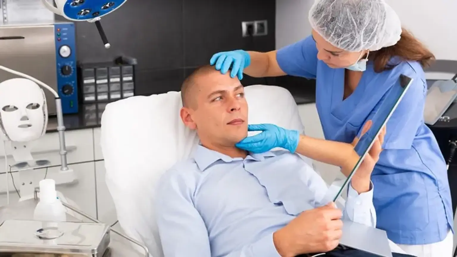
Diagnosing facial nerve paralysis typically involves a comprehensive medical evaluation, including a detailed history, physical examination, and specialized tests. A healthcare provider will first assess the symptoms and ask about the patient’s medical history to understand the potential cause of the paralysis.
Physical Examination: A clinician will assess the degree of facial weakness by asking the patient to perform various facial movements, such as smiling, raising the eyebrows, and closing their eyes. The doctor will look for asymmetry or involuntary movements (like synkinesis) that may indicate nerve damage.
Electroneurography: This test involves measuring the electrical activity in the facial nerve and its muscles. It helps determine the extent of nerve damage and can guide the decision-making process for surgery or other interventions. Electroneurography is a helpful tool for evaluating the function of the nerve and predicting the likelihood of recovery.
Imaging Tests: If a structural cause is suspected, such as a tumor or trauma, imaging tests like MRI or CT scans may be ordered to evaluate the facial nerve’s condition and identify any underlying issues. These tests can provide detailed images of the facial nerve and surrounding structures, helping doctors plan appropriate treatment.
Electromyography (EMG): EMG tests measure the electrical activity of muscles. It is often used to assess the degree of nerve damage and muscle function. An EMG can help determine whether the paralysis is caused by nerve damage or muscle dysfunction.
The diagnosis is critical for determining the appropriate treatment plan. If the paralysis is temporary and caused by something like Bell’s palsy, conservative treatments like corticosteroids and physical therapy may be sufficient. In cases where nerve damage is permanent, surgical intervention may be necessary to restore function and facial symmetry.
Initial Treatment Options for Facial Nerve Paralysis
When facial nerve paralysis is diagnosed, the first step is typically conservative treatment. In cases where the condition is temporary or caused by a condition like Bell’s palsy, the goal is to manage symptoms and promote recovery.
Medications: Corticosteroids like prednisone are commonly prescribed to reduce inflammation and swelling around the nerve, particularly in cases like Bell’s palsy. Antiviral medications may be used if a viral infection is identified as the cause.
Physical Therapy: Facial exercises and massage may be recommended to maintain muscle tone and prevent atrophy. These exercises can help patients regain facial movement over time.
Botox: In some cases, botulinum toxin injections (Botox) are used to manage unwanted muscle contractions or synkinesis that develop as a result of facial nerve paralysis.
If conservative treatments do not improve symptoms or if the paralysis is severe and permanent, more advanced surgical interventions may be required.
Surgical Intervention: When is Facial Nerve Reconstruction Necessary?
Surgical intervention becomes necessary when facial nerve paralysis causes permanent damage or when conservative treatments fail to restore function. This is often the case with trauma, tumors, or severe Bell’s palsy that does not improve over time.
Indications for Surgery: Surgery is considered when the paralysis is persistent, and the patient experiences significant facial asymmetry, impaired facial function (e.g., inability to smile, close the eyelid), or emotional distress due to the visible effects.
Timing of Surgery: The timing of reconstructive surgery is crucial. In cases like traumatic injury, immediate or early intervention may yield better results. In others, surgery might be delayed to allow the nerve sufficient time to heal naturally. If no improvement is seen after about 6-12 months, surgery is usually recommended.
Techniques for Facial Nerve Reconstruction
There are several surgical techniques used to reconstruct the facial nerve, depending on the severity of the paralysis and the cause of nerve damage.
Nerve Grafting: In cases of complete nerve injury, grafting involves taking a healthy donor nerve from another part of the body (often from the leg or neck) and transplanting it to the affected area. This technique aims to bridge the gap in the damaged nerve and promote regeneration.
Nerve Transfer: For certain types of facial paralysis, a nerve transfer may be done. A nearby healthy nerve, such as the hypoglossal nerve (from the tongue), can be rerouted to the facial nerve to restore movement.
Neurotization Surgery: This method involves using a nerve from another part of the body to stimulate facial muscles and improve their function. It is often used in cases where the facial nerve is completely severed and cannot be repaired directly.
These techniques help restore both motor function and facial appearance, though results can vary based on the extent of damage and the patient’s specific situation.
Nerve Grafting in Facial Nerve Reconstruction
Nerve grafting is one of the most common and effective methods used to treat severe facial nerve paralysis. It involves taking a healthy nerve from another part of the body and using it to replace or repair the damaged facial nerve.
Donor Nerves: The most common donor nerves for grafting are taken from the leg (sural nerve) or the neck (greater auricular nerve). These nerves are selected because they are less critical to other body functions and can be safely removed without major consequences.
Procedure: The donor nerve is carefully positioned and sutured into the damaged facial nerve to restore communication between the nerve and the facial muscles. Over time, the new nerve graft promotes the regeneration of nerve fibers and encourages the re-establishment of facial muscle movement.
Challenges: While nerve grafting can significantly improve facial function, it is a complex procedure. Nerve regeneration is a slow process, often taking several months to a year to show noticeable improvement. Additionally, nerve grafting may not fully restore fine motor control, meaning that patients may experience some asymmetry or diminished facial expressiveness.
Despite its limitations, nerve grafting remains one of the most reliable methods for restoring facial function in patients with severe nerve damage.
Benefits of Facial Nerve Reconstruction
Facial nerve reconstruction can significantly improve both the functional and aesthetic outcomes for patients with facial paralysis. The primary benefits include:
Restoring Facial Movements: Reconstructive surgeries, such as nerve grafting or transfer, can help patients regain some degree of facial muscle control, allowing them to smile, speak, and blink more naturally.
Improved Quality of Life: Restoring facial expression can improve self-esteem and social interactions, as individuals are less likely to feel self-conscious or disconnected from others due to facial asymmetry.
Psychological Impact: The psychological benefits of reconstructive surgery are profound. Many patients experience a boost in confidence and emotional well-being after treatment, as they feel more in control of their appearance and facial movements.
Despite the significant improvements, full recovery may not always be possible, and results may vary from patient to patient.
Risks and Complications of Facial Nerve Reconstruction
As with any surgical procedure, facial nerve reconstruction comes with potential risks and complications. Some of the most common include:
Infection: Any surgery carries a risk of infection, which can affect the surgical site and delay healing.
Nerve Regeneration Issues: Sometimes, the grafted or transferred nerves may fail to regenerate properly, leading to partial or no recovery of function.
Scarring: Surgical procedures involving the face can leave scars, which may affect aesthetic outcomes, especially if the incision site does not heal properly.
Facial Synkinesis: In some cases, patients may develop involuntary facial movements after surgery, where one facial movement triggers another (e.g., smiling causing the eye to twitch).
Asymmetry: Despite surgical intervention, complete symmetry may not be achieved. Patients may still experience mild to moderate facial differences.
Careful consideration of these risks, along with a detailed discussion with a qualified surgeon, can help patients make informed decisions about surgery.
Recovery After Facial Nerve Reconstruction
The recovery period following facial nerve reconstruction varies depending on the type of surgery performed and the extent of nerve damage. In general, the recovery process involves several stages:
Immediate Post-Operative Care: After surgery, patients may experience some swelling, bruising, and discomfort. Pain medications and anti-inflammatory drugs are typically prescribed to manage these symptoms. A follow-up appointment is usually scheduled within the first few days to monitor progress.
Physical Therapy: Physical therapy plays a crucial role in recovery, helping patients regain muscle strength and coordination. Facial exercises are prescribed to improve facial symmetry and prevent atrophy.
Regeneration Time: Nerve regeneration is slow, often taking several months to a year to show significant improvements. Patients must be patient and continue with rehabilitation to encourage recovery.
Long-Term Care: Once the initial recovery is complete, long-term care may involve touch-up surgeries or continued physical therapy. Regular follow-ups with the surgeon are important to monitor progress and address any complications.
While the recovery process can be lengthy and sometimes challenging, most patients experience gradual improvements that significantly enhance their facial function and appearance.
Advances in Facial Nerve Paralysis Reconstruction
Recent advances in medical technology and surgical techniques have made facial nerve reconstruction more effective than ever. Some notable advancements include:
Microsurgical Techniques: The use of advanced microscopes and fine surgical instruments allows for more precise nerve repairs and grafts. This has improved the success rate and reduced complications in facial nerve surgeries.
Stem Cell Therapy: Some studies are exploring the potential of stem cells to promote nerve regeneration and enhance the recovery process. Although this research is still in the early stages, it offers promising possibilities for patients with severe nerve damage.
Robot-Assisted Surgery: Robotic surgery is becoming more common in complex nerve repair procedures. This technology enables surgeons to perform intricate surgeries with greater accuracy, leading to better outcomes and faster recovery times.
Custom Implants for Facial Aesthetics: In addition to nerve reconstruction, some patients benefit from custom facial implants to improve the aesthetic appearance of the face. These implants are designed to restore symmetry and enhance the results of nerve reconstruction.
These advancements in surgical techniques and technologies are revolutionizing the field of facial nerve paralysis reconstruction, offering new hope to patients seeking to regain facial function and appearance.
Who is a Candidate for Facial Nerve Reconstruction?
Facial nerve reconstruction is not suitable for everyone, and the decision to undergo surgery depends on various factors. Ideal candidates typically have:
Persistent Facial Paralysis: Candidates are usually those who have had facial paralysis for 6–12 months with little to no improvement.
Sufficient General Health: Good overall health is necessary for anesthesia and to support the healing process after surgery.
Realistic Expectations: Patients should have a clear understanding of what surgery can and cannot achieve. While facial nerve reconstruction can significantly improve function, complete recovery is not guaranteed.
Non-reversible Nerve Damage: Surgery is recommended for patients with permanent nerve damage that cannot heal on its own.
After a thorough consultation, a skilled surgeon can determine if reconstruction is appropriate based on the patient’s medical condition and personal goals.
The Role of a Multidisciplinary Team in Facial Nerve Paralysis
Treating facial nerve paralysis often requires a team of specialists to ensure the best outcomes. This multidisciplinary approach may involve:
Plastic Surgeons: Surgeons specializing in facial nerve reconstruction perform the majority of surgical interventions, including nerve grafting and transfers.
Neurologists: Neurologists assess the function of the facial nerve and help manage underlying neurological conditions, such as Bell’s palsy or strokes.
Physical Therapists: After surgery, physical therapists guide patients through rehabilitation exercises to restore facial muscle function and improve symmetry.
Psychologists: Addressing the emotional and psychological effects of facial paralysis is important. Support from a psychologist can help patients cope with the changes to their appearance and regain self-confidence.
A coordinated approach ensures comprehensive care, improving both physical recovery and emotional well-being.
The Global Popularity of Facial Nerve Reconstruction
Facial nerve reconstruction has become increasingly popular worldwide, with advancements in techniques making it accessible to more patients. The procedure is particularly prevalent in countries with well-established healthcare systems, such as the United States, Germany, and South Korea.
Availability of Advanced Care: In countries like the U.S. and UK, patients have access to highly skilled surgeons and state-of-the-art medical facilities. Insurance coverage often supports the procedure for medically necessary cases, making it more accessible.
Growing Demand in Asia: In countries like South Korea, facial aesthetics are of significant cultural importance. This has driven an increase in demand for facial nerve reconstruction surgeries, as patients seek not only functional improvements but also enhanced cosmetic results.
Global Awareness: Increased awareness about facial nerve paralysis, especially through social media and patient advocacy groups, has led to more individuals seeking treatment options. As a result, facial nerve reconstruction is becoming a common solution for those affected by nerve damage.
As more people learn about the benefits of reconstructive surgery, the global popularity of facial nerve reconstruction continues to rise.
Cost of Facial Nerve Reconstruction
The cost of facial nerve reconstruction can vary greatly depending on the country, surgeon's experience, and the complexity of the procedure. On average, the cost can range from $5,000 to $25,000 or more. Several factors influence the price:
Type of Procedure: Nerve grafting, nerve transfers, and neurotization surgeries may have different costs, with more complex procedures generally being more expensive.
Geographic Location: Costs are typically higher in developed countries with advanced healthcare systems. In the U.S., for example, procedures may be more expensive than in countries with lower medical fees, like India or Mexico.
Insurance: Many health insurance plans will cover the cost of facial nerve reconstruction if it is deemed medically necessary, such as after trauma or stroke. Cosmetic procedures for aesthetic reasons may not be covered by insurance.
Patients should discuss the cost with their healthcare provider and explore financing options if necessary. The long-term benefits, including improved function and appearance, often outweigh the financial investment for those affected by facial nerve paralysis.
Post-Surgery Psychological Support
Undergoing facial nerve reconstruction can be emotionally challenging. Patients may experience a range of feelings, from anxiety about the procedure to frustration with the recovery process. Psychological support is vital for coping with these emotional challenges.
Counseling: Speaking with a psychologist or counselor before and after surgery can help manage stress and anxiety. This is especially important for patients dealing with significant changes to their appearance.
Support Groups: Many patients find comfort in connecting with others who have undergone similar procedures. Online or in-person support groups provide a space for sharing experiences and encouragement.
Realistic Expectations: A key part of psychological support is ensuring that patients understand the limitations of surgery and have realistic expectations for their recovery. This helps reduce disappointment and improves emotional outcomes.
How Facial Nerve Reconstruction Improves Confidence
For many patients, the most profound benefit of facial nerve reconstruction is the restoration of confidence. The ability to express emotions like happiness or sadness, and even engage in everyday activities such as eating or speaking without difficulty, can significantly improve one’s self-esteem.
Social Interactions: Restoring facial symmetry and movement can make it easier for patients to engage socially. Facial expressions play a critical role in non-verbal communication, and regaining this ability can reduce social isolation.
Professional Life: Many patients feel more confident returning to work or participating in social events, knowing that their appearance reflects their internal emotions and expressions.
Mental Health: The physical changes after surgery can contribute to a greater sense of emotional well-being. Feeling more "like oneself" after a successful surgery often leads to a positive shift in mental health.
Long-Term Outcomes of Facial Nerve Reconstruction
The long-term outcomes of facial nerve reconstruction vary based on the severity of nerve damage, the type of surgery performed, and the individual’s recovery process. However, most patients experience:
Improved Facial Function: Many patients regain significant muscle function, allowing for more natural facial expressions. This can be life-changing, particularly for those who have suffered from facial paralysis for years.
Partial Recovery in Some Cases: While full recovery may not be possible, many patients see a reduction in facial asymmetry, and the quality of their facial movements improves.
Ongoing Rehabilitation: Continued physical therapy and follow-up care are essential to maintain and further enhance recovery. Regular check-ups with a surgeon allow for adjustments and additional treatments if necessary.
With appropriate care and realistic expectations, most patients achieve lasting improvements in both function and appearance.
Conclusion
Facial nerve reconstruction continues to evolve, offering patients new hope for recovery and improved quality of life. The advances in microsurgery, nerve transfer techniques, and regenerative medicine are driving better outcomes.
As more research is conducted and techniques improve, the future looks promising for individuals dealing with facial nerve paralysis. With a multidisciplinary approach, patients can achieve not only functional restoration but also enhanced facial aesthetics.
Technological Advances: The future may hold breakthroughs in stem cell therapy, gene therapy, and even robotic surgery, all of which may further improve surgical outcomes.
Global Access: As awareness grows, more patients worldwide will have access to these life-changing treatments, helping to overcome the barriers of facial paralysis and restoring their confidence and quality of life.