Gingivectomy

    Last updated date: 03-Mar-2023

    Originally Written in English

    Gingivectomy

    Gingivectomy

    Overview

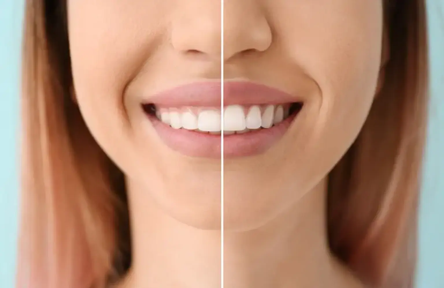
    Periodontal disease affects about 47.2 % of Americans over the age of 30. (also known as gum disease). Gingivectomy can be used to treat periodontal disease or to treat a gum issue involving the structures around the teeth. It is one of a few treatments that can aid in the treatment of periodontal disease. Continue reading to discover more about the operation, how it is performed, and whether it is a viable treatment option for restoring the health of your smile and gums.


    What is Gingivectomy?

    Gingivectomy Definition

    The surgical removal of gingiva is known as gingivectomy (gum tissue). When the gums have pushed away from the teeth, producing deep pockets, a gingivectomy is required. Plaque and calculus are difficult to remove due to the pockets. Gingivectomy is typically performed before gum disease has harmed the bone that supports the teeth.

    To eliminate pockets between the teeth and gums, loose, unhealthy gum tissue is removed and reshaped. Gingivectomy gives vision and accessibility to remove calculus and totally smooth the tooth roots by eliminating the pocket walls. This produces a favorable environment for gingival healing and gingival contour restoration. Although gingivectomy was originally created to treat periodontal disease, it is now widely used for cosmetic purposes. It is used to remove excessive gum tissue and enhance the gum's look.

    Gingivoplasty is another term for periodontal surgery. A gingivoplasty differs from a gingivectomy in that the former includes just a partial excision of the gums (plasty). The latter eliminates a complete area of gum. 


    What is Periodontal disease and how does it develop?

    Periodontal disease

    Periodontal disease starts when germs in the mouth cling to the teeth. Bacteria congregate and grow, generating a biofilm known as plaque. If plaque is allowed to accumulate on the teeth, the nearby gingival tissues can become irritated, resulting in gingivitis, an early form of gum disease. Daily flossing and twice-daily brushing with a bacteria-fighting toothpaste can help avoid gingivitis. Oral hygiene techniques remove plaque and food debris, clean the surface of the teeth, and eradicate bacterial plaque at the gum line of the teeth.

    However, if plaque and food debris are not removed and oral hygiene practices are not followed, gingivitis will worsen and the gum tissue will become more inflamed, bleeding will occur, the area between the tooth and gum tissue will deepen, forming a periodontal pocket, and periodontal disease will develop.

    As plaque bacteria accumulates and travels below the gum line, a periodontal pocket forms. At this stage, home care is insufficient for eradicating tooth plaque. If not treated, the biofilm will expand below the gum line and infect the inside of the pocket. This sort of severe periodontal disease can harm the tooth roots and cause them to get infected. The teeth may become loose or painful, necessitating gum surgery. At this time, your dentist may tell you that you require a gingivectomy.


    Who Should Undergo and Expected Results

    candidates for gingivectomy

    Patients with gingivitis are good candidates for gingivectomy. Plaque development caused by microorganisms characterizes this disorder. Over time, the plaque hardens and develops tartar or calculus. The gum gets irritated, and the connection surrounding the tooth becomes loose. Calculus can no longer be eliminated by brushing or flossing the teeth in its advanced form. Bleeding is another possibility. Gingivectomy is recommended if the condition does not improve following a manual thorough cleaning procedure known as curettage. This entails scraping off the plaque and root-planing the tooth.

    Periodontitis patients can also benefit from gingivectomy. This illness is also caused by a bacterial infection, which causes inflammation of the gums. If left untreated, periodontitis frequently results in tooth loss and damage to the underlying bone structure. In certain circumstances, the gum might get so infected that antibiotics alone will not cure it.

    Gingivectomy is a relatively safe operation that is often performed in a dentist's clinic. It also has a high percentage of success and pleasure. To promote recovery, the patient is frequently encouraged to rest for a few days. Following gingivectomy, a soft food diet is also recommended.

    The process is carried out one quadrant at a time. For people with substantial tartar accumulation, multiple gingivectomy treatments may be necessary to complete the therapy. Antibiotics are also administered to the patient to avoid infection. A regular dental checkup is also essential to monitor and prevent any additional tartar buildup. Oral hygiene should be practiced at all times.

    This surgery also aids in the prevention of tooth loss and the preservation of the underlying bone structure. Tissue healing and surface epithelial development are accomplished within a few days. 


    Contraindications to Gingivectomy?

    The patient's grin frame is influenced by dental/gingival morphologic features and peri-oral factors. They are critical in ensuring a predictable and successful smile rehabilitation for the patient.

    The typical vertical height of the maxillary central incisors in boys and females is 10.6mm and 9.8mm, respectively. The average maxillary incisor display with the lip line at rest is 1.91mm for males and 3.40mm for women (nearly double the amount). Recent research has confirmed the statistically significant sexual dimorphism in relation to the height of the visible maxillary incisor crown at rest.

    A study also clearly shows that female patients had larger smile lines and male patients have lower smile patterns. A high grin line reveals the whole crown of the tooth as well as an abundance of gingiva. Since a result, some people may perceive this operation subjectively, as some degree of gingival show may be visually pleasant and deemed youthful, and vice versa.

    Furthermore, the harmony of the gingival contour between the anterior and posterior portions may be compromised. While smiling, some persons are more likely to show the maxillary teeth from one side's second premolar to the other side's second premolar. As a result, in certain circumstances, all teeth between the first molars are included in the operation, particularly in surgical crown lengthening, to generate an aesthetically acceptable gingival architecture that blends the gingival contours of the maxillary anterior and posterior teeth harmoniously. Aside from that, "black triangles" are more prone to form in regions with labial or interproximal soft tissue recession. This results in the intended consequence.


    How is the Procedure Performed?

    Gingivectomy Procedure

    Before the surgery, the patient must have gingiva scaling and root planing to remove as much plaque as feasible. Gingivectomy can be performed using a variety of ways. All approaches entail the use of local anesthetic to make the process as painless for the patient as feasible.

    The dentist begins surgical gingivectomy by probing the pockets formed by loose gum tissue. These pockets are also identified. To assess the tooth and the underlying bone structure, small incisions are made in the gum. To expose the root of the tooth, the gingival flap is pushed away from the tooth. The diseased gum tissue is then removed, along with a border of good tissue.

    The curette is then used to remove the granulation tissue. Any deep calculus or tartar accumulation is also removed with the same device. The surgical site is cleaned with sterile saline solution and gauze is applied. A periodontal pack is put over the incision once the dentist is confident that all unhealthy gum tissue has been removed and bleeding has been stopped. This surgical garment promotes tissue regeneration and repair.

    When conducting gingivectomy, some dentists prefer to utilize a laser. A carbon dioxide or Nd:YAG laser can be used to precisely chop away the unhealthy gum tissue. Laser beams can also seal any damaged blood artery, lowering the risk of bleeding.

    Electrosurgery is another method used in gingivectomy. To transfer heat energy to the gum tissue, the dentist employs high-frequency electric current. This starts the desiccation process, which continues until all of the tissues have dried out. The cells degrade, and the unhealthy gum tissue is removed.

    Caustic chemicals known to break down cells are also used in gingivectomy. Chemosurgery is a technique that has been shown to be helpful in eliminating gingival pockets. Chemicals utilized include phenol solution and paraformaldehyde.

    This technique can also be performed using cryosurgery. Temperatures ranging from -50 to -60 degrees Celsius are introduced into the gingival pocket using a probe. The freezing temperatures cause cell death and necrosis. A scalpel is subsequently used to remove the unhealthy tissue.


    Which is Better: Gingivectomy Laser vs Scalpel?

    Gingivectomy Laser vs Scalpel

    Laser technological advancements have resulted in a less expensive and more effective surgical alternative, particularly for oral treatments. A laser allows dentists to perform more accurate work. Because of the cauterization, gingivectomy laser operations heal faster and lower the risk of infection.

    While laser operations are more expensive than scalpel gingivectomies, the price difference is closing every year. However, before proceeding, you should verify with your insurance provider to ensure that a laser surgery is covered.


    What care is required after a gingivectomy?

    gingivectomy after care

    Most patients may resume their usual dental care routine within a month after the surgery. Routine dental or periodontal checks will ensure that the procedure is a success. The dental expert who conducted the operation will most likely schedule three-month appointments, followed by at least a twice-yearly preventative health visit to clean in and around the surgical site.


    Recovery and Aftercare

    Gingivectomy Recovery and Aftercare

    You will most likely feel edema and bleeding following the surgery. This is quite normal, however doctors will advise you on how to expedite the healing process. After the anesthesia wears off, you may experience some discomfort for a few hours. Again, this is normal, and over-the-counter pain relievers will assist.

    Because just local anaesthetic will be used, you should be able to go home straight away. Please let our staff know if you need to relax following surgery or if you need help traveling home.

    You will need to change your bandages and dressings yourself, as indicated, but professionals will show you how to do so before you leave for home. If you are still unclear about the directions you were given, contact the clinic for assistance.

    Brushing, flossing, and washing your mouth should be avoided for the first 24 hours following the treatment. Following this first interval, you may continue your usual dental routine in the portions of your mouth that were not impacted by the gingivectomy. After 48 hours, rinse with saltwater to keep your gums clean and help them recover faster.

    Avoid touching the injured region during the first week of healing. This includes no contact with your fingers or tongue, as well as no forcing your lips open to look at it.


    What's the Difference Between Gingivectomy and Gingivoplasty Procedures?

    Gingivectomy and Gingivoplasty Procedures

    Gingivectomy and gingivoplasty, also known as gum contouring and gingival sculpting, are sometimes used interchangeably since they are frequently performed concurrently. The two processes, however, differ somewhat.

    A gingivectomy is the surgical excision of unhealthy gum tissue. Gingivoplasty is often used to remodel otherwise healthy gum tissues. However, one treatment is seldom performed without the other.


    How Long Does It Take to Heal after a Gingivectomy?

    This procedure begins with numbing gel applied to the gingival tissue (no injections) and is completed with a soft tissue laser that reshapes or removes the gingival tissue quickly and efficiently. Healing is almost instantaneous, with little to no postoperative sensitivity, and the tissue usually returns to its original shape in 3 to 5 days.

    Use floss and a toothbrush with gentle bristles to maintain adequate oral hygiene. To promote tissue development, rinse with an antimicrobial mouthwash.


    What to Expect?

    After the anesthesia wears off, you can resume your daily activities. The gums normally recover within a few days or weeks. Your gums' form or shape may alter.

    Most gum procedures are straightforward and quite painless. To relieve discomfort, you can take ibuprofen (Advil or Motrin) or acetaminophen (Tylenol). Take precautions when handling medications. Read and obey all label directions. It will be easier for you to keep your teeth and gums clean after a gingivectomy.


    Possible Risks and Complications

    Gingivectomy Possible Risks

    Gingivectomy is associated with the following risks and complications:

    • Bleeding, which can occur during and after the procedure
    • Pain and swelling of gums
    • The surgery site is infected. In rare situations, the infection may spread to the circulation and cause sepsis.
    • Blood Clot
    • Chemosurgery patients might have bone necrosis.
    • Abscess in the periodontium
    • When an electric current or chemicals are utilized, they cause damage to the surrounding healthy cells.
    • Nerve damage in the vicinity
    • Cold temperature sensitivity of the teeth
    • Recurrence of plaque accumulation, especially if diseased gum tissue was not completely removed


    Conclusion 

    The surgical removal of gingiva or gum tissue is known as a gingivectomy. This operation is used to treat gum disease and to remove deep pockets that form when the gums separate from the teeth. The surgery involves the removal of gingival abnormalities, which results in a better gingival contour. Most gingival smile situations may be resolved surgically by removing 1 to 2 millimeters of gingival tissue. The surgical approach provides the option of performing the incision using a standard scalpel, electrosurgery (electric scalpel), and high-frequency lasers, depending on the treatment plan and patient-professional harmony in each situation.