Gynecologic Laparoscopy

Last updated date: 02-Mar-2023

Originally Written in English

Gynecologic Laparoscopy

Overview

Laparoscopy has progressed from a restricted gynecologic surgical technique used only for diagnostic and tubal ligations to a significant surgical instrument utilized for a wide range of gynecologic and nongynecologic purposes during the last 50 years. Laparoscopy is becoming one of the most popular surgical procedures in many regions of the world.

Laparoscopy has become the treatment of choice for numerous gynecologic operations, including ectopic pregnancy removal, endometriosis therapy, ovarian cystectomy, and hysterectomy. Multiple studies have demonstrated that laparoscopy is safer, less costly, and has a quicker recovery period than laparotomy. Other treatments, such as myomectomy, sacral colpopexy, and the diagnosis and therapy of gynecologic malignancies, are benefiting from the laparoscopic technique.
 

What is Gynecological laparoscopy?

Gynecological Laparoscopy

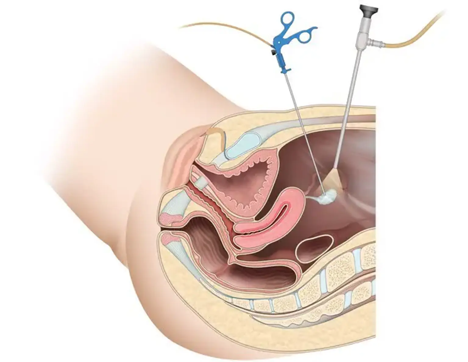
A gynecological laparoscopy (keyhole surgery) is a treatment that examines your womb (uterus), fallopian tubes, and ovaries from within your lower belly (abdomen). Gynecological laparoscopy can be used to diagnose and/or treat a problem.


Indications of Gynecological laparoscopy

Indications of Gynecological laparoscopy

  • Diagnostic laparoscopy

A clinician must often examine the pelvis for acute or persistent discomfort, ectopic pregnancy, endometriosis, adnexal torsion, or other pelvic disease. Determining tubal patency may also be a problem. A main port for the laparoscope (also known as the "lens") is usually put infraumbilically, while a secondary port is implanted suprapubically to probe and view pelvic organs methodically.

A biopsy specimen can be collected if necessary, to help in the diagnosis of endometriosis or cancer. If tubal patency is a concern, a uterine manipulator with a cannula can be used to inject a dilute dye transcervically (chromopertubation).

To reduce the danger of aspiration, diagnostic laparoscopy is frequently done under general anesthesia with endotracheal intubation. However, if the peritoneal insufflation pressure is minimized, laparoscopy can be conducted under conscious sedation.

  • Tubal sterilization

Trocar insertion is comparable to diagnostic laparoscopy. To occlude the tubes in the mid-isthmic section, roughly 2-3 cm from the cornua, bipolar electrosurgery, clips, or silastic bands may be utilized. With three contiguous passes, bipolar surgery desiccates the tube, occluding roughly 2 cm of tube. The availability of auditory tone to check entire resistance has increased the effectiveness of bipolar cautery. Pregnancy rates differ depending on patient age, ranging from 1-3% after 10 years.

  • Lysis of adhesion

Adhesions can arise as a result of a past illness, such as a burst appendix or pelvic inflammatory disease (PID), endometriosis, or surgery. Adhesions can cause infertility and severe pelvic discomfort. Most patients have a minimal likelihood of becoming pregnant following adhesion lysis, and this form of treatment has been mostly replaced by in vitro fertilization and embryo transfer. Similarly, adhesiolysis is frequently useless in treating persistent pelvic discomfort, in part because most adhesions regenerate quickly after surgery.

Blunt or sharp dissection can lyse adhesions. Prior to lysing, aquadissection may help in the creation of planes. For cutting and coagulation, any of the power tools may be utilized (see Power instruments). Although unipolar electrosurgery is safe in competent hands, gynecologist use of unipolar electrosurgery, such as with the tiny unipolar needle, is often limited to adhesions 1-2 cm from the ureter and intestine because to the unpredictable nature of current arcing. Other power approaches for adhesiolysis near the bowel may be safer.

Adhesions may reappear following lysis, however this can be minimized with excellent hemostasis and little electrocautery usage. In a recent controlled study, a 4% icodextrin solution was proven to reduce adhesion reformation. Unfortunately, the outcomes of laparoscopic adhesion lysis have been poor in terms of pain alleviation and future fertility.

  • Treatment of endometriosis

Laparoscopy is the most often used technique for diagnosing and treating endometriosis. Any of the power tools can be used to resect or ablate endometriotic lesions. Multiple well-designed studies have indicated that both of these treatments boost fertility and reduce pelvic discomfort. Franck et al. published a comprehensive evaluation that found improvements in sexual life after laparoscopic surgery for endometriosis. 

  • Treatment of ectopic pregnancy

Most ectopic pregnancies are treated surgically with laparoscopy. To remove the embryo and gestational sac, a salpingostomy or salpingectomy may be done. Although auxiliary tools (such as pretied loops and stapling devices) can be utilized for salpingectomy, a power instrument (such as bipolar electrosurgery or an ultrasonic scalpel) is most typically employed for these operations.

A cystectomy is recommended if a simple ovarian cyst measuring 6 cm or greater persists for two or more cycles in a premenopausal, nonpregnant female. Depending on the size of the cyst and the possibility of malignancy, this can be accomplished either laparoscopy or laparotomy.

A variety of procedures can be used to eliminate the cyst. If the cyst is complicated, search for evidence of ascites, excrescences on the ovary, or implantations on the peritoneal, hepatic, or diaphragmatic surfaces to rule out malignancy. If no cancer is visible, gently dissect the cyst, attempting to remove it whole. A bag can be used to convey the cyst out of the peritoneal cavity via a 10-mm port, with the cyst being drained before the bag is removed.

If there is any uncertainty, the cyst wall should be freeze sectioned to confirm a benign cyst. If cancer is discovered, a laparotomy should be performed. All cysts are permanently sectioned and pathologically diagnosed. Unless a benign cystic teratoma is a high probability, ovarian cysts with septa, internal echoes, or solid tumors are not appropriate candidates for laparoscopy.

If the cyst ruptures after removal, use Ringer lactate solution to thoroughly cleanse the peritoneal cavity. A dermoid cyst is especially dangerous because it can contaminate the peritoneal cavity with sebaceous material, resulting in chemical peritonitis. However, two case studies found that pregnant and nonpregnant women who had an intraoperative leakage of a dermoid cyst followed by prolonged peritoneal irrigation had no increased length of stay or postoperative problems. Fear of seeding the cavity with a malignant tumor has long existed, but current research suggests that spilling has little effect on prognosis provided a staging laparotomy is performed soon.

Although postmenopausal cysts can be removed with laparoscopy, an oophorectomy and laparotomy may be more judicious due to the increased risk of cancer. Physicians who conduct laparoscopies should be familiar with staging by laparoscopy or laparotomy, and malignancy should be ruled out perioperatively.

  • Salpingo-oophorectomy

In postmenopausal women with a developing or chronic cyst, salpingo-oophorectomy may be more suitable. In women who have a hereditary propensity to ovarian or tubal cancer, tubes and ovaries are also removed prophylactically. A tubal pregnancy or a big hydrosalpinx with adhesions may necessitate ovary removal as well. To safely remove the ovary, power tools, pretied loops, or stapling devices may be utilized to occlude the infundibular ligament. Because of the size of the ovary, a retrieval bag is required to remove the tissue.

  • Myomectomy

In order to maintain fertility or the uterus, many women with a symptomatic fibroid uterus prefer myomectomy versus hysterectomy. If the fibroid is pedunculated, the stalk may be readily incised. However, the risk of bleeding increases with intramural fibroids. In anemic individuals, a preoperative gonadotropin-releasing hormone (GnRH) agonist may be recommended.

However, some studies have found that using GnRH agonists in laparoscopic myomectomy results in longer surgical hours and a greater rate of conversion to laparotomy due to difficult cleavage planes. A vasopressin injection into the uterus may assist preserve hemostasis. The fibroid defect must be sutured, which might be challenging for unskilled laparoscopic practitioners. Adhesion development can be reduced via barrier approaches.

Morcellation or colpotomy might be used to remove the fibroid. The possibility of malignant tissue spreading intraperitoneally prompted the FDA to issue a black box warning against using laparoscopic power morcellation to remove uterine fibroid tumors in 2014. Laparoscopy has not been shown to be superior than laparotomy for the treatment of menorrhagia or infertility. Furthermore, there is some worry that the risk of recurrent uterine rupture during pregnancy may be higher following laparoscopic myomectomy vs laparotomy.

However, no randomized clinical trials comparing laparoscopic myomectomy to laparotomy found a higher risk of rupture or worse reproductive results. These experiments were carried out by laparoscopic suturing professionals on carefully selected patients.

To restore normal anatomy, laparoscopy was initially performed prior to vaginal hysterectomy. However, it is now often utilized for a number of purposes, including determining the feasibility of a vaginal hysterectomy (where adhesions, endometriosis, or a big fibroid uterus are suspected) and conducting some or all of the actual hysterectomy. Laparoscopic-assisted vaginal hysterectomy (LAVH), laparoscopic hysterectomy (LH), and laparoscopic supracervical hysterectomy are the three primary laparoscopic hysterectomy techniques (LSH). Although the fundamental procedures for each therapy are largely standardized, there is disagreement on the dangers, benefits, and most appropriate indications for each. 

  • Oncologic procedures

Laparoscopy has long been utilized in oncology for follow-up treatments after surgical and chemical therapy of cancer. Laparoscopy has lately been employed for staging procedures such as peritoneal washes with biopsy, partial omentectomy, and pelvic and periaortic lymphadenectomy. Some gynecologic oncologists have employed procedures such as laparoscopically assisted radical vaginal hysterectomy.

Many gynecologic cancer instances appear to be changing as a result of robotic laparoscopic surgery. The robotic technology enables surgeons to conduct treatments that would have previously required a laparotomy using modified laparoscopic methods.

The advantages of the robotic system include 3-dimensional, high-definition imaging and magnification, which improve the topography of the pelvis and the cervicovaginal plane. Fully articulated instruments simulate a surgeon's wrists and hands in their complete range of motion. This improves the surgeon's ability to conduct fine motor skills remotely, such as complex dissections and intracorporeal suturing, which are difficult to do during standard laparoscopy.


Preparing for a gynecological laparoscopy

gynecological laparoscopy preparation

Your hospital will provide instructions on how to prepare for your gynecological laparoscopy. If you need a gynecological laparoscopy to identify an issue, you will normally undergo the operation and be able to go home the same day. If the treatment is being performed to address a condition, you may be required to stay in the hospital overnight.

A gynecological laparoscopy is often performed under general anesthesia, which means that you will be sleeping during the surgery. Your hospital will advise you on when to cease eating and drinking. You should avoid eating around six hours before your gynecological laparoscopy and only drink clear fluids for up to two hours. However, listen to your anaesthetist's advise.

A nurse will test your urine (pee) at the hospital to ensure that you are not pregnant or have any conditions that might interfere with the treatment. Compression stockings may be prescribed to assist prevent blood clots from developing in your leg veins (deep vein thrombosis). You may require an anticlotting injection in addition to or instead of using compression stockings.

Your nurse or doctor will explain what will happen before, during, and after your operation, including any discomfort you may have. If you have any questions, simply ask. It is critical that you be completely informed so that you can give your consent for the treatment to proceed. You will be required to sign a permission form.


Gynecological laparoscopy procedure

Gynecological laparoscopy procedure

If you're undergoing a gynecological laparoscopy to identify a disease, it should take approximately half an hour to an hour. If your doctor needs to treat a problem, it will take longer.

One or more tiny cuts in your lower belly, including one in your tummy button, will be made by your doctor (navel). They'll next insert a tube through one of these incisions and gently inflate your stomach so they can look better. Your doctor will next insert a tiny telescope with a camera (laparoscope) into another incision. The laparoscope's camera transmits images of the inside of your belly to a display where your doctor may view them. If your doctor has to obtain tissue samples or treat a problem, he or she may pass other equipment through the wounds.

Your doctor will seal the wounds with stitches or glue at the end of the process.


What to expect afterwards?

Gynecologic Laparoscopy Expectations

You will need to relax till the anesthesia has worn off. You may have some discomfort as the anaesthesia wears off, but you will be given pain medication. When you're ready, you should be able to go within a few hours. Ask a family member or a friend to drive you home and to remain with you for a day or two while the anesthesia wears off.

A general anesthesia may be quite draining. You may discover that you are less coordinated or that it is harder to think coherently. This should be over in 24 hours. In the meanwhile, do not drive, consume alcohol, use machinery, or sign any crucial documents.

For the first day or two, you may have vaginal bleeding. You can use sanitary pads to halt the bleeding. If you have bleeding that resembles a heavy menstruation, contact your local hospital for medical help.

Your nurse will provide you with information on wound care, cleanliness, and bathing. A follow-up appointment date may be assigned to you. Dissolvable sutures or glue will dissolve on their own if you have them on your wounds. If you have non-dissolvable sutures, you will need to have them removed within five to seven days. A practice nurse at your GP clinic should be able to assist you with this.


  • Recovery after gynecological laparoscopy

You may have some discomfort for a few days following your surgery. Your hospital may provide you with pain relievers to take home, or over-the-counter pain relievers such as paracetamol or ibuprofen may be effective, but always read the patient information booklet that comes with your medicine. If you have any questions, see your pharmacist.

It's critical to rest and relax for the first 48 hours after surgery. If you have a simple laparoscopy or one to identify a disease, you should be able to resume your normal activities in approximately three days. And you might be able to return to work within a week. If you've had a more involved operation and therapy, it might take two or three weeks to feel like you're back to normal and ready to work. However, this will depend on the nature of your profession; get guidance from your doctor.

It's an excellent idea to be active. You may not feel like it, but it will help you avoid future complications, such as blood clots in your legs or lungs. Your doctor may also advise you to wear compression stockings or receive blood thinning injections for a period of time following your surgery. These also aid in the prevention of blood clots.

You should be able to have sex when you are ready, but with your doctor first. If you feel ill or have any of the following symptoms after returning home, consult your doctor or a hospital.

  1. A burning or stinging feeling when you pass urine, or passing urine often – this could mean you have a urine infection
  2. Sore, red skin around your scars, which could be a sign of infection
  3. Tummy pain that’s getting worse, which could be a sign of damage to your bladder or bowel
  4. A red, swollen and painful lower leg, which could be symptoms of a blood clot in your vein (deep vein thrombosis, DVT)


Complications of gynecological laparoscopy

Complications of gynecological laparoscopy

Complications occur when issues arise during or after your surgery. The following are some of the risks associated with a gynecological laparoscopy.

  • Other organs in your tummy, such as your bowel, bladder, womb or major blood vessels, may be damaged during the procedure.
  • You may develop an infection.
  • You may have bleeding or an incisional hernia around a wound. This is a lump beneath your skin. It can occur when a wound is not adequately healed and tissue pokes through.

If a problem arises during your laparoscopy, your doctor may need to switch to open surgery. This implies they'll have to create a larger incision in your belly.

If you're having a laparoscopy to determine what's causing your symptoms, your doctor may not notice an obvious cause during the surgery. So, even if you have a laparoscopy, you may still not know what is causing your symptoms.


What are the alternatives to a gynecological laparoscopy?

Gynecological laparoscopy isn't suitable for everyone. Your doctor may suggest alternative tests or treatment if you:

  • Are very overweight
  • Are older
  • Have had surgery on your tummy before
  • Have a heart or lung condition

Other tests or treatments may be available depending on what is causing your symptoms. If you have endometriosis, your doctor may recommend pain relievers and hormone therapy.

You may undergo an ultrasound scan before being given a gynecological laparoscopy. Some gynecological disorders, such as endometriosis and fibroids, can be diagnosed with this method. To check for fibroids, you can get an ultrasound scan that looks at your womb from the outside, via your lower abdomen. You can also get an ultrasound that examines within your womb with a probe placed into your vagina. Before a gynecological laparoscopy, you may undergo another form of scan, such as a CT or MRI scan. An MRI scan can also be used to diagnose diseases like fibroids.


Frequently asked questions about gynecological laparoscopy

gynecological laparoscopy

  • How long does a gynecological laparoscopy take?
    If you're undergoing a gynecological laparoscopy to identify a disease, it should take approximately half an hour to an hour. If your doctor needs to treat a problem, it will take longer.

  • Will I bleed after a gynecological laparoscopy?
    You may experience vaginal bleeding for a day or two after your laparoscopy. You can use sanitary pads to halt the bleeding. If you have bleeding that resembles a heavy menstruation, contact your local hospital for medical help.

  • What is a gynecological laparoscopy for sterilization?
    Sterilization is a surgical procedure that prevents you from becoming pregnant. It is a long-term method of contraception. It may be a choice for you if you do not want to become pregnant in the future. Because the operation is permanent, you must be as certain as possible that you will not be disappointed. Counseling should be given to you, ideally with your partner if you have one, to help you consider and discuss all of the advantages and downsides. You might also request that your doctor discuss additional possibilities to you.

  • What happens during gynecological laparoscopy for sterilization?
    If you select gynecological laparoscopy for sterilization, your doctor will insert a laparoscope through a slit in your stomach button (navel). They'll next make another incision and insert a tool through your fallopian tubes to shut them with clamps. Instead of clips, some doctors may use heat from an electric current or another equipment to shut your fallopian tubes. Even if you have unprotected intercourse, sperm cannot reach an egg to fertilize it once your fallopian tubes have been blocked.


Conclusion 

Laparoscopy is a surgical procedure that has been regularly utilized in medicine for more than 30 years. Faster recovery time, less discomfort, less hospitalization, and superior cosmetic results are just a few of the benefits that have made laparoscopy popular among patients and surgeons alike.

Laparoscopy has taken the lead and appears to be the gold standard method for a wide range of gynecologic procedures such as tubal ligation, removal of ovarian cyst or adnexa, treatment of ectopic pregnancy, hemorrhagic rupture of a cyst, exploration of chronic pelvic pain, sterility, treatment of endometriosis, removal of fibromyomata, hysterectomy, and recently for treatment of pelvic organ prolapses, urinary incontinence and even in gynecologic cancers.