Micro Tese (2 testicle)

    Last updated date: 03-Mar-2023

    Originally Written in English

    Micro Tese (2 testicle)

    Micro Tese

    Overview

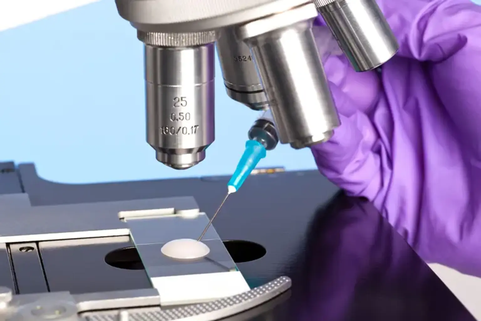
    MicroTESE (microscopic testicular sperm extraction) is a method of extracting sperm directly from the testicular tissue of a man's reproductive system. This medical therapy may be administered for reproductive difficulties if a man is unable to create or release enough quality sperm on his own. The testicular tissue is found in the two testes, which are responsible for sperm production. The testes are housed within the scrotum, a little sac behind and under the penis.

    The aims of the microTESE process are to collect the highest quality sperm possible, to obtain enough sperm to fertilize a woman's egg, and to limit harm to the reproductive organs.


    What is Micro TEZE?

    Micro TEZE

    Microsurgical testicular sperm extraction (microTESE) is a surgical procedure that removes sperm from the seminiferous tubules of a male's testes. It is used to treat males with non-obstructive azoospermia, a condition in which a man cannot produce enough sperm to have a detectable quantity in his sperm - a prevalent cause of male infertility. If a man has enough testosterone in his blood after a hormone test and subsequent tests reveal that his testicles are not generating normal quantities of sperm, physicians will typically recommend microTESE.

    If a person continues to be azoospermic following treatment and testosterone levels have been normal for at least four months. MicroTESE has a high success rate in men. Doctors can identify sperm 60% of the time using microTESE treatments.


    What is Male Infertility?

    Male Infertility

    Infertility is a reproductive system condition that makes it difficult for the body to perform fundamental reproductive tasks. Men and women are both impacted. The majority of cases of infertility are treated with medication or surgery. Male infertility can be caused by either nonobstructive (the male does not produce sperm) or obstructive azoospermia (sperm is created but obstructed and cannot be released from the body).

    The causes of infertility are frequently unknown. Sometimes the challenges are the consequence of a hereditary condition, such as cystic fibrosis, a birth defect, a medical problem, or a previous therapy that resulted in infertility (such as certain cancer treatments). In some circumstances, we don't know what's causing the infertility, but there are therapies that might assist.


    What Makes Micro TESE Successful?

    Male Infertility

    MicroTESE requires a knowledgeable surgeon and an excellent andrology technician to look for sperm. During your microTESE procedure, an andrology lab technician will be present in the operating room to analyze your seminiferous tubules for sperm. If sperm is found during your microTESE, it will be collected and conserved for future reproductive treatments such as in-vitro fertilization (IVF) with intracytoplasmic sperm injection (ICSI).

    Current study suggests that frozen sperm may outperform fresh sperm during IVF (in-vitro fertilization). Please remember that any sperm found during this therapy must be used for IVF (in-vitro fertilization). Because the sperm in your testis has not yet learned to swim, it will not be able to fertilize an egg if placed inside the uterus.


    How is male infertility diagnosed?

    infertility diagnosed

    If a couple has been trying for several months without taking birth control and is still unable to conceive, they should see a doctor. If the lady is over the age of 35, the couple should seek medical attention as soon as feasible. Your doctor will evaluate both partners to rule out any physical issues that might be causing infertility. The doctor will also ask numerous questions and review each person's medical history.

    To examine male fertility, the following tests may be advised or ordered:

    • Semen analysis: It determines the quantity and quality of sperm. Semen is a bodily fluid produced by the male reproductive organs. It transports sperm and other nutrients that help the sperm survive and fertilize successfully.
    • Blood test: Examines for genetic or hormonal issues. (Hormone levels affect both male and female fertility.)
    • Ultrasound of the scrotum: Examines the veins that transport blood from the testicles to the remainder of the body for abnormalities.

    In-home testing kits for sperm analysis may be accessible. Inquire with your doctor for further information.


    What will the infertility tests determine?

    Infertility tests determine

    Semen analysis will provide the doctor with the information he or she needs to determine fertility and develop a treatment strategy. The testing should reveal the following:

    • Amount of semen: A minimum of 1.5 milliliters is deemed acceptable. A smaller quantity may indicate an internal problem with a section of the reproductive system, such as the seminal vesicles or a prostate gland, which is preventing the discharge of sperm.
    • Sperm count: A usual range is fifteen million to 300 million per milliliter. A population of less than 15 million is considered abnormal.
    • Morphology: The sperm's size and form. Using the "strict" criterion, 4% of normal-shaped sperm is optimum for fertilizing an egg. Some doctors see a smaller proportion as normal.
    • Motility: The sperm's movement. Around 40% of the sperm should be migrating. The movement quality is scored from 0 to 4, with a score of 3 deemed satisfactory.


    How does Micro-TESE work?

    Micro-TESE work

    Micro-TESE is typically performed in conjunction with ICSI since the concentration of sperm retrieved is frequently low and insufficient for IVF. Once you have opted to have micro-TESE, you and your doctor will agree on a date for the procedure. This will only happen after you have had an in-depth consultation with a member of your health-providing team and have undergone a series of tests to see if this is the best option for you. This is usually done before to beginning IVF treatment.

    The surgeon opens the testicles on both sides and examines the tissues under a microscope, eliminating any sections he feels are likely to contain sperm - he can tell by looking at the seminiferous tubules (the portions of the testicles that make and transport sperm) and detecting which ones are dilated.

    Once all of these regions have been retrieved, they are brought to the lab and tested to determine whether there is any sperm present. Any discovered sperm will be used for therapy or saved until the point in the IVF treatment cycle when it is required. At that point, the sperm will be defrosted and the strongest specimens will be chosen to be injected straight into the collected eggs.


    Optimization before Micro TESE

    Optimization before Micro TESE


    Medical management:

    Most men who appear with azoospermia owing to hypothalamic-pituitary axis dysfunction will benefit greatly from medical care with gonadotropin releasing hormone (GnRH) or gonadotropins (i.e., hCG, hMG, recombinant FSH). Pulsatile GnRH 25-600 ng/kg subcutaneously (SC), intravenously (IV), or via pump every 120 minutes, hCG 1,000-2,500 IU administered intramuscularly (IM)/SC twice per week, or hMG 75-150 IU injected three times per week, and recombinant FSH 75 IU every 1-2 days are common regimens. If testicular function is enhanced with hormone treatment, most of these men will not require sperm retrieval.

    Medical optimization techniques for individuals with azoospermia owing to testicular dysfunction have been examined, with the objective of boosting both testosterone and FSH levels to increase spermatogenesis. In a multi-institutional trial, 612 males with non-obstructive azoospermia (NOA) were given a combination of clomiphene citrate (CC), hCG, and/or hMG to elevate blood testosterone to 600-800 ng/dL and serum FSH to 1.5 times baseline.

    In this study, the intervention group underwent hormonal optimization according to a predefined protocol, but the control group did not, resulting in a considerably higher sperm retrieval rate (SRR) for the intervention group (SRR 57% vs. 34% for intervention group vs. control group). While this study supports medical hormonal adjustment of FSH and endogenous testicular testosterone levels to enhance SRR, it is crucial to highlight that the control group had a lower SRR than prior literature findings.

    Furthermore, a major retrospective analysis of 1,054 men found that hormone treatment had little effect in males receiving micro TESE. In this study, no change in SRR was seen between men with baseline testosterone >300 ng/dL and not receiving medical hormonal therapy, or those taking aromatase inhibitors, selective estrogen receptor modulators, or any combination with hCG.

    Aromatase inhibitors, such as anastrozole and letrozole, have also been utilized to improve hormonal parameters in NOA and cryptozoospermia patients. Although the majority of research used anastrozole, which appears to have less adverse effects, one study comparing letrozole to placebo in cryptozoospermia and NOA patients found that letrozole may assist improve spermatogenesis with sperm return to the ejaculate in certain NOA patients. Men with low blood testosterone (300 ng/dL) and low testosterone/estradiol ratios (10) are candidates for aromatase inhibitor medication, since aromatase therapy has been indicated to increase intratesticular testosterone levels and promote spermatogenesis.


    Varicocele repair (VR):

    VR has been shown to restore sperm to the ejaculate in 10% of patients, eliminating the requirement for surgical sperm retrieval. When compared to more severe NOA histologies, VR is more likely to result in sperm return to the ejaculate in individuals with hypospermatogenesis or late maturation arrest on testicular histology. Of course, they are the same people who are likely to have sperm found in their ejaculate during a more extensive or repeat semen examination.

    According to a meta-analysis, VR enhances the chance of successful sperm retrieval during microTESE by an odds ratio of 2.65. Other major studies, which appear to have been removed from the meta-analysis, reveal that previous VR had no influence on SRRs. In one study, there was no increase in post-varicocele microTESE SRR and sperm returned to the ejaculate in 22% of males, but only 9.6% have enough motile sperm for ICSI. 

    Because the potential advantages of VR are not seen until at least 3-6 months following the repair, both the female partner's age and the number of children sought are essential factors when addressing VR in the treatment of the azoospermic male.


    What Happens During Micro TEZE?

    Microteze performed

    MicroTESE is best performed under general anesthesia. An incision in the median raphe is made to gain easy access to both testes. The largest of two testicles is delivered first by incising through the dartos muscle and tunica vaginalis.

    The tunica albuginea of the testicle is then transversely incised with a 15-degree micro-knife under the operating microscope, taking care to avoid equatorial testicular vessels. To minimize tissue avulsion, mosquito clamps are put on either side of the tunical incision, including the edge of the seminiferous tubules, while the testicle is bivalved using the surgeon's fingers.

    Right-handed surgeons should stand to the left of the patient, with their left hand resting between the patient's knees rather than on the abdomen. A three-finger approach is employed with the left hand to support and expose the testicle. The third digit here supports the posterior side of the testis, while the thumb and index finger provide access to the cut surface of the testis. The seminiferous tubules are next meticulously examined for dilated, opaque tubules.

    Excellent visibility of the seminiferous tubules is critical, which is achieved by maintaining the seminiferous tubules inside the microscope's focus distance and ensuring sufficient hemostasis by bipolar electrocautery. Following the identification of dilated, opaque tubules, the whole length of the centrifugally-oriented tubule is retrieved using tissue micro-forceps and placed in a small petri dish with sperm transport buffer.

    After exploring the whole cranial or caudal half of the testis, the tissue is prepared for transmission to an embryologist in the operating room, who will look for sperm in the removed testicular tissue. The contralateral testicle is not dissected if sperm is detected; however, if sperm is not found, the contralateral side is dissected.

    Tissue processing of excised tubules is essential for identifying sperm present inside the tissue. Excised tubules are minced with scissors until the suspension is thin enough to be sucked in and out of a 24-gauge angio-catheter prior to embryologist examination. This method has boosted sperm recovery by a factor of 300.

    Hemostasis is rigorously maintained during dissection using bipolar electrocautery. Following complete dissection of a testis, a series of mosquito clamps is used to re-approximate the tunica albuginea borders, which are then closed in a running fashion with a 5-0 non-absorbable monofilament suture. This assists to identify the location of the dissection in the event that a repeat operation is required in the future. The testicle is returned to its normal anatomical position within the tunica vaginalis, which is then closed in a running fashion using an absorbable monofilament suture. If the contralateral testicle must be dissected, this is also done at this time.

    Otherwise, the tunica vaginalis is anesthetized and the dartos layer is closed in a running fashion with an absorbable suture, taking care to include the whole cut edge in the closure for maximal hemostasis. Each hemi-scrotum is given 5 mL of local anesthetic before the knot is tied. The knots are concealed by the dartos layer. Finally, an appropriate dressing is applied, and the skin is stitched up with interrupted horizontal mattress stitches.


    What are the possible complications of MicroTESE?

    Complication of Microtese

    To assess for sperm, a sperm analysis should be obtained prior to the scheduled microTESE. Because 5-10% of men with NOA will have viable sperm in their ejaculate for ICSI, surgical sperm retrieval is unnecessary. Another crucial factor to consider before undergoing microTESE is whether to use fresh or frozen sperm for IVF-ICSI.

    Only 33% of sperm collected, frozen, and later thawed from males with NOA will be viable for use with ICSI. In NOA patients, we advocate doing simultaneous ICSI with fresh testicular sperm obtained by microTESE. Clinical pregnancy rates with IVF-ICSI utilizing sperm from microTESE range from 20 to 50%.

    According to studies, serum testosterone levels after microTESE decline from 316 to 251 ng/dL, but rebound to 95% of baseline within 18 months; 5-10% of men will have a testosterone reduction large enough to necessitate further androgen replacement. At one month, ultrasound alterations in the testes are found in 18.3% of microTESE testes, 10-44% of testes after three months, but just 3.3-10% of testes at six months.

    Early results after microTESE include hypoechoic alterations, but late findings at 6 months are often restricted to isolated echogenic lesions of fibrosis and calcification. Because it takes time for men with NOA to recover from their reduced sperm production, at least 6-12 months should elapse following microTESE before contemplating repeat microTESE operations if additional efforts are necessary.

    MicroTESE had lower complication rates, fewer hematomas, less testicular fibrosis, and less frequent testicular atrophy with greater SRRs than standard TESE. Although some anecdotal accounts exist among extremely tiny testes, we have not seen testicular loss as a result of microTESE.


    Will the MicroTESE procedure need to be repeated?

    MicroTESE procedure

    Extra sperm obtained after testicular sperm extraction should be frozen wherever feasible to preserve the sperm and avoid further procedures. Although freezing sperm has several disadvantages, medical experts feel that the outcome is often the same as using non-frozen sperm in reproductive techniques such as intracytoplasmic sperm injection (ICSI), a kind of in vitro fertilization in which one sperm is injected directly into one egg.

    In vitro fertilization is a laboratory procedure that involves fertilizing a sperm and an egg outside of the body. The fertilized egg is then put in the female's uterus. It is normally advisable to wait six to twelve months before repeating the microTESE operation.


    Conclusion

    Nonobstructive azoospermia

    Nonobstructive azoospermia is the absence of sperm in the ejaculate as a result of spermatogenesis failure (NOA). It is the most severe form of male infertility caused by intrinsic testicular failure or a lack of gonadotropin production. However, for males with NOA, microsurgical testicular sperm extraction (microTESE) may pave the way to biological parenting.

    According to the most recent American Urological Association and American Society for Reproductive Medicine male infertility recommendations, males with NOA should be given microTESE. MicroTESE is a surgical procedure that harvests sperm from the seminiferous tubules of the testis.