Nephrectomy

Last updated date: 14-May-2023

Originally Written in English

Nephrectomy

Overview

The kidneys are in the back of the abdomen, sheltered by the lower ribs. Their purpose is to filter the blood that flows past them numerous times every day. The kidneys filter waste, manage fluid balance and maintain electrolyte equilibrium. The kidneys produce urine when they filter blood, which is ultimately expelled through the urinary tract. Because each kidney cell (nephron) is a tiny filter, a patient can function normally after partial or total nephrectomy.

A nephrectomy is a surgical procedure that involves the removal of all or part of a kidney. A nephrectomy includes removing only the damaged or diseased portion of one kidney, all of one kidney, or the whole kidney, together with the surrounding adrenal gland and lymph nodes, depending on the underlying cause for the procedure. General anesthesia is used for all nephrectomies.

A nephrectomy may be required to treat the following conditions, kidney damage (due to kidney stones or cyst), kidney cancer, traumatic injury, kidney donation, or extremely high blood pressure, and its consequences may necessitate a nephrectomy.
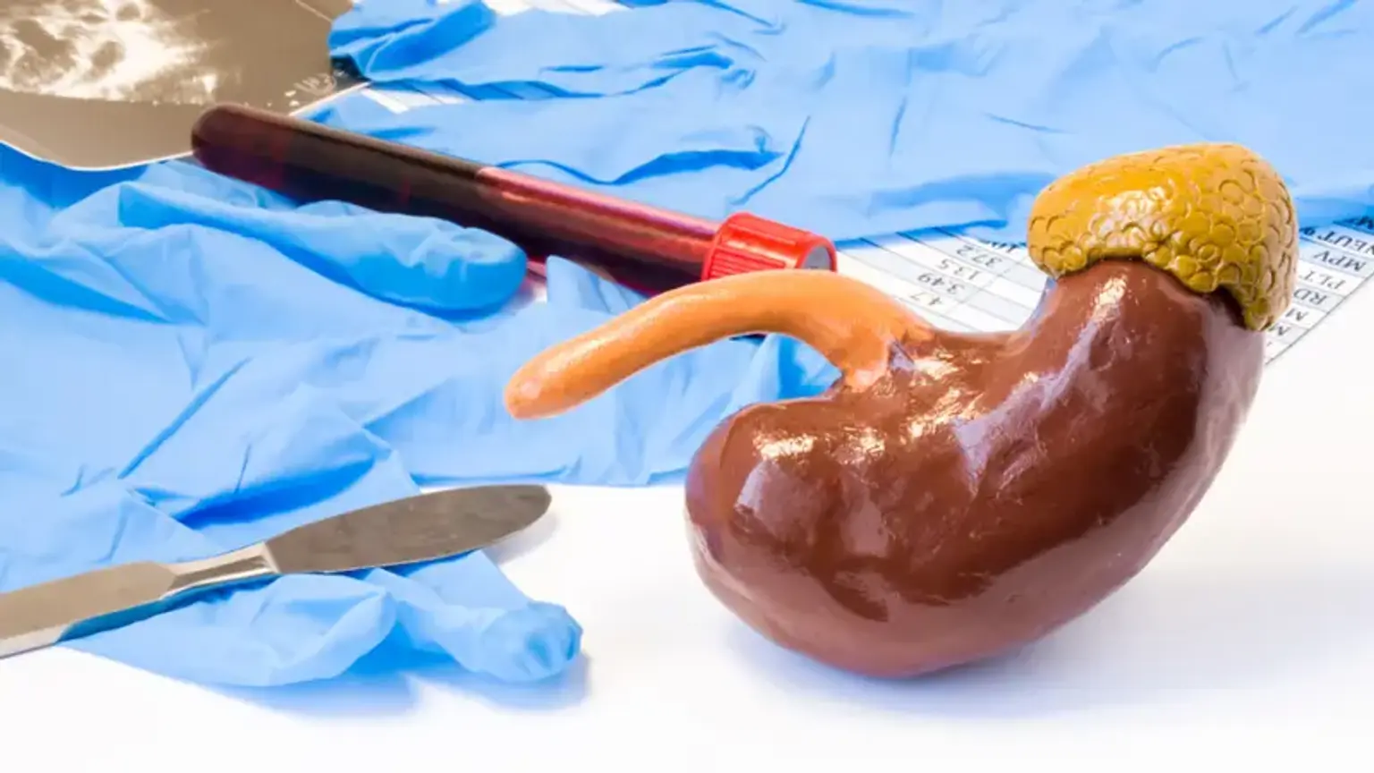
There are two different forms of nephrectomy, partial nephrectomy or radical nephrectomy.


What is Nephrectomy?

Nephrectomy Definition

The surgical removal of a kidney is known as a nephrectomy (nephro = kidney, ectomy = removal). The technique is performed to treat kidney cancer and other kidney disorders and injuries. Nephrectomy is also used to take a healthy kidney from a donor (living or deceased) for transplantation. Every year, thousands of nephrectomies are done in the United States.


Structure & Functions of the Kidneys

Functions of the Kidneys

The kidneys are paired, bean-shaped organs placed below the chest on either side of the backbone, between the 12th thoracic (T12) and 3rd lumbar (L3) vertebrae. Normally, the left kidney is somewhat higher than the right kidney. The kidneys measure roughly 10-12 cm long, 5-7 cm wide, and 2-3 cm thick.

The adrenal glands, also known as suprarenal glands, are little glands placed on top of each kidney. The renal fascia or Gerota's fascia covers the kidneys and adrenal glands on the outside, with the adipose or fat capsule of the kidney beneath it. The kidney has two layers: the outer cortex and the inner medulla. The renal artery, vein, and ureter enter and exit the kidney through its medial or inner aspect known as the hilum. The renal artery is an aortic branch, and the renal vein connects to the inferior vena cava. The urine produced by the kidney exits the body via the ureter, which transports it to the urinary bladder for brief storage before expulsion.


Types of Nephrectomy

Types of Nephrectomy

There are two forms of nephrectomy based on the degree of kidney removal:

Complete Nephrectomy: This is sometimes referred to as a radical nephrectomy. The entire kidney, as well as a piece of the ureter (the tube linking the kidney to the urine bladder), the adrenal gland, and the fatty tissue around the kidney, are removed with this form of nephrectomy. Bilateral nephrectomy refers to the removal of both kidneys at the same time.

Partial Nephrectomy: In this procedure, a diseased portion of the kidney is surgically removed while the healthy tissue remains intact.


Why is Nephrectomy Done?

Kidney Cancer

Nephrectomy is typically performed for the following reasons:

  • Kidney Cancer Treatment: Kidney cancer, such as nephroblastoma, renal cell carcinoma in adults or Wilms' tumor in children, can be treated surgically by removing the malignant tissue by a partial or radical nephrectomy.


  • Excision of a Damaged or Diseased Kidney: A radical nephrectomy can be used to remove a severely damaged, scarred, or non-functioning kidney caused by a traumatic injury, kidney infections or due to nephropathies. Partial nephrectomy can also be used to remove damaged kidney tissue selectively.


  • Kidney Transplantation: A nephrectomy is performed to harvest a healthy renal from a donor for transplantation into a patient with kidney failure. A healthy individual with a good functional kidney who does not have hypertension (high blood pressure) or diabetes or is at risk of developing these conditions in the future is an ideal candidate for donating a kidney to a patient in need of a transplant.


Surgical Options For Nephrectomy

Surgical Options For Nephrectomy

  • Laparoscopic Surgery:

Some patients who need a nephrectomy are candidates for laparoscopic surgery (also known as minimally invasive surgery) to remove the kidney. A laparoscope (wand-like camera) is inserted through a series of small openings or "ports" in the abdominal wall to perform laparoscopic surgery. It is used to see into the abdomen and remove the kidney through a small incision. The operation is performed under general anesthesia (you are asleep and do not feel any pain). You will need a bladder catheter, which will be inserted while you are asleep and withdrawn several hours later.

Laparoscopy accomplishes the same goals as traditional surgical procedures and can be utilized for both major and minor surgery. It is also the recommended procedure for nephrectomy of kidney transplant donors.

The advantages of laparoscopic surgery include:

  • Shorter recovery time.
  • Shorter hospital stay.
  • Smaller incisions.
  • Fewer post-operative complications:

Laparoscopy achieves the same objectives as traditional surgical methods and can be used for major and minor surgery. It is also the preferred method for nephrectomy in kidney transplant donors.


  • Open Nephrectomy:

Although open nephrectomy is seldom necessary, it is also performed under general anesthesia. The surgeon creates an incision in the abdomen or on one of its sides (flank area). To perform the surgery, a rib may need to be removed. The ureter (the tube that transports urine from the kidney to the bladder) and blood vessels around the kidney are cut, and the kidney is removed. Stitches are then used to seal the incision.


Difference Between Laparoscopic & Open Surgery

Surgical Options For Nephrectomy

Nephrectomies are performed using two distinct procedures by surgeons. A small instrument with a camera (laparoscope) and a few tiny incisions are used in laparoscopic nephrectomy. A surgical robot is operated by the surgeon in a robotic-assisted laparoscopic case to manage these microscopic instruments. Open nephrectomy is performed by a single big incision. Surgeons do not utilize cameras and instead see directly into the body.

Shorter hospital stays and quicker recovery periods may result from laparoscopic nephrectomy.

If laparoscopic surgery is not an option, surgeons may turn to open surgery. Laparoscopic surgery, for example, frequently necessitates extended periods of anesthesia. Some people may not respond well to lengthy periods of anesthesia. Others may have a big kidney tumor for which these minimally invasive methods are not appropriate. In some circumstances, an open approach may be preferable.


What are the Contraindications for Nephrectomy?

Contraindications for Nephrectomy

Nephrectomy is contraindicated in the following conditions:

  1. Risk of Kidney Failure: A person with only one healthy kidney or a history of either chronic of acute renal failure should not donate a kidney for transplantation.
  2. Bleeding Disorders: Nephrectomy might result in severe uncontrolled bleeding if the patient has a major bleeding problem such as hemophilia. If nephrectomy is inevitable, it necessitates substantial planning and preparation, as well as provision for substitute treatment for the missing coagulation factor (for example, factor IX or Christmas factor for hemophilia B). A multidisciplinary team in a complete tertiary care facility should execute such a surgery, which can address any complications that may emerge during the procedure.
  3. Decompensated Heart Failure (DHF): A abrupt decline of the heart's pumping capacity occurs in this form of heart failure. Nephrectomy is not recommended in people with DHF.
  4. Decompensated Diabetes Mellitus: In this case, high blood sugar levels are uncontrollable by drugs, resulting in serious organ damage. The emergence of such a dangerous ailment need immediate medical intervention. Nephrectomy is not recommended in patients with decompensated diabetes mellitus.
  5. Anticoagulant Medications: Nephrectomy is not recommended if the patient is using blood thinners. In such cases, the doctor will assess the situation and carefully compare the risks (of discontinuing anticoagulant therapy) vs the advantages (of a nephrectomy), which may be a difficult decision. Based on this, the doctor will advise when and how to quit the drugs if the operation is scheduled.


How Do You Prepare Before Nephrectomy?

Routine Tests

  • Routine Tests: 

Prior to nephrectomy, you will need to undergo some routine tests, which include the following:

  1. Urinalysis: This test examines your urine for the presence of pus cells, which suggest infection, as well as other aberrant elements such as glucose, protein, ketones, red blood cells, and casts.
  2. Kidney Function Test (KFT): This blood test examines whether or not your kidneys are working properly. Blood urea nitrogen (BUN), serum creatinine, serum electrolytes (sodium, potassium, chloride, bicarbonate), and estimated glomerular filtration rate are among the parameters examined (eGFR).
  3. Complete Blood Count (CBC): This blood test offers a complete view of your blood. Hemoglobin concentration, red blood cell (RBC) count, white blood cell (WBC) counts such as total count (TC) and differential count (DC), platelet count, and erythrocyte sedimentation rate are all frequent characteristics examined in this test.
  4. Blood Grouping, Cross-matching, and Tissue Typing: If you need a blood transfusion, your blood group will be identified. If you are a donor for transplantation, your blood will be cross-matched with the recipient's. Tissue typing will also be performed between your tissues and those of the receiver to ensure that the tissues are compatible and that the kidney transplant is less likely to be rejected.
  5. Coagulation Profile: A blood test will be performed to provide information on blood clotting. The prothrombin time (PT), partial thromboplastin time (PTT), and international normalized ratio (INR) are all measured by the test (INR).
  6. Chest X-ray: As part of the usual preoperative screening, you will get a chest X-ray.
  7. ECG (Electrocardiogram): An ECG will be performed on you to study the electrical activity of your heart. If you are of advanced age, a more comprehensive cardiac check may be required to ensure that you are fit for surgery.


  • Specific Tests: In addition to the above tests, you may also have to undergo some specialized tests:
  1. CT Scan: A computed tomography (CT) scan is extremely effective in evaluating kidney cancers. It provides precise information about tumor size, location, and indicators of metastasis (spread) in cancer patients. You will be required to lie down motionless on a special table for a few minutes throughout this operation. Images will be taken while a doughnut-shaped device passes over the component to be inspected. Because a contrast dye may be injected into your vein, you should notify the radiologist if you have any allergies.
  2. MRI Scan: If your doctor detects a problem with the inferior vena cava (the blood vessel carrying deoxygenated blood to the heart), you may be submitted to a magnetic resonance imaging (MRI) scan. MRA gives reliable pictures of vascular architecture and aids in surgical planning. If you have a phobia of enclosed places, this process will place you within a tiny tube, which can be uncomfortable at times. The MRI machine produces loud clicking noises, therefore it is best to request earplugs to help block them out.


What Happens During Nephrectomy?

open kidney removal

This procedure is performed in the hospital while you are sleeping and without discomfort (general anesthesia). The operation might take 3 hours or more.

Simple nephrectomy or open kidney removal:

  1. You will be positioned on your side. Your surgeon will make an incision (cut) that is up to 12 inches (30 cm) long. This cut will be on your side, either slightly below or over the lowest ribs.
  2. Incisions and manipulations are made to muscle, fat, and tissue. To do the operation, your surgeon may need to remove a rib.
  3. The ureter (the tube that transports urine from the kidney to the bladder) and blood arteries are severed from the kidney. The kidney is then extracted.
  4. Sometimes only a portion of the kidney is removed (partial nephrectomy).
  5. Stitches or staples are used to close the wound.


Radical nephrectomy or open kidney removal:

  1. Your surgeon will create an 8 to 12-inch (20 to 30 cm) long incision. This cut will be made on the front of your stomach, slightly below your ribs. It is also possible to accomplish it through your side.
  2. Muscle, fat, and tissue are cut and moved. The ureter (the tube that transports urine from the kidney to the bladder) and blood vessels are detached from the kidney. The kidney is then extracted.
  3. Your surgeon will also remove the surrounding fat, as well as the adrenal gland and maybe some lymph nodes.
  4. Stitches or staples are used to close the wound.


Laparoscopic kidney removal:

  1. Your surgeon will make three or four little cuts in your abdomen and side, each no more than one inch (2.5 cm) long. The surgeon will do the procedure using small probes and a camera.
  2. At the conclusion of the surgery, your surgeon will make a bigger cut (approximately 4 inches or 10 cm) to remove the kidney.
  3. The ureter will be cut, a bag will be placed around the kidney, and it will be pulled through the bigger cut.
  4. This procedure may take more time than an open kidney removal. However, as compared to the discomfort and recovery period following open surgery, most patients recover faster and have less pain after this form of surgery.
  5. Your surgeon may occasionally make an incision at a location other than the one mentioned above.


Aftercare & Recovery

Aftercare & Recovery

Your health care team will closely monitor your blood pressure, electrolytes, and fluid balance immediately following surgery. The kidneys regulate several of these bodily activities. During your recovery, you will most likely have a urinary catheter (a tube that drains pee) in your bladder.

You may have pain and numbness at the incision site as a result of nerve damage. Pain medications are used following the surgical operation and as needed during the recovery period. Because the incision is near to the diaphragm, heavy breathing and coughing may be uncomfortable, but breathing exercises are necessary to prevent pneumonia.

Depending on the technique of operation, you will most likely be in the hospital for 1 to 7 days. You will be encouraged to resume mild activities as soon as you are able. Heavy lifting and strenuous activities should be avoided for 6 weeks after the operation.

Your doctor will provide you with additional specific instructions regarding your post-operative activities, restrictions, and diet.


Risks/Complications of Nephrectomy Surgery

Complications of Nephrectomy Surgery

Every procedure involves risks and complications. Nephrectomy surgery may result in the following complications:


Conclusion

nephrectomy

A nephrectomy is a surgical operation that involves the removal of all or part of a kidney.

Complete nephrectomy, the urologic surgeon removes the entire kidney as well as certain extra tissues, such as a portion of the tube that links the kidney to the bladder (ureter) or other neighbouring structures such as the adrenal gland or lymph nodes, during radical nephrectomy.

A partial nephrectomy, also known as kidney-sparing (nephron-sparing) surgery, involves the removal of diseased tissue from a kidney while leaving healthy tissue in place.

A nephrectomy is typically performed to treat kidney cancer or to remove a noncancerous (benign) tumor. In some situations, a nephrectomy is performed to treat a sick or severely damaged kidney. In the instance of a donor nephrectomy, the urologic surgeon takes a healthy kidney from a donor for transplant into a person who need a working kidney.

The urologic surgeon may do a nephrectomy with a single incision in the abdomen or side (open nephrectomy) or through a series of tiny incisions in the abdomen using a camera and small instruments (laparoscopic nephrectomy).

In certain situations, these laparoscopic operations are carried out with the assistance of a robotic system. The surgeon performs robotic surgery while sitting at a computer console beside the operating table. He or she is in charge of the camera arm and mechanical arms, which are attached to surgical tools that are functioning within the patient's body.