Percutaneous endoscopic laser annuloplasty (PELA)
Last updated date: 03-Mar-2023
Originally Written in English
Percutaneous Endoscopic Laser Annuloplasty (PELA)
Overview
Patients with persistent low back pain who do not respond to conservative therapy have a significant influence on their economic, psychological, and social activities. Conservative therapy, local treatments, microscopic surgery, and fusion surgery are all alternatives for persistent low back pain that has lasted more than 6 months.
The laser-assisted spinal endoscopy (LASE) kit was utilized for percutaneous intradiscal decompression to evaporate and reduce the posterior and central nucleus in order to alleviate leg and radicular discomfort caused by confined disc herniation. LASE is used to directly coagulate the inflammatory disc granulation tissue associated with annular tears in percutaneous endoscopic laser annuloplasty (PELA), a revolutionary minimally invasive method. The endoscope's tiny diameter, which includes a YAG laser, irrigation, and illumination, as well as the extreme posterolateral approach into the posterior annulus, allow for little injury to normal nuclear tissue.
The clinical effects of PELA were studied in individuals with discogenic low back pain (DLBP) caused by an annulus-torn degenerative disc or a confined disc herniation.
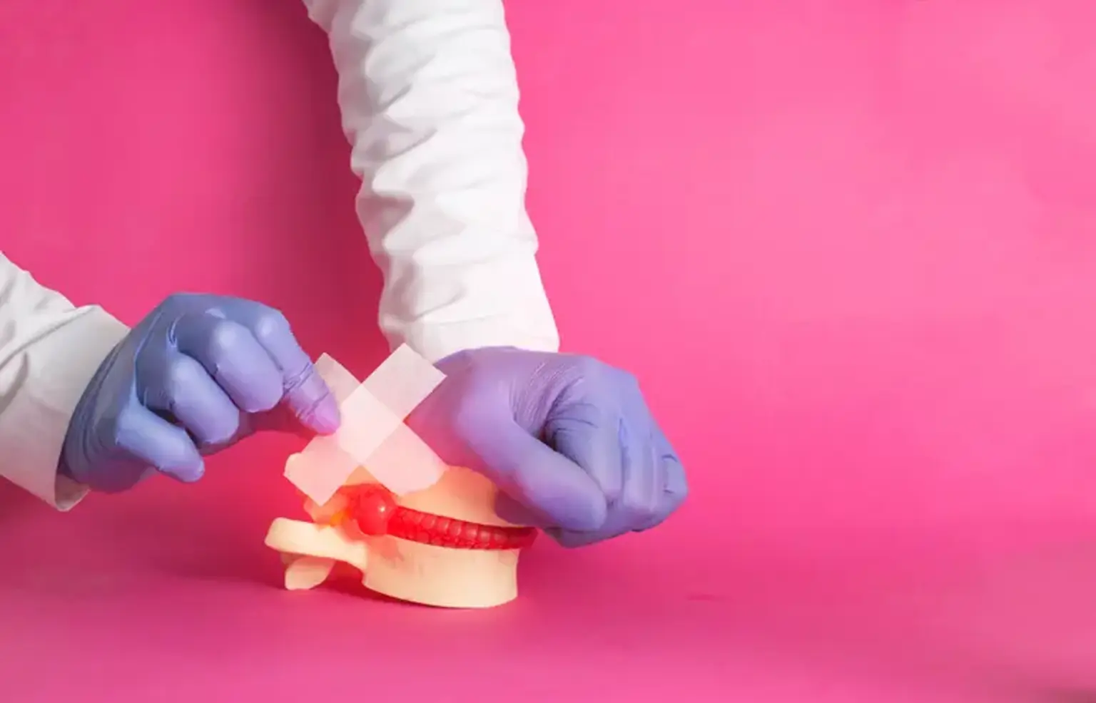
Discogenic low back pain (LBP)
It is believed that discogenic low back pain (LBP) caused by internal disc disturbance accounts for around 40% of chronic low back pain patients. Internal disc disruption is distinguished by matrix deterioration in the nucleus pulposus and the formation of radial and circumferential fissures in the annulus fibrosus. Pain is thought to be caused by a combination of increased mechanical stress in the posterior annulus and chemical irritation of nociceptors by inflammatory mediators in the annulus fibrosus tears.
The pain is mediated by nociceptors found in the outer third of the annulus as well as nociceptors that develop into fissures. Although sitting intolerance, an extension catch, difficulty lifting, or an inability to maintain the same posture are sometimes associated with discogenic LBP, no clinical features are diagnostic of the condition, and its features are difficult to distinguish from those of pain originating from the lumbar zygapophysial joints or the sacroiliac joint. Certain MRI findings, such as Modic lesions or a high-intensity zone, are associated with the damaged disc being the cause of pain, however, these abnormalities occur in only a minority of individuals with low back pain.
Provocation discography is recommended by certain sources as the ultimate diagnostic technique for discogenic LBP. In response to concerns regarding the false-positive rate of discography, research has revealed that the rate is less than 10%. However, there are limited data on the discography false-negative rate, or how frequently a disc may be symptomatic despite discography failing to replicate pain.
There has been no research into any sort of conservative treatment for discogenic LBP. Conservative therapy is often used in the absence of a discogenic pain diagnosis. Spinal fusion and disc arthroplasty are surgical procedures. However, even if the operations are performed appropriately, spinal surgery is quite intrusive and can result in consequences that cause lasting pain and functional impairment.
As an alternative to surgery, a number of minimally invasive, percutaneous intradiscal techniques, such as intradiscal electrothermal therapy, transdiscal biacuplasty, and nucleoplasty, have been investigated. However, by design or practice, these treatments target the nucleus pulposus or the inner annulus and either ignore or fail to capture the outer annulus, which is the primary source of nociception. Furthermore, these methods rely on fluoroscopic guidance to position instruments in the estimated location of target tissues, which are not visibly visible.
Minimally Invasive (Interventional) Treatment for Back Pain
1. Intradiscal Thermal Lesioning
Percutaneous intradiscal radiofrequency thermocoagulation (90 seconds at 70 degrees Celsius) is ineffective in treating persistent discogenic low back pain. Although the mechanism of pain alleviation through heating for discogenic back pain is unknown, two possibilities are commonly proposed.
- The first is denervation of the disc tissue or elimination of nociceptors overgrowth.
- The second is that the structure of the collagen fibers in the annulus is altering and remodeling, resulting in increased annular stability. These two possibilities, however, were not supported by histology investigations employing Intradiscal Electrothermal Therapy (IDET).
2. Intradiscal electrothermal therapy (IDET)
Intradiscal electrothermal therapy (IDET) is still debated in the treatment of discogenic back pain. In a randomized, double-blind, controlled experiment, Freeman et colleagues found no difference between the IDET group and the sham group, with no improvement in either group. The use of IDET in the treatment of discogenic back pain remains contentious, and a recommendation for its usage cannot be made without excellent, long-term outcomes.
Some researchers believe that the most common reason of IDET failure is improper target lesioning since most discogenic pain generators are situated in interposed disc tissue in the posterior annulus. The thickness of the annulus following thermal modulation determines the length of therapeutic success.
3. Laser Annuloplasty
PELA is a minimally invasive treatment that employs LASE (Laser-assisted spinal endoscopy) to directly shrink and coagulate the intervening disc tissue in the posterior annulus associated with annular tears.
- Mechanism
- Targeted removal of granulation tissues in the posterior annulus
- Laser during PELA blocks the sensory nerve surrounding the annulus
- Continuous saline irrigation removes chemical irritants
4. Percutaneous Disc Decompression Device (L’DISQ)
The Nucleoplasty device, like most other minimally invasive devices and methods, has the problem of being difficult to access the herniated nucleus. In contrast to the Nucleoplasty device, the L'DISQ has a navigational tip that can be curled and steered into a disc herniation by twisting a control wheel.
Percutaneous removal by a very tiny bore introducer cannula put directly into the herniation or into the posterior lateral annulus may theoretically better protect the integrity of the outer annulus and perhaps minimize the 7 to 25% re-herniation rate following open discectomy.
Because the distance between the two electrodes on the L'DISQ tip is 2 mm, a nerve root that is more than 2 mm from the tip is theoretically safe from electric harm.
5. PELDA (Percutaneous Endoscopic Lumbar Discectomy and Annuloplasty)
The comparatively high failure rate in the IDET method may be related to blind thermo-coagulation and indirect decompression. PELDA, on the other hand, immediately removed the compressing extradural fragment and anchoring disc fragment in the annular fissure. It could succeed.
- Decompression by disc fragment removal and intradiscal pressure reduction; and thermal ablation using RF and laser, which rectified the annular defect of neoinnervation and neovascularization.
- A well-designed study, however, should be explored to confirm the clinically effective outcomes.
6. Percutaneous Adhesiolysis
Dr. Gabor Racz invented percutaneous lysis of adhesions (also known as decompressive epidural adhesiolysis) in 1989 for patients with CLBP who had not responded to conservative therapies. This interventional procedure's goal is to break down fibrous adhesion scar tissues in the epidural space and supply appropriate medicine (i.e. local anesthetics and corticosteroids). However, whether adhesion scars in the epidural space induce CLBP is still debated.
Fibrous epidural scars can form after a surgical laminectomy or as a result of an annular rupture, hemorrhage, or infection. Adhesion scars can restrict neurovascular bundle mobility in the intervertebral foramen and central spinal canal. Medication can be delivered directly to specified structures via epidural adhesiolysis.
7. Trans-Sacral Epiduroscopic Laser Decompression (SELD)
In recent years, great progress has been achieved in the treatment of spinal lesions with an epiduroscopic laser as a less intrusive surgical alternative to standard open surgery. Epiduroscopy allows direct observation of lumbar epidural lesions through the sacral hiatus. This steerable flexible epiduroscopic laser device is utilized to validate epidural pathologic findings while also providing accurate and targeted treatment. Many surgeons have recently done trans-sacral epiduroscopic laser decompression (SELD) for discogenic low back pain or ruptured disc lesion.
A steerable flexible epiduroscopic laser device is employed in this surgery to validate epidural pathologic findings while also providing precise and guided therapy. Epiduroscopy enters the body through a natural entrance, the sacral hiatus.
- Surgical Technique
The surgery was carried out while the patient was sedated. Over the sacral hiatus, a 5 mm skin incision was created. Under fluoroscopic supervision, the sacrococcygeal ligament was pierced with a Tuohy needle, followed by a guide wire and dilator. A 3.0 mm steerable Video Guided Catheter (VGC) was then introduced. The VGC was used to insert an epiduroscope and the YAG laser into the catheter's end to see the epidural space and execute HNP ablation. Fluoroscopy and epiduroscopy were utilized after inserting the VGC to confirm that the catheter tip was placed in the most inferior portion of the targeted disc, covering the PLL.
An epidurogram was obtained using radio-opaque dye to illustrate the shape of herniation and flow blockage caused by HNP and adhesion at the pathologic level. The YAG laser decreased adhesiolysis and the size of the herniated disc lying below the PLL under direct view with epiduroscopy. Using 1mm forceps, the floating and sequestered HNP was extracted. At the prior pathologic level, a repeat epidurogram should demonstrate a flattened contour of herniation and free flow.
8. Transforaminal epiduroscopic laser annuloplasty (TELA)
The essential purpose of this technique is quite similar to that of SELD. Each, however, has a completely distinct targeted target in the lumbar spine epidural area and access to it. The posterior annulus and the intervertebral foramen serve as the target and access, respectively.
TELA is a procedure that is comparable to the outside-in approach of percutaneous endoscopic lumbar discectomy (PELD). This method, however, has a distinct entrance point with PELD. Because the typical skin entrance site is less than 12 cm from the midline, a curved scope can be employed.
What is Percutaneous endoscopic lumbar annuloplasty (PELA)?
Percutaneous endoscopic lumbar annuloplasty (PELA) is a minimally invasive treatment designed specifically to decompress the posterior nucleus or granulation tissue in the torn annulus. PELA is unique in that it allows physicians to remove tissues under direct visualization by endoscopy and fluoroscopy using laser energy or forceps. This characteristic enables the excision of specific tissues while conserving as much of the remaining disc as feasible. The purpose of this study was to investigate the clinical results of PELA.
Types of Endoscopic Annuloplasty
- Endoscopic thermal annuloplasty: A specific quantity of heat is applied directly to the afflicted disc area throughout the treatment. The surgery is usually done following discectomy with an endoscope and a transforaminal approach.
- Percutaneous endoscopic laser annuloplasty: This minimally invasive method employs Laser-Assisted Spinal Endoscopy to directly condense and stiffen the inflammatory granulation tissue associated with annulus tears.
Indications of the endoscopic annuloplasty
Endoscopic annuloplasty may be indicated in the following conditions:
- Discogenic lower back pain
- Annulus tears
- Disc herniation
- Inflamed disc granulation tissue
- Disc derangement due to annular tears
- Radiculopathy
- Predominant leg and radicular pain due to disc herniation
- Percutaneous intradiscal compression
- Abnormal disc tissues, new vessels are nerves in the central torn posterior annulus
Advantages of the thermal and laser-assisted endoscopic annuloplasty
- Smaller size incisions
- Easy access and visualization due to the small diameter of the endoscope
- Minimal damage to normal nuclear tissue
- Reduced risk of serious complications
- 90% success rate with favorable outcomes for selected groups of discogenic lower back pain patients
- Preservation of back muscles
Recovery
Discectomy followed by endoscopic annuloplasty often results in significant reductions in leg discomfort, back pain, and disability within a few days to months following surgery. If no postoperative complications develop, your doctor will follow you and you may be permitted to go home within 24 hours. You will be given instructions on how to walk, as well as physical therapy and follow-up consultations to check your rehabilitation process.
Conclusion
Disc herniation is defined generically as a localized or focused displacement of disc material beyond the boundaries of the intervertebral disc space. The disc material might be the nucleus, cartilage, apophyseal bone fragments, annular tissue, or any mix of these. Laser surgery is one of the therapeutic options for lumbar disc herniation patients.