Pituitary Surgery
To remove pituitary tumors while preserving as many of the healthy gland's tissues as feasible, many surgeons perform pituitary surgery. This makes it possible for the gland to continue secreting the appropriate amount of hormones, which are essential for carrying out various body activities. When pituitary tumors cause Cushing's disease, acromegaly, secondary hyperthyroidism, hypopituitarism, visual issues, or malignant tumors, surgery is frequently the initial course of treatment. Rare emergencies such as the destruction or hemorrhage into the pituitary gland may also call for surgery.
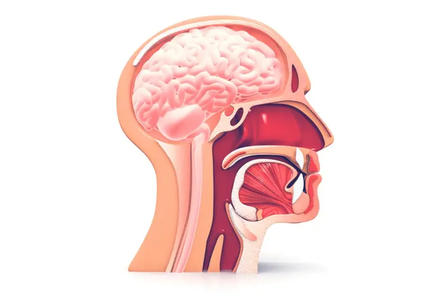
Pituitary Gland
The pituitary gland is located directly beneath the brain and behind the eyes at the base of the skull and it is a hormonal gland the size of a pea. Although it is not a component of the brain, a stalk links it to the brain. Special blood vessels found in the pituitary stalk allow the brain to transmit hormonal signals to the gland to control its activity. The nerves (also known as optic nerves) that connect the brain to the back of the eyes traverse the region between the pituitary gland and the brain. The optic nerves may be touched, pressed against, or stretched by a pituitary tumor. This may impact your vision and the signal that travels from your eyes to your brain.
The pituitary gland regulates a variety of hormones (chemicals) that have an impact on many physiological systems. These include cortisol, a steroid hormone that regulates several vital processes, thyroid hormone, which regulates metabolic rate, sex hormones, which are used for sex and reproduction, prolactin, which is used by women to produce breast milk, growth hormone, which is used to promote growth during childhood and adolescence, and anti-diuretic hormone (for water balance).
Pituitary Tumor Symptoms
The type of tumor and the extent of the pituitary gland that is damaged determine the symptoms. These tumors may result in symptoms caused by excessive or insufficient pituitary hormone production. Symptoms can differ from person to person. The symptoms could potentially resemble those of other medical conditions. For a diagnosis, consult your healthcare provider at all times.
Pituitary Gland Surgery
In a procedure known as pituitary surgery, tumors that are on or close to the pituitary gland are removed. Three inches behind the bridge of the nose, at the base of the brain, the pituitary is a pea-sized gland. It releases a variety of hormones that regulate various bodily functions. There are several methods for doing pituitary surgery, the majority of which don't require any obvious incisions.
The method a neurosurgeon chooses to use is primarily determined by where a tumor is located in the brain. Some pituitary tumors extend into the nasal cavity's sphenoid sinus, which is where they originate. Therefore, a neurosurgeon can reach and remove these tumors by performing surgery through the nose or the top of the skull.
Operating through the sphenoid sinus in the nose, also known as transsphenoidal surgery, is the method that is used the most frequently. A neurosurgeon enters through the nostril or the gums without making a skin incision to perform transsphenoidal surgery. This is also known as endoscopic endonasal surgery or endoscopic transsphenoidal surgery. An endoscope, a small, flexible instrument with a tiny camera and light on the tip that may be inserted through the nostrils and guided to the sphenoid sinus, is used to accomplish this procedure. Incisions within the mouth are not required with an endoscope.
An alternative procedure that a neurosurgeon may opt for is a craniotomy if the tumor is especially big or has deep brain penetration. To access the brain during this treatment, a neurosurgeon makes an incision on the scalp and then removes a portion of the skull.
Pituitary Surgery Indications
About 10 to 15 percent of brain tumors are pituitary tumors, which are rather common. Pituitary tumors can be asymptomatic in some patients, meaning they go their entire lives without ever noticing the tumor. Surgery might not even be required if imaging detects the tumor.
Pituitary tumors that do cause symptoms, however, frequently impair a person's quality of life, necessitating medical attention. These tumors can harm adjacent structures, particularly the optic nerve, which is essential for vision, and can change hormone levels, resulting in several diseases.
Surgical tumor resection is frequently the first step in treatment, with total excision being the final goal. There are other therapeutic alternatives for the remaining tumor when only subtotal resection is possible while maintaining patient safety. Prolactinomas are the main exceptions to this general trend since they may frequently be managed without surgery with long-term treatment.
Pituitary Surgery Preparation
Inform your doctor about any drugs or dietary supplements you are taking, including any herbs or vitamins. Your doctor will likely ask you to stop taking certain medications or dietary supplements, such as aspirin and warfarin, which might increase bleeding during surgery, so you must let them know. Any food or drug allergies you may have should also be mentioned to your doctor.
Your doctor will ask you to stop eating and drinking at midnight the night before the procedure because general anesthesia is utilized during surgery. If you have been told by your doctor to keep taking a certain prescription on the day of surgery, do so with a little sip of water.
Be sure to dress comfortably and loosely on the day of the procedure. Wear your glasses; avoid cosmetics, jewelry, body piercings, or contact lenses. Pack lightly for your weekend or weeklong stay. Toiletries, dentures, and a change of clothing after hospital discharge are a few items people frequently want to bring. You cannot drive yourself home after surgery, so make sure you have transportation arranged.
Pituitary Tumor Treatment
General anesthesia is administered before transsphenoidal surgery and a craniotomy to keep the patient pain-free throughout the treatment. Following that, the steps vary:
Transsphenoidal Surgery
The sphenoid sinus at the back of the nasal passages is first opened by passing the endoscope's tip through one of the nostrils during this minimally invasive treatment. For the surgeon to have a clear view of the surgical field located deep inside the nose, an endoscope or microscope offers light and magnification. To access the tumor, instruments are inserted through the nose.
When the neurosurgeon gets to the pituitary tumor, he or she utilizes devices to carefully separate the tumor from the pituitary gland and any further structures it may be connected to, such as the optic nerve. The neurosurgeon can construct a 3-dimensional map of these and other structures in a patient's brain using cutting-edge computer technology and imaging technologies, such as computed tomography (CT) and magnetic resonance imaging (MRI), to attain the highest degree of precision.
The neurosurgeon then delicately separates and aspirates portions of the pituitary tumor using cutting-edge instruments. The pituitary gland and other structures and tissue of the brain are preserved while as much of the tumor as possible is removed. The neurosurgeon also takes a biopsy of the tumor during tumor removal and sends it to a pathologist for diagnosis confirmation.
The neurosurgeon might need to replace the space left by the tumor after it has been removed. If so, a tissue graft from the patient's abdomen or thigh may occasionally be taken by the neurosurgeon and inserted where the tumor was. Instead of creating an incision in the abdomen or thigh, artificial grafts and biologic glue are frequently an option. Stitches are used to close any incisions. The procedure is then finished.
Transcranial Pituitary Surgery
The alternative surgical technique entails entering the skull rather than the nasal cavity. To access the skull bone, the neurosurgeon first makes an incision on the front or side of the head and then peels back the skin and muscle. To expose the dura mater, the neurosurgeon then removes a little portion of the skull bone. The skull piece is retained in case it needs to be replaced in the future. The neurosurgeon carefully cuts through the dura mater with surgical scissors. The neurosurgeon utilizes an operating microscope and other cutting-edge technologies to operate through the available space and approach the pituitary tumor because the skull has limited extra space and the brain tissue, blood vessels, and nerves are delicate.
The neurosurgeon additionally employs stereotactic methods, which create three-dimensional images of the brain using computer technology and imaging devices like MRI and CT scans. These scans help the neurosurgeon locate the tumor with pinpoint accuracy while protecting healthy brain tissue.
The neurosurgeon carefully separates the pituitary tumor from the pituitary gland and other vital structures including the optic nerve as soon as they reach it. The neurosurgeon takes care to remove as much of the tumor as they can while minimizing damage to healthy tissue. To confirm the diagnosis, a pathologist receives a biopsy of the removed tumor for investigation.
A section of the skull bone is replaced and fixed with titanium plates and screws after the neurosurgeon has removed as much tumor as is practicable. The dura mater incision is then sewn up. The neurosurgeon then closes the scalp wound with sutures. The procedure is then completed.
Pituitary Surgery Recovery
Patients who undergo transsphenoidal surgery, the most common procedure, typically stay in the hospital for one to two days before being discharged and sent home. People who have a craniotomy stay for a little longer (about a week).
Patients undergoing transsphenoidal surgery are given painkillers and occasionally antiemetics, and those undergoing craniotomies may also be prescribed drugs to avoid seizures, brain swelling, and stomach ulcers.
Additionally, patients who have low levels of one or more pituitary hormones may be recommended long-term hormone replacement therapy.
When Can I Resume Exercise?
Transsphenoidal surgery patients can anticipate starting light exercise, such as jogging or swimming, around two weeks following surgery, and regular exercise around four weeks after surgery. The day after surgery, patients may usually move, but they might discover that they get tired quickly and need to rest frequently. It's normal to feel this fatigue.
Patients who have had a craniotomy will require more time to heal before starting their usual exercise program again, and if they do, they should exercise with a partner or under supervision until they are fully recovered. Once the neurosurgeon approves that level of activity, patients can start doing modest exercise and then progress to more demanding exercise. If patients have the energy, it is advised that they get up and go for a walk every day.
Pituitary Surgery Follow-Up
The neurosurgeon and endocrinologist will schedule many follow-up appointments for the patients. A consultation with their ophthalmologist will also be scheduled for patients who had vision issues before surgery or presently experience visual issues.
The neurosurgeon will assess the patient's progress throughout these visits and will also request an MRI to look for any residual tumor. A patient's neurosurgeon may advise radiotherapy to remove any residual tumor if it is found. If the pituitary gland is not producing enough of a certain hormone, the endocrinologist will schedule blood tests to check hormone levels and prescribe drugs for hormone replacement therapy. A patient's vision will be evaluated by the ophthalmologist.
To have the best recovery possible, patients must keep all of these appointments. Patients should also mention any new or deteriorating symptoms to their doctor. Physical therapy is often not necessary for patients.
Pituitary Surgery Risks
Before the procedure, you will also be informed about the dangers associated with surgery. There are many measures doctors take to try to prevent problems from occurring, as well as those they take to lessen the burden of such complications when they do. If issues do arise, they are typically manageable. These issues could arise following surgery:
- Nose pain or obstruction, loss of smell, crusting requiring nasal douching, and septal perforation.
- Bleeding, infections, including meningitis, CSF leaks from the surgical site, and hormonal problems such as water imbalance or inadequate steroid production necessitating lifelong hormone replacement.
- If a CSF leak does occur, doctors sometimes use a piece of fat to cover the opening before installing a drain in the spine to temporarily redirect the fluid while the hole is being repaired. This can call for more care, which might entail a longer hospital stay.
- Less than 1 in 1000 times, any brain operation poses a very modest risk of complication-related death.
- Additionally, there is a chance of stroke, double vision, and blindness.
Pituitary Surgery Long-Term Effects
There is a small chance that patients who had a particularly large or aggressive pituitary tumor removed might experience diminished brain function or a shorter life span. The adjacent brain tissue was either affected by the tumor or surgery-related tissue damage is the cause of these long-term problems. Many highly skilled neurosurgeons take great care to prevent any injury during operation.
Patients typically experience diabetes insipidus for a few days following surgery, which is characterized by extreme thirst or frequent urination. When the pituitary gland produces too little antidiuretic hormone, this disease develops. Without this hormone, the kidneys produce too much water because the antidiuretic hormone tells the kidneys to limit the amount of water in the urine. The majority of the time, diabetes insipidus resolves within a few days after patients are recommended to drink lots of water. Rarely, diabetes insipidus might persist even for the rest of a patient's life, in which case hormone replacement therapy is required to replace the antidiuretic hormone's depleted levels in these patients.
Conclusion
Pituitary tumors are a diverse group of benign and malignant tumors that often manifest with mass effect and recognizable hypersecretory symptoms. Transsphenoidal surgery continues to offer the best results for non-prolactin secreting tumors with a low incidence of severe morbidity, even though medical and radiotherapy are successful treatments for specific functional tumors in some situations.