Rotational Atherectomy

Last updated date: 27-Aug-2023

Originally Written in English

Rotational Atherectomy

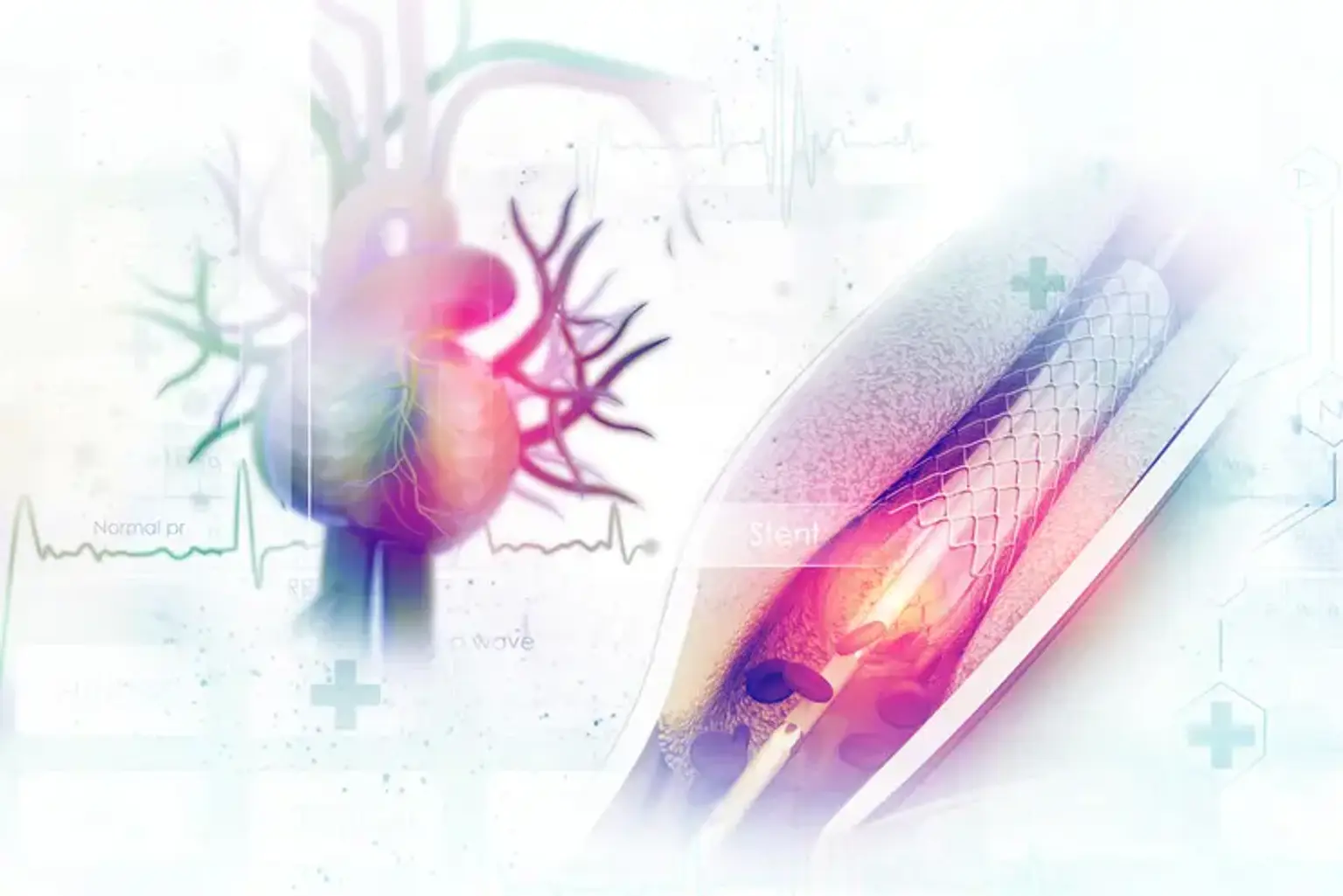
A coronary artery's calcium deposits are a sign of prior inflammation, healing, and scarring. Considerable atherosclerotic coronary artery disease (CAD) is correlated with significant calcification. Coronary calcification can be diffusely distributed throughout coronary arteries, and when the vessel is imaged, considerable calcification can completely encircle it. When using standard balloon angioplasty, coronary stenosis with circumferential or substantial vascular calcification is stiff and generally can't be dilated. In severely calcified coronary lesions, stent dilatation and maximum vessel wall apposition are frequently compromised. Stents placed in severely calcified vessels without atherectomy often thrombose, produce restenosis, and even fracture. Balloon angioplasty still has significant calcification as a major drawback, as does successfully delivering stents to severely damaged arteries. High-pressure, non-compliant balloon inflations may still fail to sufficiently dilate and prepare a substantially calcified artery for stent delivery in situations with heavily calcified lesions.

In this example, calcium is the blocking material, and atherectomy refers to its removal. The vessel wall compliance in calcified or fibrotic lesions is increased by removing significant calcification or changing the calcified atherosclerotic plaque, and the lumen diameter obtained from using this device will be significantly better than that obtained from using straightforward balloon angioplasty. One of the several techniques for doing an atherectomy in a coronary vessel is rotational atherectomy. It is the most popular atherectomy tool and eliminates atheromatous plaque via differential cutting, which involves removing the inelastic calcified plaque with microscopic diamond chips implanted on the surface of a fast-revolving olive-shaped burr (150,000 to 200,000 rpm). The reticuloendothelial system removes the 2 to 5-micrometer microparticles that are produced as a result of this abrasion after they have traveled through the coronary microcirculation. The burr has a diameter of 1.25 to 2.50 mm and travels on a specialized guidewire. Smaller burr sizes should be used initially in cases with severe calcification, followed by bigger burrs in 0.25 to 0.50-mm increments up to 70% of the reference vessel diameter. In the early 1980s, David Auth began looking into the potential of employing a rotary tool to debulk atherosclerotic plaque. In 1988, Fourier et al. performed the first RA procedure on a human coronary artery.


What is Rotational Atherectomy

Rotational atherectomy is a highly specialized catheterization operation used to remove plaque from an artery that has calcified to such an extent that it has narrowed or obstructed. When balloon angioplasty cannot or is not appropriate to unblock a clogged artery, this procedure is typically used. Rotational atherectomy is a procedure that unclogs calcified plaque from a coronary artery by dislodging it with a rotating tool called a Rotablator. To the right patients, doctors provide this therapy as a component of complex coronary intervention.


Rotational Atherectomy Indications

Rotational Atherectomy Indications

The main indication for RA is the modification of substantially calcified de novo coronary stenosis that is unlikely to be sufficiently widened by balloon angioplasty to enable full stent expansion. Fluoroscopy was previously used to detect significant coronary calcification, but it has been shown that this method is less sensitive than intravascular imaging for calcium detection. On fluoroscopy, severe coronary calcification has been reported as radio-opacities seen on both sides of the artery wall before contrast injection but without cardiac motion.

With intravascular ultrasound, severe calcification is described as a broad arc of superficial calcium involving 3 quadrants, which is indicative of bright echodensity attenuating deeper structures. Areas of coronary calcification on optical coherence tomography (OCT) look highly defined and signal-poor. OCT coronary calcification measurements with length >5 mm, arc >180°, and maximum thickness >0.5 mm are indicators of stent under-expansion. When there is severe calcification, RA can be employed as a primary technique or as a backup plan if coronary angioplasty fails to widen a lesion. Although the long-term results of planned RA and bailout RA are similar, planned RA is linked to the shorter operation and fluoroscopy times, less contrast, and fewer in-hospital MACE. Because of this, operators should have a low threshold for performing a planned RA approach in heavily calcified lesions if there is a high preprocedural possibility of its utilization. There is also a possibility of unrecognized intimal dissection when doing RA in a lesion that has received aggressive balloon angioplasty, albeit this is uncommon.


Rotational Atherectomy Contraindications

Rotational atherectomy works best on calcified, inelastic lesions; it is not recommended for use on soft, thrombus-containing lesions like those found in acute myocardial infarction or saphenous vein graft lesions with a significant thrombotic burden.


Rotational Atherectomy Equipment

Rotational Atherectomy Equipment

A long catheter with an oval-shaped burr and a surface tip covered with small diamond particles makes up the rotational atherectomy device. A lubricious solution is pushed into the catheter to lessen heat generation and burr entrapment throughout the process. The burr has a smooth, flat end at the proximal end. The operator can extend and retract the burr inside the vessel by extending and retracting the catheter's back end, which is attached to an advancer. The advancer is attached to an external console, and air or nitrogen is pushed into it via a pneumatic hose, spinning the advancer's internal turbine, which in turn spins the driving shaft and burr. The operator can turn on the burr and start spinning it with a foot pedal. At the external console, the burr's spinning speed is modified to the desired value.

The coronary guidewire that the rotablation catheter travels over has a 0.014-inch tip and a 0.009-inch body diameter. The bigger diameter wire tip prevents the burr from moving forward once it crosses the 0.009-inch section of the wire. A wire clip is attached to the back of the wire when the burr is spinning actively to stop the guide wire from spinning and potentially damaging the vessel. When conducting rotational atherectomy, the flush bag is lubricated with a lubricious lipid emulsion to lessen friction between the burr and guidewire. Olive oil, phospholipids, sodium deoxycholate, disodium EDTA, sodium hydroxide, and water make up the rotating atherectomy lubricant.


Rotational Atherectomy Preparation

Rotational Atherectomy Preparation

Similar pharmacological treatments are given to patients having rotational atherectomy and balloon angioplasty. It takes the administration of heparin or bivalirudin to keep the activated clotting time over 300 seconds. The emergence of delayed coronary flow or no flow phenomena is one of the rotational atherectomy's potentially dangerous side effects. In the absence of an evident occlusive dissection or spasm, this is characterized as a reduction or halt of blood flow. It is believed that rotational atherectomy-induced distal microparticle embolization causes slow flow and coronary no-flow abnormalities. Usually, verapamil, diltiazem, nicardipine, adenosine, or nitroprusside are administered intracoronary. Microcirculation is where these drugs work. A combination of nitroglycerin, verapamil, and heparin is frequently used by catheterization labs since it has been demonstrated to lessen the likelihood of spasms and slow/no flow.


Rotational Atherectomy Procedure

Rotational Atherectomy Procedure

Plaque ablation and pulverization by the abrasive diamond-coated burr are the cornerstones of high-speed mechanical rotational atherectomy. Due to the idea of differential cutting, the rotational atherectomy device can selectively remove inelastic tissue while preserving the integrity of elastic tissue. Differential cutting involves selectively removing one material while retaining the integrity of another. This results in a polished, smooth lumen and is based on a distinct substrate composition.

The inclusion of verapamil and nitroglycerin in the flush solution, sluggish burr advancement, a burr that pecks back and forth, shorter burr runtimes (15 to 20 seconds), lower burr speeds (150,000 rpm to 160,000 rpm and careful avoidance of significant rpm drops are examples of improvements in technique. The incidence of no-reflow and coronary artery spasms has significantly decreased as a result of these advancements and adjunctive treatments.


Rotational Atherectomy Recovery

Rotational Atherectomy Recovery

You will be transported to a recovery area for observation and monitoring after the surgery. To stop bleeding, it's crucial to lie flat for several hours. Pressure may be applied to the area to stop bleeding if the surgery was performed at the groin. If the catheter was introduced through the wrist, you will be able to stand up after the treatment. The insertion site will be covered with a bandage.

You might spend one or more days in the hospital, depending on your health. You may take a shower, go back to work, and resume your normal activities hours after the procedure. Your puncture wound will be painful for some time. It can have a tiny bump and a mild bruise. Most likely, your doctor will recommend drugs to prevent blood clots. It's crucial to take the blood-thinning medications as prescribed by your doctor.

Coronary artery disease will not be cured by this procedure, but it will help open blocked arteries. You must be committed to leading a heart-healthy lifestyle. You can modify your diet, stop smoking, exercise frequently, follow all of your visits, and participate actively in your treatment.


Rotational Atherectomy Risks

Rotational Atherectomy Risks


No Reflow and Slow Flow

Slow-flow/no-reflow is a complication of periprocedural MI and is caused by microvascular embolization of atherosclerotic debris and thrombi, platelet activation, and vasoactive mediators. According to a theory, slow flow and lack of reflow are also caused by micro-cavitations produced by the revolving RA burr across the lesion. The improved cardiac echo contrast effect has been used to demonstrate this temporary micro-cavitation phenomenon in vivo. Rotablation speed, burr size, and duration are all closely correlated with this phenomenon. According to recent estimates, this problem can occur up to 2.5% of the time. The use of larger burrs, longer runs, and abrupt decelerations are technical aspects that increase this problem. Preventive measures include using the best technique, a continuous flush solution, and antithrombotic treatment. Intracoronary vasodilator delivery, ideally distally via a microcatheter, is the cornerstone of treatment. Additionally, it's essential to maintain a healthy coronary perfusion pressure through hydration, the use of vasopressors, or, if necessary, IABP.


Perforation and Dissection

In the drug-eluting stent (DES) era, the prevalence of coronary dissection with RA ranges from 2% to 6%. When a major dissection is discovered, RA should be discontinued, and the focus should instead be placed on keeping the wire in the correct lumen and finishing PCI with balloon angioplasty and stenting. The procedure could be paused if the antegrade flow is kept intact, and it could be resumed three to four weeks after the dissection has healed.

Coronary perforation as a consequence of rotablation is uncommon, with a reported frequency of up to 2%, and is typically caused by poor technique, such as burr oversizing or forceful advancement. However, myocardial perforation rates following PCI in patients with RA are higher than those following PCI in patients without RA (around 0.4% higher). RA was found to be a standalone predictor of coronary perforation after PCI of native arteries in patients with prior CABG in a report from the British Cardiovascular Intervention Society (BCIS) database. Extreme tortuosity, angulation, long lesion length, and position in RCA or left circumflex artery are lesion-specific predictors of perforation during RA. By using the floppier RA wire, wire bias caused by vessel angulation or tortuosity, which leads to both dissection and perforation, can be lessened. The use of covered stents, coil embolization, balloon tamponade, and anticoagulation cessation are a few treatment options. With pericardial tamponade, urgent pericardiocentesis may also be necessary.


Burr Entrapment

A significant RA complication called burr lodging within a lesion may call for surgical treatment. Retrograde ablation is not possible once the RA burr has been stuck inside a lesion because there are diamond chips on the front of the RA burr but not on the back. Due to its fusiform shape, which might cause an abrupt forward movement if the tension in the system was not released before beginning RA, this issue is more likely with 1.25 mm burrs. Burr entrapment can be avoided by using a careful, conservative technique, tiny burr sizes, short runs, brief segment ablation, and pecking motion. Options for percutaneous salvage include pushing and pulling on the drive shaft while pulling the RA wire's 0.014-inch spring tip, balloon dilation proximal to the entrapped burr with various guide catheters from a different access site, deep intubation of the guiding catheter, cutting the burr drive shaft with a mother-in-child catheter, and subintimal tracking and re-entry with balloon dilation next to the trapped burr. A cardiac surgery consultation will be required if retrieval is failed using the aforementioned catheter-based techniques.


Rotational Atherectomy Success Rate

Fat-covered plaque that accumulates in the artery's walls as it deteriorates can cause the artery to harden, constrict, or experience persistent complete occlusions. The amount of plaque collected determines the likelihood of success, which can be determined by a computed tomography scan (computed tomography coronary calcium scoring).


Conclusion

There is growing interest in the use of atheroablative methods for the best lesion preparation before stent deployment due to the complexity of the patient group presenting for PCI and the improved detection of significant coronary calcification with the use of intravascular imaging. The best practices in an optimal method that minimizes related problems without compromising efficacy have been informed by the collective experience in the use of RA over the past few decades. Randomized data have demonstrated that RA is related to improved procedural success in the treatment of substantially calcified lesions, despite the absence of conclusive evidence supporting superior clinical results in the modern DES era. As a result, RA continues to be an important element for complex PCI in the present day.