Ureteral Diseases

Last updated date: 13-May-2023

Originally Written in English

Ureteral Diseases

Ureteral Diseases

Overview

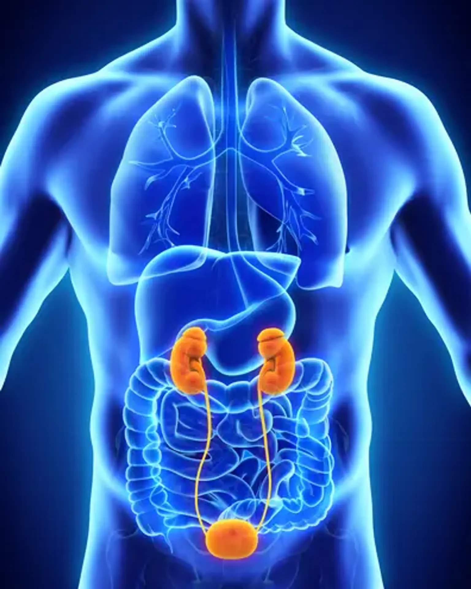
Urine is produced by your kidneys by filtering wastes and excess water from your blood. Urine flows from the kidneys to the bladder via two narrow tubes known as ureters. Ureters range in length from 8 to 10 inches. To drive urine down and away from the kidneys, muscles in the ureter walls tense and release. Every 10 to 15 seconds, little volumes of pee flow from the ureters into the bladder. The ureters can become clogged or damaged at times. This can obstruct urine passage to the bladder.


Anatomy of the Ureter 

Anatomy of the Ureter 

The ureter is a 22–30 cm long muscular tube, which connects renal pelvis and bladder. On radiographs, the ureter is divided into three sections 

  • Upper third: from renal pelvis to the top edge of the sacrum
  • Middle third: from the top edge to the lower edge of the sacrum
  • Distal third: lower edge of the sacrum to the urinary bladder.

The ureter's upper portion is located anterior to the psoas muscle. The upper physiologic constriction of the ureter is caused by the transition between the renal pelvis and the ureter. The ureter sits beneath the gonadal vessels before reaching the iliac vessels. The ureter crosses the iliac vessels ventrally, causing the ureter to narrow in the center. The lower portion of the ureter in men is located dorsally of the vas deferens, medial umbilical ligament, and superior vesical artery. In women, the ureter is located dorsally of the round ligament, uterine artery, and the tissues listed above. The lower physiologic narrowing is caused by the passage of the ureters into the bladder.


Congenital Diseases of the Ureter

Ureteropelvic junction obstruction

Ureteropelvic junction

A ureteropelvic junction (UPJ) blockage occurs when a portion of the kidney is obstructed. It is usually occluded at the renal pelvis. This is the point at which the kidney connects to one of the ureters (the tubes that carry urine to the bladder). The obstruction delays or prevents urine flow out of the kidney. Urine can then accumulate and cause kidney injury. Sometimes surgery is required to increase urine flow, while other times the condition resolves on its own.


Symptoms of UPJ Obstruction

Most instances are discovered well before delivery using ultrasonography. Infants and children show the following indications after birth:

  • Abdominal mass
  • Urinary tract infection with fever
  • Flank pain (pain in the upper abdomen or back, mostly with fluid intake)
  • Kidney stones
  • Bloody urine
  • Vomiting
  • Poor growth in infants


UPJ obstruction may also cause pain without an infection

Some instances of UPJ blockage remain unclear. Urine may drain properly at times and get clogged at others. This produces intermittent discomfort. Most youngsters, it is assumed, are not in discomfort unless the urine becomes contaminated or the obstruction worsens.


Diagnosis of UPJ obstruction

While ultrasonography allows your doctor to examine your kidneys, more tests are required to establish UPJ occlusion. Your urologist will examine how properly urine is generated and drained to get an accurate diagnosis. There are several tests that may be performed. Blood and urine samples may be collected. The blood urea nitrogen (BUN) and creatinine tests determine how efficiently the kidney filters the blood.

In the past, an intravenous pyelogram (IVP) was often employed. A dye is injected into the circulation during this procedure. To examine how the kidneys eliminate the dye from the blood, an X-ray is utilized. Your doctor can examine if the kidney, renal pelvis, and ureter look normal when the dye flows via urine.

An IVP is comparable to a nuclear renal scan, although it is more current. Instead of dye, this test use radioactive material. A specific camera may be used to view the substance. This test provides the clinician with valuable information about how the kidney is functioning and how much obstruction there is.

CT scans are occasionally utilized in the emergency room to determine the cause of acute pain in youngsters. If a blocked kidney is the source of the discomfort, a CT scan can easily reveal it. The kidneys, ureters, and bladder are also examined using magnetic resonance imaging (MRI). However, MRI is pricey and not widely utilized.

Siblings should be screened for UPJ blockage only if they exhibit symptoms. There have been few occasions when many members of a family have been denied UPJ, but the vast majority of cases are individual.


Treatment UPJ obstruction

Treatment is not always essential, and specialists vary on this point. It is critical to understand that impaired drainage in babies and children under the age of 18 months may be transitory. Many infants with strong renal function but poor drainage can improve significantly after a few months. On the other hand, in some infants, the blockage will worsen rather than improve.

Repeat ultrasounds and, if necessary, nuclear scans are performed on young individuals with an enlarged kidney. Improvement can happen quickly. If so, it usually happens during the first 18 months of life. If an infant's urine flow does not improve and the blockage persists, surgery is required. Adults may be able to obtain therapy in various ways.


Vesicoureteral reflux

Vesicoureteral reflux

VUR is described as the retrograde regurgitation of urine from the urinary bladder up the ureter and into the kidney collecting system. It is the outcome of various irregularities relating to the ureter's functional integrity, bladder dynamics, and the anatomic makeup of the ureterovesical junction (UVJ)

VUR affects 1% to 2% of all children, and up to one-third of these children will have a urinary tract infection (UTI). Acute pyelonephritis caused by VUR can result in renal scarring and, eventually, chronic kidney disease known as reflux nephropathy. 10%-25% of people with severe reflux nephropathy will develop end-stage renal disease requiring dialysis or kidney transplantation.

However, the severity of VUR varies significantly, as does the clinical presentation; most individuals are either asymptomatic or have hydronephrosis or pyelonephritis. Spontaneous resolution is rather common. Conservative treatment consists of two major approaches: active surveillance and antibiotic prophylaxis to limit the risk of bacterial infection of the bladder when reflux is present. Endoscopic therapy or open/laparoscopic/robotic surgical procedures can both be used to treat VUR.

The pillars of VUR care are early diagnosis and diligent monitoring, although the beginning point for VUR evaluation is controversial. Guidelines differ on whether to focus on assessing kidney status ("top-down") or recognizing the existence of VUR.


Ureteral stones

Ureteral stones

Kidney stones are rock-like accumulations of naturally occurring mineral salts in urine that can become trapped in the ureter (kidney tube). Stones that obstruct the flow of urine can cause major problems such as kidney damage, uremic poisoning, severe infection, and even death. In many situations, modest lifestyle modifications such as drinking more water or avoiding particular foods can prevent the creation of new kidney stones.


Ureteral stones are rock-like deposits formed by naturally occurring mineral salts in urine. The majority of kidney stones originate in the kidney and move into the renal tubes or ureters. A typical urinary stone patient is a 40- to 50-year-old Caucasian guy with a family history of urinary stones.

Urinary stones can be spherical or uneven, smooth or jagged, with jagged edges or protrusion. Urinary stones vary in color depending on their chemical makeup. Stones in the kidneys can remain there for years without creating symptoms.

Causes and Risk Factors of Ureteral stones

When naturally occurring mineral salts in urine aggregate and become trapped in the ureter, stones develop. Urinary stones are caused by a variety of factors. As an example:

  • Calcium stones: Calcium stones, the most frequent kind, arise when there is an excess of calcium and/or oxalate in the blood. Calcium concentrations can be increased by taking some medicines or taking too much vitamin D. Oxalate levels can be raised by genetics or a diet heavy in oxalate-rich foods like spinach.
  • Uric acid stones: Excess uric acid, a byproduct of protein metabolism, can crystallize to form stones.
  • Struvite stones: Struvite stones are created by bacteria that infect the urinary tract more commonly in women.
  • Cystine stones: These rare stones occur in people with a hereditary condition that causes the kidneys to excrete large amounts of certain amino acids.

By one estimate, 1 in 10 people will develop urinary stones sometime during their life. Risk factors include:

  • Gender (stones typically occur in men);
  • Age (most stones occur in men between the ages of 40 and 60);
  • Lifestyle: (factors include being sedentary, eating a diet high in animal protein, and not drinking enough water);
  • Family history of stones; and
  • Chronic urinary tract infections.


Symptoms of Ureter Stones

A tiny ureter stone will not produce any symptoms. If it becomes large enough to form an obstruction, it will hinder the passage of urine and cause excruciating discomfort in the kidney area and lower abdomen, which may eventually migrate to the groin. Urine may appear pink from blood as the body strives to release the stone, and urination may become more frequent, unpleasant, and cause burning. The presence of fever and chills indicates the existence of infection.


Diagnosis of Ureter Stones

Stones that are too tiny to cause symptoms may show up on an X-ray. Blood and urine testing can reveal substances that may cause stones to develop. A urinary CT scan may also be advised. To identify ureter stones, an intravenous pyelogram (IVP), a test that utilizes iodine as a contrast agent with X-rays, is performed in some circumstances.


Treatment of Ureter Stones

Treatment of Ureter Stones

Treatments that do not involve surgical procedures are always desirable. A daily routine of the patient drinking two to three quarts of water can induce a stone to flow spontaneously out of the body if the stone is tiny enough. Pain medication, as well as follow-up drugs to manage acid or alkali in the urine (the essential ingredients of crystals), or to reduce calcium released into urine by the kidneys and aid in calcium retention in the bones, may be advised.

It is critical to keep the urine free of germs after removing infected (struvite) stones in order to prevent subsequent infection. Antibiotics may be administered, as well as frequent urinalyses.

Surgery is frequently required when stones cause discomfort, impede urine flow, cause infection, or develop. There are new non-surgical and endoscopic alternatives available.


Extracorporeal Shock Wave Lithotripsy (ESWL)

Extracorporeal Shock Wave Lithotripsy (ESWL) is the most generally recommended procedure due to its ability to be performed as an outpatient procedure and fast recovery time, if acceptable. There are several types of ESWL, some of which employ X-rays and others which employ ultrasound. Shock waves from outside the body go via the skin and tissues to the stone, breaking it down into particles. The remaining particles are naturally expelled from the body. A stent is put through the bladder into the ureter if necessary to help with urine flow. This procedure is done under anaesthetic.


Percutaneous Nephrolithotomy

Purcutaeneous nephrolithotomy is used mostly to remove big stones or stones that will not respond to ESWL. Purcutaeneous nephrolithotomy involves making a tiny incision directly into the kidney and using an instrument called a nephroscope to detect and remove the stone. Ultrasound or an electrohydraulic device is used to break apart larger stones. This operation necessitates a multi-day hospital stay.


Ureteroscopic Stone Removal

Ureteroscopic stone removal, which is exclusively used for stones in the mid and lower ureters, does not need an incision. Instead, a fiberoptic ureterscope is passed through the bladder into the ureter to find the stone, which is then either removed or fractured using shock waves. A stent may be utilized to help with urine flow.


Ureteral Stricture

Ureteral Stricture

Ureteral stricture is a constriction of the ureter caused by a variety of disorders that results in an upper urinary tract blockage.


Causes of Ureteral Stricture

Diseases of the ureter (intrinsic causes):

  • Ureteral stones
  • Infections (e.g. urogenital tuberculosis and schistosomiasis)
  • Ureteral cancer
  • Idiopathic or congenital diseases


Iatrogenic causes

Ureteral injury may be caused by radiation or intra-operative injury (e.g. ureteroscopy, rectal surgery, hysterectomy or vascular surgery).


Extraureteral diseases (extrinsic causes)

A variety of diseases may lead to obstruction of the ureter by compression or infiltration,


Signs and Symptoms of Ureteral Stricture

Unnoticed ureteral stricture can develop. Asymptomatic slowly progressing ureteral stricture Depending on the underlying condition or infectious complications, flank discomfort, fever, and hematuria may ensue.


Diagnostic Work-Up

Imaging of Ureteral stricture:

  • Renal ultrasound: Grade of hydronephrosis
  • Intravenous urography or abdominal CT
  • Renal scintigraphy: Renal function

Endoscopy:

To confirm the diagnosis and determine the length of the stricture, retrograde pyelography and ureterorenoscopy are used. In the case of an unknown cause, a stricture biopsy is required.


Treatment of Ureteral Stricture

Treatment of Ureteral Stricture

  • Ureteral Stenting:

After an endoscopic diagnosis of symptomatic hydronephrosis, ureteral stenting is the first line of therapy. In cases when spontaneous healing and clearance of the ureteral stricture is conceivable, the ureteral stent can be removed within a few weeks (e.g. infections).

  • Endoscopic Balloon Dilatation:

A balloon dilatator is injected after retrograde (or antegrade) pyelography and the insertion of a guide wire. Dilatation to a diameter of 4-6 mm is performed. A ureteral stent (DJ) is implanted for 2-4 weeks. The success rate ranges between 50 and 75%. Balloon dilatation is notably useful for short segment strictures of less than 2 cm and strictures after ureteral stone lithotripsy.

  • Endoscopic Ureterotomy:

The stricture is cut under endoscopic vision after retrograde (or antegrade) pyelography and the introduction of a guide wire. A full thickness incision is made through the ureteral wall till the periureteral fat is visible. Visualization can be accomplished using either retrograde (URS) or antegrade (nephroscope) approaches. There are several technical alternatives for ureterotomy, including cold knife (without cauterization) and laser fibers (holmium or Neodym:YAG).

Care must be taken with vessels near the ureter. Latero-dorsally cut proximal strictures (above the iliac arteries). Strictures above and below the iliac vessels should be clipped anterior-medially. A thick ureteral stent is implanted for 8-10 weeks after ureterotomy. The success rate is somewhat greater than for balloon dilatation, especially following ureteral anastomosis strictures. The most important prognostic factor is the length of the stricture.

  • Ureteroureterostomy:

Ureteroureterostomy is a straightforward procedure used to treat proximal and middle ureter strictures of 3 cm or less. See the section ureteroureterostomy for further information. Short distal ureter strictures are best treated with reimplantation.

  • Ureteroneocystostomy:

Ureteroneocystostomy can be used to treat distal ureteral strictures up to 4-5 cm long [see section ureteroneocystostomy]. The psoas hitch procedure can be used to replace 6-10 cm of ureter. 12-15 cm of ureter can be replaced using the Boari-flap procedure, depending on bladder capacity. An further 6 cm can be bridged by mobilizing the kidney and attaching the lower pole to the psoas muscle.

  • Transureteroureterostomy:

Transureteroureterostomy can be used to treat mid-ureter strictures in conjunction with poor bladder capacity. Contraindications include a short donor ureter (proximal stricture) and a sick ureter on the opposite side. Furthermore, transureteroureterostomy is contraindicated in individuals with Ormond disease, following retroperitoneal radiotherapy, upper tract transitional cell carcinoma, and recurrent nephrolithiasis.


Tumors of the Ureter

Tumors of the Ureter

Ureteric neoplasms are uncommon tumors. The yearly incidence was 0.95-1.15/100.000 person-year. They are virtually invariably urothelial tumors, particularly papillary transitional cell carcinoma, as seen in the figure above, and are less prevalent than renal pelvic tumors and ten times less common than urinary bladder tumors. Concomitance with bladder tumors has also been seen, either synchronously or synchronously. Bladder cancers precede Ureteric tumor in 10% of metachronous cases and 49% of synchronous cases. Both ureters are equally implicated, and both ureters were found to be engaged in 15 of 930 instances.

Although ureteral tumors are most commonly found in the distal ureter, and multifocal implants can occur, tumor site should not be utilized to predict outcomes or make treatment choices. Because tiny Ureteric tumor might elicit early symptoms, ureteral neoplasms are more prone to present with organ-confined illness. When these tumors spread, they typically affect the lymph nodes, lungs, liver, bone, and brain. Males are more likely to have Ureteric tumor (M:F = 2:1). It is anticipated that 3000 new instances arise in the United States each year, with the number gradually increasing each year.

The typical age of diagnosis is 71 years, and higher-grade tumors are more common among the elderly. The environment has been implicated.

Arsenic exposure was linked to the development of upper urinary tract cancer in Taiwan. Aristolochic acid-containing Chinese medicines may induce progressive renal fibrosis and a considerable rise in the incidence of urothelial malignancies of the renal pelvis or ureter. Balkan endemic nephropathy and Lynch syndrome are other risk factors. It is worth noting that, unlike bladder cancer, cigarette smoking was not linked to ureteric neoplasms.

Clinically, the most common sign is hematuria, which occurs in 75% - 90% of cases, followed by obstruction symptoms, which are typically shown as flank pain (8% - 40% of cases), bladder irritation, and constitutional symptoms. The picture also demonstrates progressive hydroureter and hydronephrosis, as well as significant renal parenchymal atrophy.

Some individuals are asymptomatic, and the diagnosis is confirmed after a screening urinalysis reveals microhematuria or when abdominal ultrasonography is performed for another cause. Physical examinations are frequently unrevealing. Because of the degradation of tumor cells in the urine, cytologic examinations of the urine might be difficult to interpret, but imaging scans often show the tumor as well as the hydronephrosis.


Ureter Injury and Ureteral Trauma

Ureter Injury

Ureteral damage is uncommon and is mostly caused by penetrating trauma (gunshot or stab wounds). A ureteral ripping caused by deceleration stress is another uncommon mechanism of damage.

The mechanism of damage typically dominates the symptoms of ureteral injury. Abdominal discomfort, fever, upper urinary tract blockage, urinoma, hematuria, peritonitis, or urine discharge from the incision are some symptoms of ureter damage.


Diagnostic Work-up in Suspected Ureteral Injury

  • Abdominal and renal sonography
  • Intravenous urography, or better an abdominal computed tomography
  • Cystoscopy, retrograde pyelography and ureteroscopy, if endoscopic treatment is deamed possible


Treatment of Ureteral Injury

Endoscopic therapy can be used to treat minor ureteral injuries (Insertion of a ureteral stent, percutaneous nephrostomy). Major ureteral damage (suture repair, ureteroureterostomy, transureteroureterostomy, or ureteral reimplantation) or long-segment ureteral stricture necessitate surgical treatment.


Conclusion 

A ureter stone is a mineral mass in the ureter that began in the kidney and migrated down into the ureter. A stone forms when mineral particles in stagnant urine solidify and form a bulk. A tiny enough stone can detach and flow undetected through the ureter and out of the body. If it is excessively big, it may become lodged in the ureter and restrict urine flow.