Uterine Myoma

Last updated date: 04-Mar-2023

Originally Written in English

Uterine Myoma

Uterine Myoma

Overview

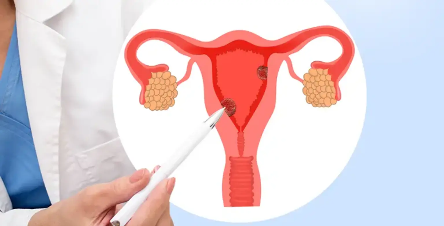
Uterine Myomas are non-cancerous smooth tumors that can grow in or around the uterus. Myomas, which are partially made of muscle tissue, seldom form in the cervix, but when they occur, they are generally accompanied by myomas in the larger, upper region of the uterus. Fibroids and leiomyomas are other names for myomas in this area of the uterus.


What is Uterine Myoma?

Uterine Myoma

Uterine Myoma are noncancerous uterine growths that commonly arise during reproductive years. Uterine myomas, also known as leiomyomas or uterine myomas, are not connected with an elevated risk of uterine cancer and usually never develop into malignancy. Myomas range in size from invisible seedlings to bulky masses that can deform and expand the uterus. You might have a single myoma or many. Multiple myomas can cause the uterus to enlarge so much that it approaches the rib cage and adds weight in severe situations. Many women get uterine myomas at some point in their lives. However, because uterine myomas seldom cause symptoms, you may be unaware that you have them. During a pelvic check or prenatal care. During a pelvic exam or prenatal ultrasound, your doctor may uncover myomas by chance. In women of reproductive age, myomas are the most prevalent benign tumor of the reproductive organs. They might be single or many, producing severe morbidity and degradation of quality of life, and can have a deleterious influence on the reproductive system. According to the relevant literature, myomas account for 40-60% of all hysterectomies done. The most prevalent reason for the hysterectomy is myomas.

In 1793, Matthew Baille was the first to characterize myomas. Myomas are mostly composed of smooth muscle cells with varying levels of fibrous tissue. A myoma compresses the surrounding tissues (the myometrium and connective tissue) as it grows, resulting in the gradual creation of a pseudocapsule rich in collagen fibers, neurofibers, and blood arteries The continuous surface of the pseudocapsule is occasionally disrupted by collagen fiber and vascular bridges that attach the myoma to the myometrium. As a result, a distinct cleavage plane forms between the myoma and the pseudocapsule, as well as between the pseudocapsule and the surrounding myometrium. This pseudocapsule exerts a non-destructive displacement effect on the myometrium; yet, the uterine structure's integrity and contractility are preserved.  


Epidemiology

myomas

Many epidemiologic variables have been associated with the development of myomas, however, many are still unknown. Age, race, heritage, reproductive variables, and sex hormones are examples of these factors. Some epidemiological data is contradictory.

  • Age

During the reproductive years, the chance of developing a myoma increases with age. Myomas do not appear before puberty, and their occurrence diminishes after menopause. Myomas are identified in 20-25% of reproductive-age women and 30-40% of women after the age of 40. Women who reach menarche at a younger age are more likely to develop uterine myoma. Because of the lengthier exposure to gonadal steroids, late-onset menopause increases the likelihood of myoma development.

  • Race

Myomas are more frequent in African-American women and rare in Asian-American women. Other than Caucasian and African American women, data on race disparities are few. determined the prevalence of 18% among black women, 8% among white women, 10% among Hispanic women, and 13% among "others," primarily Asian women. Black women are more likely to be diagnosed at a younger age, with myomas that are frequently many, bigger, and associated with more severe symptoms than other ethnic groups.

  • Genetics 

Genetic factors can play an important role in the development of myoma. The presence of several myomas in the same uterus suggests that genetics play a role in myoma formation, with some women being more prone than others. The existence of so-called "myoma families'' described in the literature demonstrates a familial predisposition to myoma formation. In cases of familial myomas, women were diagnosed at a younger age and more frequently with multiple myomas, so they tended to undergo hysterectomies at a younger age as well. Twin studies have shown that monozygotic twins had a higher risk of myoma development than dizygotic twins.

  • Reproductive elements

The inverse relationship between myoma risk and parity is widely recognized, and having more term pregnancies lowers myoma risk. This link might be explained by both hormonal and non-hormonal processes. Parity results in reduced menstrual cycles and term pregnancies alter ovarian hormones, growth factors, and estrogen receptor levels, as well as alterations in uterine tissue. Thus, myomas are more prevalent in nulliparous women, however, obesity and excess weight appear to reduce the adverse connection with parity. The chance of myoma formation decreases as the woman's age increases throughout her last-term pregnancy. According to the findings of the Nurses' Health Study II, the risk of myoma decreases with the woman's age at the first and final delivery.

  • Endogenous hormones

Myomas appear solely during the reproductive phase, demonstrating their reliance on ovarian steroids. Both clinical and experimental investigations show that estrogen and progesterone have a role in myoma development and growth. It is unclear exactly how they impact myoma development and growth. Early menarche raises the incidence of myomas owing to lifetime exposure to circulating ovarian hormones. Estrogen is thought to increase the development of myomas. According to recent studies, progesterone may also be significant for the formation of myomas since it works synergistically with estrogen to induce myoma growth. Because of this, selective progesterone receptor modulators (SPRMs) including asoprisnil, ulipristal, and telapristone have been studied as possible treatment medicines for uterine myomas. Ulipristal acetate (UPA) has shown good results as a potential treatment medication for uterine myomas. 

  • Use of Exogenous 

The link between oral contraceptives and myomas has been extensively studied. The epidemiological research on the association between oral contraceptive usage and myomas is inconclusive. Due to detection bias, the use of oral contraceptives may improve diagnosis. Published research indicates that the use of combination oral contraceptives reduces or eliminates the risk of myomas. As a result, there is no relationship between oral contraceptive usage and the risk of myoma among African American women.


What are the Signs and Symptoms of Uterine Myoma?

Symptoms of Uterine Myoma

Many women with myomas exhibit no symptoms. Symptoms can be altered by the location, size, and quantity of myomas in persons who have them. The following are the most prevalent indications and symptoms of uterine myoma in women who have symptoms:

  • Heavy menstrual flow
  • Menstrual cycles that last more than a week
  • Pelvic discomfort or pressure
  • Urine frequency
  • Having trouble emptying the bladder
  • Constipation
  • Back pain or leg pain
  • A myoma can occasionally produce intense discomfort when it outgrows its blood supply and begins to die.

Myomas are categorized based on their location. Intramural myomas develop within the uterine muscle wall. Submucosal myomas protrude from the uterine cavity. Subserosal myomas protrude from the uterus to the exterior.

Consult your doctor if you experience any of the following symptoms:

  • Pelvic discomfort that is persistent
  • Periods that are excessively heavy, lengthy, or painful
  • Bleeding or spotting between periods
  • Having trouble emptying your bladder
  • Unknown cause of decreased red blood cell count (anemia)


What factors contribute to uterine myoma?

factors contribute to uterine myoma

Although doctors do not know what causes uterine myomas, research and clinical experience point to the following factors:

  • Genetic alterations: Many myomas have gene alterations that vary from those found in normal uterine muscle cells.
  • Hormones. Estrogen and progesterone, two hormones that drive the formation of the uterine lining in preparation for pregnancy throughout each menstrual cycle, appear to encourage the growth of myomas.

Myomas have more estrogen and progesterone receptors than uterine muscle cells in general. Because of a decrease in hormone synthesis, myomas tend to diminish after menopause.

  • Other elements that influence development. Insulin-like growth factor and other substances that aid in tissue maintenance may influence myoma development.
  • Matrix extracellular (ECM). ECM is the substance that holds cells together, similar to mortar between bricks. ECM also stores growth factors and induces physiological changes in cells.

Doctors think that uterine myomas arise from a stem cell in the uterus' smooth muscle tissue (myometrium). A single cell divides repeatedly, resulting in a solid, rubbery mass separate from surrounding tissue. Uterine myomas grow in a variety of ways; they might grow slowly or quickly, or they can stay the same size. Some myomas develop rapidly, while others diminish on their own. Many myomas that were present throughout pregnancy diminish or vanish after delivery when the uterus returns to its normal size. 


What are the side effects of uterine myoma?

side effects of uterine myoma

Although uterine myomas are not generally harmful, they can cause discomfort and may lead to problems such as a decline in red blood cells (anemia), which causes exhaustion, as a result of excessive blood loss. A transfusion is only required in rare cases owing to blood loss.


Myomas and pregnancy

Myomas do not frequently prevent women from becoming pregnant. However, myomas, particularly submucosal myomas, may cause infertility or pregnancy loss. Myomas can also increase the risk of pregnancy problems such as placental abruption, fetal growth restriction, and premature labor.


How are uterine myomas diagnosed?

uterine myomas diagnosis

Uterine myomas are commonly discovered by chance during a normal pelvic exam. Your doctor may see anomalies in the shape of your uterus, which might indicate the existence of myomas.

If you have uterine myoma symptoms, your doctor may request the following tests:

  • Ultrasound. If more proof is required, your doctor may request an ultrasound. It takes an image of your uterus using sound waves to confirm the diagnosis and map and quantify myomas. To get pictures of your uterus, a doctor or technician slides the ultrasound equipment (transducer) across your abdomen (transabdominal) or within your vagina (transvaginal).
  • Laboratory tests .If you experience unusual monthly bleeding, your doctor may prescribe further testing to rule out possible reasons. A complete blood count (CBC) to see if you have anemia from continuous blood loss, as well as other blood tests to rule out bleeding disorders or thyroid issues, may be included.


Other imaging procedures

If regular ultrasound is insufficient, your doctor may request further imaging investigations, such as :

  • Magnetic resonance imaging (MRI). This imaging examination can show the size and location of myomas in greater detail, detect different types of tumors, and assist in determining appropriate treatment options. An MRI is most commonly utilized in women who have a bigger uterus or who are nearing menopause (perimenopause).
  • Hysterosonography. Hysterosonography also known as a saline infusion sonogram, employs sterile salt water (saline) to enlarge the uterine cavity, making it simpler to obtain photos of submucosal myomas and the uterine lining in women trying pregnancy or experiencing heavy monthly blood
  • Hysterosalpingography. A dye is used to highlight the uterine cavity and fallopian tubes on X-ray pictures during hysterosalpingography. If infertility is an issue, your doctor may advise you to try it. This test can tell your doctor whether your fallopian tubes are open or obstructed, and it can also detect submucosal myomas.
  • Hysteroscopy. Your doctor will put a tiny, illuminated telescope called a hysteroscope through your cervix into your uterus for this inspection. Your doctor will next inject saline into your uterus, widening the uterine cavity and allowing your doctor to view the uterine walls and fallopian tube openings.


What is the treatment for uterine myomas?

There is no one optimum technique to uterine myoma therapy; there are several possibilities. If you are experiencing symptoms, see your doctor about symptom alleviation alternatives.


Medications

treatment for uterine myomas

Uterine myoma medications target hormones that control your menstrual cycle, alleviating symptoms including heavy monthly bleeding and pelvic discomfort. They do not remove myomas, although they may diminish them. Among the medications are:

  • Agonists of gonadotropin-releasing hormone (GnRH). GnRH agonists are medications that treat myomas by suppressing the synthesis of estrogen and progesterone, causing a brief menopause-like condition. Menstruation ceases, myomas decrease, and anemia frequently improves as a result.

Leuprolide (Lupron Depot, Eligard, and others) is a GnRH agonist, as is goserelin (Zoladex) and triptorelin (Trelstar, Trip To Door Kit). When utilizing GnRH agonists, many women have substantial heat flashes. GnRH agonists are usually only taken for three to six months since symptoms recur when the medicine is withdrawn, and long-term usage can cause bone loss. A GnRH agonist may be prescribed by your doctor to lower the size of your myomas prior to surgery or to aid in the transition to menopause.

  • Intrauterine progestin-releasing device (IUD). Heavy myomas-related bleeding can be relieved with a progestin-releasing IUD. A progestin-releasing IUD merely relieves symptoms; it does not shrink or remove myomas. It also helps to avoid pregnancy.
  • Tranexamic acid (Lysteda, Cyklokapron). This non-hormonal medicine is used to relieve painful menstrual periods. It is only used when there is a lot of bleeding.
  • Other medicines. Other drugs may be suggested by your doctor. Oral contraceptives, for example, can help manage menstrual flow but do not diminish myoma growth.
  • Nonsteroidal anti-inflammatory medicines (NSAIDs), which are not hormonal pharmaceuticals, may be useful in alleviating myomas-related discomfort, but they may not diminish myomas-related bleeding. If you have excessive monthly bleeding and anemia, your doctor may also advise you to take vitamins and iron.


Noninvasive treatment

Noninvasive treatment

MRI-guided focused ultrasound surgery (FUS) is a non-invasive treatment. 

  • A noninvasive treatment option for uterine myomas that does not need an incision and is performed as an outpatient procedure.
  • Treatment is administered while you are inside an MRI scanner outfitted with a high-energy ultrasound transducer. The photos show your doctor exactly where the uterine myomas are. When the myoma's position is determined, the ultrasound transducer directs sound waves (sonications) into the myoma, heating and destroying tiny sections of myoma tissue.
  • Researchers are learning more about the long-term safety and efficacy of newer technology. However, evidence obtained thus far indicates that FUS for uterine myomas is both safe and effective.


Minimally invasive procedures 

Minimally invasive procedures

Certain methods can kill uterine myomas without removing them surgically. They are as follows:

  • Embolism of the uterine artery. Small particles (embolic agents) are injected into the uterine arteries, cutting off blood flow to myomas and causing them to shrink and die.

This approach has the potential to be useful in reducing myomas and alleviating the problems they cause. If the blood flow to your ovaries or other organs is disturbed, complications may ensue. However, studies suggest that the consequences are comparable to surgical myoma treatments, and the danger of transfusion is significantly decreased.

  • Ablation with radiofrequency. Radiofrequency radiation is used in this therapy to eliminate uterine myomas and decrease the blood arteries that feed them. This is possible during a laparoscopic or transcervical operation. Cryomyolysis, a similar treatment, freezes the myomas.

Laparoscopic radiofrequency ablation (Acessa), also known as Lap-RFA, is performed by inserting slim viewing equipment (laparoscope) with a camera at the tip through two tiny incisions in the belly. Your doctor locates myomas to be treated using a laparoscopic camera and a laparoscopic ultrasound instrument.

After finding a myoma, your doctor will insert multiple little needles into it with specialized equipment. The needles heat up and kill the myoma tissue. The damaged filament instantly changes consistency, for example, from hard like a golf ball too soft like a marshmallow. The myoma will continue to diminish over the following three to twelve months, alleviating discomfort. Lap-RFA is considered a less invasive option to hysterectomy and myomectomy since no uterine tissue is cut. After 5 to 7 days of recuperation, most women who had the surgery resume their normal activities. To detect myomas, the transcervical — or via the cervix — technique to radiofrequency ablation (Sonata) also employs ultrasound guidance.

  • Myomectomy, either laparoscopic or robotic. Your surgeon will remove the myomas while leaving the uterus in situ during a myomectomy.

If the myomas are low in number, you and your doctor may choose a laparoscopic or robotic operation that removes the myomas from your uterus using slender devices entered via small incisions in your belly. Larger myomas can be removed through smaller incisions by breaking them up (morcellation) within a surgical bag or by extending one incision to remove the myomas. A tiny camera linked to one of the equipment allows your doctor to observe your abdominal area on a monitor. Robotic myomectomy provides your surgeon with a magnified, 3D image of your uterus, allowing for greater precision, flexibility, and dexterity than previous procedures.

  • Myomectomy through hysteroscopic surgery. If the myomas are confined within the uterus, this treatment may be a possibility (submucosal). Myomas are accessed and removed by your surgeon utilizing devices put via your vagina and cervix into your uterus.
  • Ablation of the endometrium. This therapy, which is done with a specialized tool placed into your uterus, uses heat, microwave radiation, hot water, or electric current to damage the lining of your uterus, either terminating or lowering menstruation.

Endometrial ablation is usually efficient at stopping irregular bleeding. Submucosal myomas can be removed during hysteroscopy for endometrial ablation, although this does not impact myomas outside the uterine lining. Although women are unlikely to become pregnant after endometrial ablation, birth control is required to prevent a pregnancy from forming in a fallopian tube (ectopic pregnancy).

There is a danger of new myomas growing and causing problems with any treatment that does not remove the uterus.


Typical surgical procedures

Typical surgical procedures

Traditional surgical treatments have the following options:

  • Myomectomy of the abdomen. If you have many myomas, very big myomas, or very deep myomas, your doctor may remove them using an open abdominal surgical operation.

Many women who have been advised that a hysterectomy is their sole choice might instead have an abdominal myomectomy. However, scarring from surgery can have an impact on future fertility.

  • Hysterectomy. The uterus is removed during this procedure. It is still the only permanent therapy for uterine myomas.

Your capacity to bear children is terminated after a hysterectomy. If you also choose to have your ovaries removed, you will experience menopause and must decide whether to need hormone replacement treatment. The majority of women with uterine myomas may be able to maintain their ovaries.


Conclusion 

Uterine Myomas are a form of noncancerous tumor that grows in and on the uterus. Myomas do not always create symptoms, but when they do, they might include heavy menstrual flow, back pain, frequent urination, and pain during sex. Small myomas may not require treatment, while bigger myomas may require medication or surgery.