Endoscopic Cranial Surgery

Last updated date: 13-Mar-2023

Originally Written in English

Endoscopic Cranial Surgery

Overview

Endoscopic cranial surgery involves passing equipment and a tiny camera via small holes in the skull. Once the camera and equipment are in place, the neurosurgeon can utilize them to repair or remove structures in or around the brain.

Endoscopic brain surgery is very beneficial for tumors, aneurysms, and other skull base abnormalities. The skull base contains essential structures such as the cranial nerves, carotid arteries, and the basilar artery, which are located where the brain sits on the floor of the skull. Skull base lesions can be difficult to approach with typical open surgery due to the close proximity of so many essential structures.

To access the skull base, endoscopic cranial surgery through the nose or sinuses might be employed. This method may result in fewer problems, less blood loss, and quicker recovery times. 
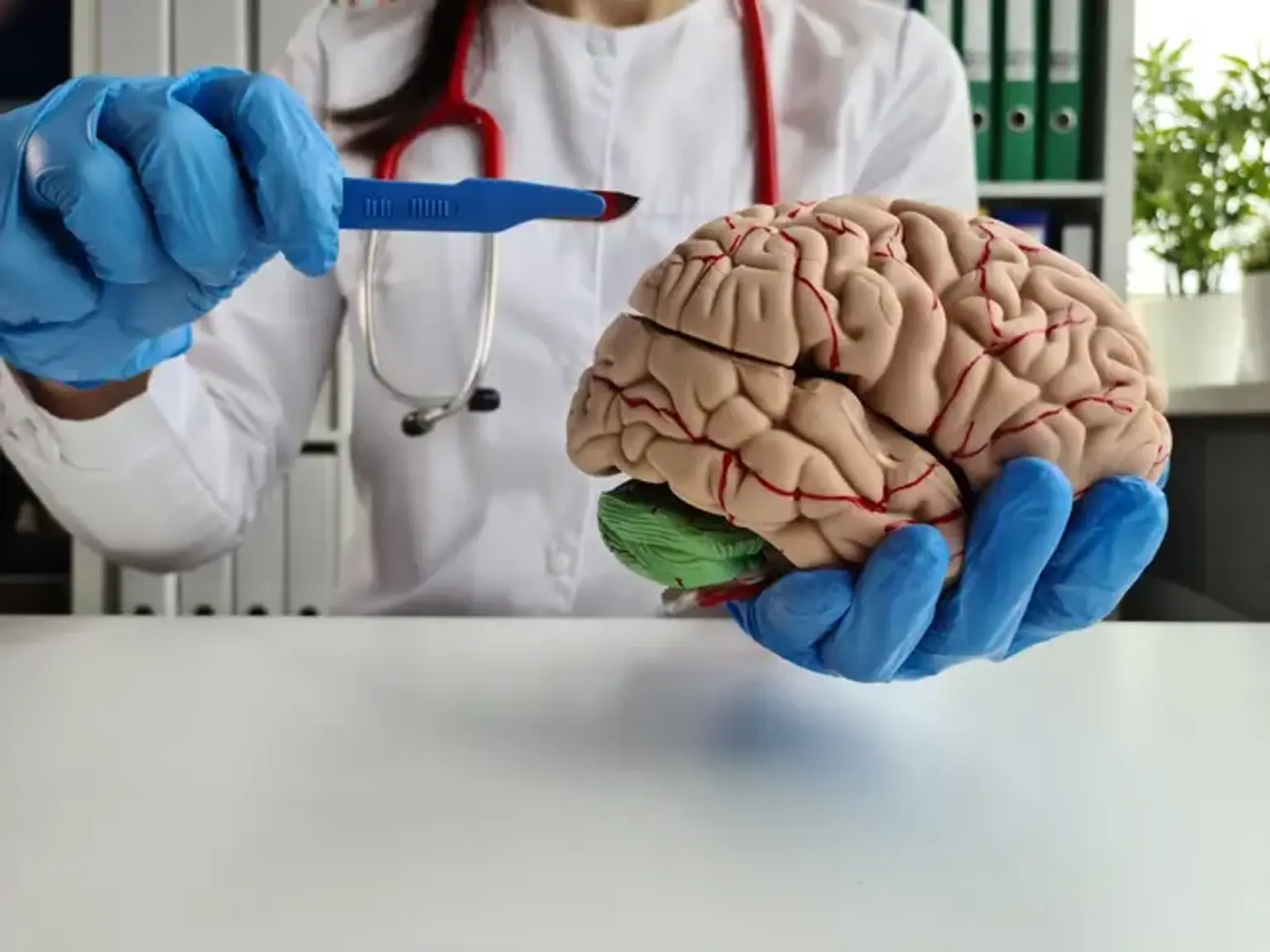
 

Benefits of Endoscopic Cranial Surgery

Benefits of Endoscopic Cranial Surgery

Endoscopic cranial surgery has several advantages for patients. This form of surgery is less aggressive than other open surgical treatments, allowing the patient to recover faster and with less discomfort.

While each patient's medical history, age, condition, and other factors influence the success of endoscopic brain surgery, the following are some of the most typical advantages of the procedure:

  • Minimally invasive (results in less pain for the patient).
  • Faster recovery time than open brain surgery.
  • Reduced risk of brain trauma.
  • Reduced risk of side effects.
  • Reduced hospital stay.


Indications For Endoscopic Cranial Surgery

Indications For Endoscopic Cranial Surgery

Clear visibility of the anatomy is required for safe endoscopic operations. Endoscopes perform best in preformed cavities filled with crystal-clear CSF, such as the ventricular system, subarachnoid space, and certain cystic lesions. As a result, hydrocephalus, intraventricular lesions, and space-occupying arachnoid or parenchymal cysts are excellent candidates for endoscopic surgery. Even highly vascularized tumors can be removed thanks to additional advancements in endoscopic hemostasis (the introduction of bipolar coagulation probes and appropriate laser instruments). Additionally, neuroendoscopes can be utilized in conjunction with an operating microscope to get more comprehensive information for dissection.

  • Management of Hydrocephalus

The typical indication for a neuroendoscopic technique is hydrocephalus. Hydrocephalus is still the most common intracranial illness treated endoscopically. Endoscopic third ventriculostomy has become a standard treatment option for non-communicating hydrocephalus. The third ventriculostomy has been shown to be effective in the treatment of obstructive hydrocephalus caused by tumors, aqueductal stenoses, hemorrhages, and infarctions. Although the treatment is generally seen to be safe and simple, severe and, in rare cases, deadly consequences can develop.

The correct placement of the fenestration in the third ventricle floor is critical to avoiding vascular and neurological damage. The floor should be perforated midway between the infundibular recess and the mammillary bodies in the midline, slightly behind the dorsum sellae. Hypothalamic damage, oculomotor palsy, and vascular injury are therefore improbable.

A careful examination of a CT scan or sagittal MR image is recommended to examine the individual relationship between the basilar artery and the floor of the third ventricle.

Small, weakly vascularized tumors of the lateral or third ventricle that generate ventricular enlargement by occluding CSF routes are suitable candidates for endoscopic treatment. The ventricular dilatation allows enough room to operate the endoscope and manipulate the devices.

Intraventricular lesions, on the other hand, maybe accessed precisely via tiny ventricles utilizing computerized neuronavigation. The tumor size is a key limiting factor in endoscopic tumor resections. Most neuroendoscopes on the market today have a maximum working channel diameter of 2.4 mm. The employment of the equipment to remove even minor tumors is clearly a time-consuming technique.

The advantages of the minimally invasive method, such as reduced brain retraction and a small burr-hole approach, are therefore balanced by the length of the procedure. As a result, our operational endoscope does not have a distinct working channel. For the excision of big tumor fragments, the entire inner diameter (> 6 mm) of the operating sheath through which the endoscope is inserted is available.

It is difficult to estimate a tumor's size limit for efficient endoscopic removal. Endoscopic piecemeal resection may become time consuming and ineffectual if the tumor is too big. As a result, a solid tumor should not be more than 2 cm in diameter.

Furthermore, the tumor's consistency and vasculature must be examined. A soft tumor may be removed more easily and quickly than a hard lesion. Even bigger lesions can be removed endoscopically if the tumor is cystic or contains significant cystic components. Cavernomas and hemangiomas, which are highly vascularized lesions, can also be safely eliminated with a Nd:YAG laser and bipolar diathermy.

Larger tumors (with concomitant hydrocephalus) that are not amenable to endoscopic excision may require a third ventriculostomy or aqueductal stent insertion to restore CSF flow. If the tumor occupies the whole third ventricle, the stent should be placed through the entire third ventricle and aqueduct linking the lateral, third, and fourth ventricles. A septostomy of the septum pellucidum may be used to treat unilateral blockage of the foramen of Monro.

To restore CSF flow, both stent insertion and septostomy may be required. Any apparent tumor on the ventricular surface can be sampled for biopsy.

  • Colloid Cysts

For the treatment of colloid cysts, transcallosal-transventricular, transcortical-transventricular, transcallosal-interfornicial, transcortical-transventricular stereotactic methods, and stereotactic aspiration have been proposed. Because of the significant risk of complications, shunting should not be considered as a therapeutic option.

The rule in microsurgical operations is that the cyst must be completely removed. However, the hazards are widely understood. Complications of the transcallosal technique include venous infarction, sagittal sinus thrombosis, disconnection syndromes, fornicial damage, and thalamic and basal ganglia infarctions. The transcortical method has been linked to an increased risk of seizures. CT-guided stereotactic aspiration of colloid cysts has been recommended due to its ease of use and minimal risk.

However, stereotactic aspiration has frequently failed in hyperdense cysts with solid substance. An endoscopic or microsurgical operation is then required. Furthermore, attempting "blind" stereotactic aspiration risks damaging the fornix and inducing ependymal vein hemorrhage.

Most critically, a high recurrence rate (up to 80%) following colloid cyst aspiration has been documented in a long-term follow-up research. The surgery failed during the first two months and after more than eight years. Tentorial herniation caused unconsciousness in some cases.

These findings highlight the reality that just aspiration is insufficient. The key appears to be creating a broad cyst opening and complete or near-complete capsule excision. Because colloid cysts have been linked to unexpected mortality, even asymptomatic cysts with evidence of CSF route blockage should be surgically addressed.

  • Arachnoid Cysts

The CSF-like composition of congenital arachnoid cysts makes it ideal for the use of an endoscope. Arachnoid cysts are most commonly found in the sylvian fissure. Cysts in different areas, such as the anterior and posterior cerebral fossa, quadrigeminal cistern, interhemispheric fissure, suprasellar region, or intraventricular space, can be treated endoscopically. Microsurgical cyst excision or fenestration, stereotactic aspiration, cyst fenestration with arachnoidplasty, cystocisternostomies, ventriculocystostomies, cystosubdural shunting, cystoperitoneal shunting, and endoscopic fenestration have all been advocated for the treatment of arachnoid cysts.

The optimum therapeutic choice, however, has yet to be discovered. Meningitis, hemiparesis, oculomotor palsy, subdural hematomas, new grand mal seizures, and even mortality have been documented following microsurgical cyst fenestration/resection operations, while shunt dysfunction and infection have been described following the latter. Although shunt implantation is clearly safer, it is associated with a greater frequency of further surgical procedures as well as the drawback of life-long shunt reliance.

Endoscopic procedures reduce the risks involved with microsurgery while producing comparable or even superior results. In the case of symptomatic space-occupying arachnoid cysts, surgical surgery is advised. If MR imaging reveals no mass impact or the relationship between symptoms and arachnoid cyst is unclear, the ICP should be monitored for elevated ICP and/or abnormal pressure waves. 

Arachnoid cyst surgery for asymptomatic patients is debatable. Despite the significant susceptibility of arachnoid cysts in individuals with modest head trauma, we believe surgery for asymptomatic arachnoid cysts in adults is unnecessary. Surgery, on the other hand, should be undertaken in children who have asymptomatic cysts that have a mass effect, despite the fact that spontaneous regression of arachnoid cysts has been documented on occasion.

Because cyst expansion may jeopardize normal development and function of the adjacent brain in children, this potentially harmful effect outweighs the risk of the operative procedure.

  • Pituitary Surgery

For decades, the microsurgical transseptal transsphenoidal approach to pituitary tumors has been the gold standard. In the transseptal technique, endoscopes were primarily employed as an auxiliary to the operating microscope

However, breathing issues, septum perforations and deviations, and numbness of the maxillary dentation following sublabial incision have all been documented as possible consequences of transseptal dissection. To get enough working room for the endoscope and equipment within one nostril, the middle turbinate must be outfractured or resected, and the nasal septum must be displaced.

For decades, the microsurgical transseptal transsphenoidal approach to pituitary tumors has been the gold standard. In the transseptal technique, endoscopes were primarily employed as an adjunct to the operating microscope

However, breathing issues, septum perforations and deviations, and numbness of the maxillary dentation following sublabial incision have all been documented as possible consequences of transseptal dissection. To get enough working room for the endoscope and equipment within one nostril, the middle turbinate must be outfractured or resected, and the nasal septum must be displaced

Nasal packing is not required, or, if it is, only for a short time. Endoscopes provide an excellent panoramic view in the depth of the sphenoid sinus and sella. The ability to inspect supra- and parasellar tumor extensions, not visible when using the operating microscope, increases the completeness of tumor removal.


How Do I Get Ready for Endoscopic Cranial Surgery?

Endoscopic Cranial Surgery preparation

This operation will be performed while you are sedated. That implies you'll be sleeping and won't be able to feel anything. If you have any concerns regarding the anesthetic or any other element of the treatment, please consult with your medical team ahead of time.

By properly planning, you can boost your chances of success. In general, pre-surgery preparation consists of the following steps:

  • If you smoke, you should stop at least two weeks before your operation. This will improve the success of anesthetic and aid in recovery.
  • Take no herbs or over-the-counter pain medicines for at least two weeks before your operation. This includes basic medications such as aspirin and ibuprofen.
  • Take no vitamin E for two weeks before surgery.
  • Inform your healthcare practitioner about any medications you are taking, any other health issues you are experiencing, and any past surgical complications.
  • Eat or drink nothing for at least 8 hours before your procedure, or as directed by your healthcare practitioner.
  • You should arrive at the surgical center at least an hour before your operation, or as directed by the institution. Check in at the admissions desk to fill out any documents that are required, including a permission form that must be completed before the operation can begin.
  • In preparation of the surgery, your surgical team will provide you with additional detailed instructions to follow. Some of these may change significantly from those mentioned above.


Procedure and Equipments

Endoscopic Cranial Surgery Procedure

A sophisticated and complicated neuroendoscopic equipment, including multiple rigid, semiflexible, and flexible scopes and brilliant cold light sources, as well as a high-resolution video camera system, effective tools, and irrigation devices, is required for intracranial neuroendoscopy.

Combination with a guidance system is beneficial, and in certain cases, required. The endoscopes are delivered through an operating sheath that is first implanted with the use of a trocar, allowing for intraoperative interchange of various scopes without reinserting scopes into brain tissue, hence avoiding needless harm to the surrounding healthy brain. Rigid rod-lens scopes are preferred because to their excellent optical quality and wide-angle vision, as well as their simplicity of guidance and orientation.

These endoscopes allow a thorough examination of the intraventricular anatomy. Although tiny hemorrhages may cause the image to blur, the surgeon will remain orientated, which is exceedingly difficult with a fiberscope's weak optics. Rigid scopes with four distinct viewing angles (0, 30, 70, and 120°) are offered. The 0° and 30° scopes are for inspection and manipulation, while the 70° and 120° scopes are just for examination ("looking around a corner"). Because there is no distinct working channel on the operational endoscope (wide-angle straight-forward scope with slanted eyepiece), the whole inner diameter (approximately 6 mm) of the endoscopic sheath may be employed, allowing for successful tissue removal and implant placement of devices such as stents.

The 3.2-mm outer diameter tiny endoscope was designed for use in pediatric patients. This semirigid minifiber endoscope (10,000 fibers/mm2) has an instrument channel as well as two independent irrigation in- and outflow channels. When performing "around the corner" operations, steerable fiberscopes with an instrument channel of 1.2 mm (outside diameter 2.5 mm and 3.5 mm) are employed.

However, due of their better optical qualities, rigid rod-lens scopes are used for most endoscopic operations. Mechanical devices of various sizes, such as scissors, biopsy and gripping forceps, hooks, and puncture needles, are available. Both the operational endoscope and the micro scope allow manipulations to be conducted in a straight line with stiff instruments, which offers strong tactile input from the tissue and allows for straightforward tool guiding.

Hemostasis and dissection are accomplished using bipolar and monopolar diathermy probes, as well as a laser guide. We prefer a 1.064-m Nd:YAG laser. Balloon catheters are used to increase ventriculostomies or other fenestrations. Lactated Ringer's solution at 36 to 37°C is preferred to saline because postoperative elevations in body temperature, which are sometimes noticed after extensive saline irrigation, are uncommon.

It is critical to keep the outflow channel open in order to avoid hazardous rises in intracranial pressure (ICP). Because the color temperature of xenon light is similar to that of sunshine, it provides the optimum lighting (6000 K). The light is delivered from the light fountain to the endoscope through fiberglass or fluid wires. A sterile optical bridge connects digital 1- or 3-chip tiny video cameras to the endoscope. Because the camera and bridge are wrapped in a sterile covering, sterile intraoperative endoscope interchange is possible without sanitizing the delicate electronics.

High-resolution video monitor screens display the endoscopic picture. Each endoscopic procedure is taped with a S-VHS recorder. Analog video recordings can be processed using a digital processing unit to enhance contrast as required. Finally, the documentation equipment includes a video printer and digital still recorder. 

For some endoscopic procedures, the simultaneous use of two endoscopes is beneficial. The images from both scopes can be displayed on one video monitor with the aid of a digital picture-in-picture device In this way, the surgeon obtains information provided by both scopes while looking at only one screen.


What Happens After Endoscopic Cranial Surgery?

Endoscopic Cranial After-Surgery

Your nose and sinuses may be bandaged after your procedure. These are often removed within a week of surgery. You will also be given medications to help you fight infection. Inform your healthcare professionals if you are in pain or discomfort. Most patients stay in the hospital for 1 to 2 days before heading home.

Take any medicines you are given as directed and follow all postoperative instructions once you are released.

Call your healthcare provider if you are confused about how to take the recommended medicines or if any of the following occur:

  • Pain that can't be managed with prescribed medications.
  • Fever of more than 100.4˚F (38˚C).
  • Shaking chills.
  • Redness, tenderness, heat, or pus, which are signs of infection, at the surgery site.
  • Swelling in the area around the nose.
  • Shortness of breath or trouble breathing.
  • Chest pain.
  • Any clear drainage from the nose. 

Your healthcare team may give you other instructions about what you should do after your procedure.


Is Endoscopic Cranial Surgery Risky?

Any major surgery carries some risk. Most people get through this surgery and heal without problems, but complications can develop. Possible risks include:

  • Reactions to the anesthesia.
  • Excessive bleeding.
  • Hematoma (a pooling of blood in the wound site).
  • Damage to veins, arteries, nerves, and other structures in the area.
  • Cerebrospinal fluid leaking from the nose.
  • Infection.
  • Slow healing.
  • Blood clots.
  • Pneumonia.

The procedure may carry other risks, depending on your specific medical condition. Be sure to discuss any concerns with your surgeon before the procedure.


Conclusion

Endoscopic Cranial Surgery

Endoscopic cranial surgery is mostly used to treat brain tumors. It is a minimally invasive brain surgery that allows neurosurgeons to discover and treat deep-seated brain diseases.

This sort of surgery allows us to treat brain tumors in a less intrusive manner than standard open brain surgery while yet providing an in-depth look of the brain.

Thin tubing that transmits video pictures of the brain is placed through one or two tiny incisions in the skull or through an entrance in the body during this surgery. An endoscope is a tube-like tool that incorporates a tiny camera that allows the neurosurgeon to see detailed pictures of the problem location in the brain.

The neurosurgeon will use the images transmitted by the endoscope as a guide for removing the tumor or repair the affected area of the patient’s brain. The removal of the tumor or damaged area is performed with specialized surgical instruments.