Endovascular Therapy
Last updated date: 03-Mar-2023
Originally Written in English
Endovascular Therapy
Overview
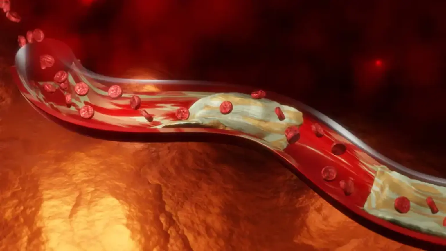
Peripheral artery disease develops when blood arteries thin or get clogged by plaque over time, a condition known as atherosclerosis.
An ischemic stroke happens when a blood artery in the brain becomes blocked or clots. When plaque builds up on the interior wall of an artery, it can cause a blockage or clot. As the cells move through the artery, they adhere to the plaque, causing the blockage or clot (also known as a thrombus) to develop large enough to obstruct normal blood flow. Stroke symptoms include a drooping face, difficulty to lift both arms, and slurred or confused speech.
Rehabilitation and drugs, such as tissue plasminogen activator, blood thinners, and blood pressure medications, can be used to treat stroke. Physical changes (ability to communicate, ability to move, and bowel and bladder issues), relationship changes, legal and financial issues (ability to earn a living, return to work), and less visible changes (emotional changes, fatigue, changes in perception) can all result from a stroke (e.g., vision, sensation, spatial relations, time awareness, unilateral body neglect, visual neglect).
Stroke will account for one in every six fatalities from cardiovascular disease in 2020. A stroke occurs every 40 seconds in the United States. Someone dies from a stroke every 3.5 minutes. Every year, around 795,000 people in the United States suffer from a stroke. Approximately 610,000 are first or new strokes. Approximately 185,000 strokes, or approximately one in every four, occur among adults who have previously had a stroke. Ischemic strokes account for approximately 87% of all strokes, in which blood flow to the brain is impeded.
Between 2017 and 2018, the cost of stroke in the United States was over $53 billion. This sum includes the expense of health care services, stroke medications, and missed work days. Stroke is the most common cause of major long-term impairment. More than half of stroke survivors aged 65 and older have reduced mobility as a result of their stroke.
What is Endovascular Therapy?
Endovascular stroke therapy is a non-surgical treatment for abrupt loss of brain function caused by blood clots. Microcatheters (thin tubes visible through X-rays) are introduced into the blood clot from the groin or the arm. A thrombectomy is a technique that removes a blood clot from a blood artery. If the blood clot cannot be removed, it is dissolved with medications administered via catheter in a technique called as thrombolysis.
Endovascular therapy (EVT) is a treatment for people suffering from acute ischemic strokes that involves removing the clot or thrombus from the brain that caused the stroke. EVT includes thrombectomy, or the mechanical disintegration of vessel-occluding thrombi or blood clots, with or without intra-arterial thrombolytic drug delivery. EVT is becoming a part of the current standard of therapy for strokes, which is often a pharmacological intervention including tissue plasminogen activator (tPA; also called alteplase, a thrombolytic drug).
Endovascular therapies are minimally invasive operations performed inside blood vessels that can be used to treat stroke and peripheral arterial disease, or PAD, a frequent kind of vascular disorder in the leg, aorta, or carotid arteries.
Interventional therapy is required for PAD when patients acquire symptoms such as discomfort or tissue loss owing to a lack of circulation. Endovascular therapies are typically employed when patients have failed conservative therapy, such as medication and supervised exercise, and are facing a constraint in their lifestyle as a result of their condition, such as being unable to work.
Endovascular therapy for acute ischemic stroke
Early recanalization of the occluded artery improves clinical outcomes in individuals suffering from acute ischemic stroke (AIS) by protecting time-sensitive ischemic tissue (known as Penumbra). Intravenous injection of pharmacologic thrombolytic drugs has long been used to treat AIS.
80% of all acute ischemic strokes (AISs) are caused by intracranial artery occlusions, for which reperfusion therapy is the basis of treatment, with the clot as the target. Thrombolysis with intravenous recombinant tissue plasminogen activator (rt-PA) is only effective if administered within 3 or 4.5 hours. Furthermore, the rate of recanalization is less than 50%. Patients with large-vessel occlusions and patients in the early postoperative phase who do not want the systemic effects of IV rt-PA are candidates for endovascular therapy in AIS via intra-arterial thrombolysis or mechanical thrombectomy.
Also, cerebral bleeding following fibrinolytic drug administration, delayed recanalization periods, and low recanalization rates in proximal major artery occlusions with substantial thrombus load, such as the ICA or M1 segment, might impair patients' outcomes. As a result, the significance of mechanical thrombectomy has expanded dramatically, and several devices have been adopted in recent years for endovascular stroke therapy.
Endovascular thrombectomy was initiated to improve recanalization rates, extend the time window, and reduce the risk of cerebral bleeding. The Merci clot retriever, a corkscrew-like equipment, was initially approved, followed by the Penumbra thrombo-aspiration system. Both devices have a high recanalization rate.
On the other hand, the average duration to recanalization was 45 minutes, with most patients achieving just partial recanalization. Recently, retrievable stents have showed promise in terms of reducing time to recanalization and achieving a higher rate of full clot resolution. The retrievable stent can be deployed into the clot to engage it within the struts of the stent and then pulled back under flow arrest. As evidenced by the introduction of various significant treatments, neuro-interventional strategies continue to play an ever-increasing and exciting role in the therapy of AIS. Stent retrievers have the potential to be the most important technique to endovascular stroke therapy.
What are the types of stroke endovascular therapy?
Proximal endovascular thrombectomy:
Mechanical thrombectomy devices use a catheter to remove occluding thrombi from the target vessel. Subgroups include (1) suction thrombectomy devices, which aspirate occlusive material from cerebral vessels (Proximal Thrombectomy), and (2) clot removal devices, which physically grasp cerebral thrombi and draw them out of the cerebral vessels (Distal Thrombectomy).
Manual suction thrombectomy is performed by advancing an aspiration catheter near the thrombus's proximal (near) surface. The aspiration catheter is then returned under continuous negative pressure after manual aspiration.
The Penumbra Device is an adaptation of the manual proximal aspiration approach that consists of a specialized reperfusion catheter connected to a pumping system that uses continual aspiration. A second retriever device, similar to a stent, is used to remove a recalcitrant clot. In patients who are not eligible for IV thrombolysis or whose intravenous thrombolysis fails, the time window for neuroradiological intervention is 8 hours following stroke start.
In 2007, the approach was authorized for AIS therapy. Many experiments have been conducted to evaluate the Penumbra System. One experiment was a prospective, multicenter research that enrolled 125 stroke patients within 8 hours of symptom onset and found that 81.6% of treated arteries were effective. However, only 25% of patients had a good clinical result after 90 days, while 29% of patients with successful recanalization of the target artery had a good clinical outcome. Despite comparably superior recanalization rates, clinical outcomes were dismal, with a death rate of 32.8%.
Distal endovascular thrombectomy:
Distal thrombectomy is technically more challenging than proximal thrombectomy. Many clinical trials have been conducted with the Merci device, which was the first distal thrombectomy device to get FDA approval in 2004. The occlusion location must first be penetrated with a microcatheter in order to deploy the device beyond the thrombus. The gadget is drawn back into the thrombus and placed into the clot. The Merci Retriever and the trapped clot are then removed, first into the positioning catheter and then out of the patient's body.
The target vessel was successfully recanalized in 57.3% of cases utilizing only the Merci retriever and in 69.5% of cases using supplemental recanalization modalities. In 36% of cases, a favorable clinical outcome was obtained. The average surgery time was 1.6 hours, with clinically significant procedural problems occurring in 5.5% of patients.
Stent retrievers (retrievable thrombectomy stents):
Stent retrievers, which are the most recently launched mechanical treatment approaches, have been developed. These are stent-like thrombectomy devices that are self-expandable, re-sheathable, and re-constrainable, combining the benefits of intracranial stent deployment with rapid reperfusion and subsequent retrieval with definitive clot removal from the occluded artery. Because it is retrievable, it does not become a permanent implant, and it also serves as a thrombectomy device while being recovered. Mechanical thrombectomy using stent retrievers is a potential therapy for AIS.
The complete removal of the device avoids the most significant disadvantages associated with permanent stent implantation, such as the demand for double anti-platelet medication, which significantly increases the risk of hemorrhagic complications and the possibility of in-stent thrombosis or stenosis.
Under general anesthesia, a guide catheter is positioned in the proximal internal carotid artery via a transfemoral approach. A guide wire is inserted through the obstructed cerebral artery using a microcatheter and guided beyond the thrombus. The microcatheter is then inserted through the clot over the wire, and the guide wire is used in place of the embolectomy device. The revascularization device is positioned with the center third of the device resting within the thrombus formation.
In the majority of situations, the radial force of the stent retriever is capable of rapidly creating a channel by compressing the thrombus and partially restoring blood flow to the distal area, generating a channel for a temporary bypass. The second angiography is performed to check that the damaged artery's flow has been restored. The device is often kept in place for up to 10 minutes, allowing the thrombus to become entrapped inside the stent struts. To retrieve the thrombus, the unfolded stent and microcatheter are progressively drawn into the guide catheter with flow reversal by continuous aspiration from the guide catheter with a 50-ml syringe.
Many single-center trials using stent retrievers have demonstrated the ability to reduce operation time (42-55 minutes) and enhance recanalization rates in big cerebral arteries by more than 80 to 90%, with satisfactory clinical outcomes in a high proportion of patients (42-54%).
Endovascular therapy for peripheral arterial disease
The formation of plaque (fats and cholesterol) in the arteries of your legs or arms is known as peripheral arterial disease, or PAD. This makes it more difficult for your blood to transport oxygen and nutrients to those tissues. PAD is a chronic illness that can be improved by exercising, eating less fat, and quitting smoking. Peripheral artery disease refers to arterial illnesses that do not include the coronary vasculature but is usually restricted to lower aortic branches.
Peripheral artery disease can be treated with several heart disease therapies. Endovascular (minimally invasive) or surgical therapy may be required for more advanced PAD that is causing significant discomfort and restricted mobility despite adequate medication care and lifestyle adjustments.
Endovascular therapy advancements during the last decade have expanded the possibilities for treating peripheral vascular disease percutaneously (through the skin). In many patients with many comorbidities, endovascular therapy provides a safer option to open surgery.
Noninvasive physiological testing and artery imaging assist to pinpoint the disease and organize the operation before an endovascular intervention. Claudication, critical limb ischemia, and acute limb ischemia are the three primary clinical presentations that influence the time and necessity for revascularization.
Exercise and medication treatment can help many claudication sufferers. When standard treatments fail to enhance the quality of life and function, endovascular procedures are explored. Critical limb ischemia and acute limb ischemia, on the other hand, endanger the limb and necessitate more immediate revascularization. Endovascular therapies for aortoiliac disease have longer long-term durability than femoral-popliteal disease. In general, infra-popliteal revascularization is reserved for critical and acute limb ischemia.
Endovascular treatment relies heavily on balloon angioplasty and stenting. Drug-eluting stents and drug-coated balloons are two new, well-tested inventions. Adjunctive devices for crossing chronic complete occlusions or debulking plaque with atherectomy have received less serious research and have limited roles.
Patients undergoing endovascular operations require a planned follow-up care strategy. This involves aggressive treatment of cardiovascular risk factors in order to avoid myocardial infarction and stroke, the two leading causes of mortality. Limb surveillance seeks to detect restenosis and new illness outside the intervening segments, both of which might compromise patency and result in recurring symptoms, functional impairment, or a jeopardized limb.
Selection of patients with PAD for peripheral vascular intervention (PVI):
The ankle-brachial index (ABI) is the approved screening test for suspected PAD and establishes the diagnosis of PAD in individuals with signs and symptoms of lower limb ischemia. PAD is defined as an ABI of 0.9, with values ranging from 0.91 to 1.0 classified as borderline abnormal. ABI >1.0 is considered normal.
Claudication commonly manifests as leg cramps during exertion that is eased by rest. Many patients, however, report with unusual symptoms such as leg tiredness, numbness, or the leg giving way when walking, as well as aberrant findings on physiological or imaging testing. Even in the absence of symptoms, an ABI of 0.9 necessitates strict clinical monitoring since it signals an increased risk of other cardiovascular illness, necessitating extensive secondary preventive measures as well as monitoring for the emergence of symptoms, which may be gradual.
Preprocedural assessment of PAD patients:
Noninvasive anatomic imaging of PAD in symptomatic patients prior to endovascular treatment can help with preprocedural planning and increase procedural success.
- Duplex ultrasound. The use of high-frequency sound waves to examine the pace of blood flow and the anatomy of the leg vessels. It also tells you where the stenosis is and how severe it is physiologically.
- High-resolution computed tomography angiography (CTA) or magnetic resonance angiography (MRA). Depending on the modalities utilized, provides a thorough assessment of vascular architecture, disease load and location, and lesion nature. This type of imaging can enhance physiological data from segmental leg pressures and pulse volume measurements.
The overall condition determines whether to use duplex ultrasound, CTA, or MRA. Duplex ultrasound necessitates educated personnel and scan reading skills, yet it avoids contrast and radiation exposure. CTA is quicker and can detect flow within stents, but MRA does not require x-rays and is less affected by arterial calcification. Contrast enhancement is used in both CTA and MRA, including iodinated contrast for CTA, which might induce kidney damage, and gadolinium for MRA, which can cause nephrogenic systemic fibrosis in individuals with stage 5 chronic kidney disease.
Strategies for peripheral vascular intervention in PAD:
Revascularization is usually considered in people with PAD who have one of three unique clinical manifestations:
- Conservative treatment is no longer effective for intermittent claudication (IC).
- Ischemia of the extremities (CLI).
- Immediate limb ischemia (ALI).
ALI is typically caused by peripheral thromboembolism rather than occlusive PAD, despite the fact that the first two clinical symptoms reflect distinct but linked phases of progressive PAD (caused by atherosclerosis). The therapeutic urgency and aims are determined by the presenting illness, comorbidities, and anatomy.
The clinical appearance and morphology of the stenosed arteries define the kind of peripheral vascular intervention for PAD. Among these interventions are:
- Balloon angioplasty: In this process, your healthcare professional inserts a small balloon into your arteries using a catheter. As the balloon grows inside your artery, it pushes against the plaque and creates more room.
- Stents: These are tiny metal support coils that your healthcare professional inserts into your arteries through catheters through small openings (long, thin tubes). Stents extend against the inner blood vessel wall after they're in place to maintain and keep it open. Some stents, known as drug-eluting stents, are covered with medication that slowly dissolves into your artery. This maintains your artery smooth and open, preventing future blockages.
- Atherectomy: To eliminate plaque accumulation in your blood artery, your healthcare practitioner will use a catheter with a blade at the end.
What happens after Endovascular therapy for PAD?
Medications:
Following PVI, blood pressure management, statin medication to lower lipid levels, smoking cessation, and regular exercise are all effective secondary preventive approaches to help reduce future cardiovascular risk. After all endovascular operations, lifelong monotherapy with low-dose aspirin is commonly administered. Aspirin is justified in reducing cardiovascular events in general and may aid in the prevention of thrombosis at the site of a peripheral intervention.
Because there are no randomized outcomes data to guide therapy, treatment should be tailored to individual needs, such as bleeding risk. Furthermore, clinical investigations indicate that cilostazol may have a significant effect on lowering in-stent restenosis (ISR). Routine use of cilostazol following PVI has not yet been included in any guideline statements, however, it may be justified in selected patients with recalcitrant ISR.
Clinical surveillance:
Routine ABI is advised promptly after the intervention to establish a baseline for future observation. After surgical revascularization, duplex ultrasonography is used for surveillance. There is debate on the best and most cost-effective way of surveillance. Although the time of assessment following peripheral revascularization has been addressed retrospectively, there are currently no prospective randomized data to advise therapy.
According to the American Heart Association, restenosis most commonly occurs between 3 and 6 months after an intervention, so clinical follow-up for recurrent signs or symptoms of restenosis is typically scheduled for 1 to 3 months after the intervention, then every 6 months for 1 year, then at longer intervals. The precise timing and frequency of surveillance are not consistent and should be adapted to significant factors such as the presenting clinical illness, the complexity of the intervention done, and the anatomy addressed.
Surveillance includes asking about recurring symptoms, adhering to medical therapy and a supervised walking program, as well as optimizing general cardiovascular risk factors. The physical examination focuses on the quality of all pulses, the perfusion of the lower extremities, and tissue integrity. The ABI offers a rapid in-office assessment of infra-inguinal patency, although more advanced procedures such as segmental leg pressures, pulse volume recordings, and duplex ultrasonography may also aid in the detection of early restenosis.
Conclusion
Endovascular procedures for the treatment of acute ischemic stroke (AIS) are a rapidly emerging area with constantly improving instruments and expanding indications. In comparison to prior research, recanalization rates appear to be increasing in more recent investigations. The existing data on stent retrievers reveal promising results, including better and faster recanalization rates, maybe superior short-term outcomes when compared to other reported endovascular devices, and mortality and Intra Cerebral Hemorrhage rates comparable to prior neuro-interventional trials.
Over the last several decades, amazing technological advancements have led to an increase in the utilization of endovascular treatment to treat lower extremities peripheral occlusive artery disease of increasing severity. As a result, open surgical revascularization has given place to percutaneous endovascular treatment as a first option. Although multiple potential treatment devices and procedures exist in various stages of clinical validation, the usage of a preliminary endovascular approach is anticipated to grow as clinical results from such a strategy improve and best practices become more widely communicated.