Gynecological surgery

Last updated date: 03-Mar-2023

Originally Written in English

Gynecological surgery

Overview

Obstetrics and gynecology are two disciplines that deal with the female reproductive system. While obstetrics deals with pregnancy and the procedures and issues that come with it, gynecology treats women who are not pregnant. Gynecology encompasses both medical and surgical specialties. While many gynecological disorders require hormonal and other pharmaceutical treatment, malignancies, fibroids, and other gynecological ailments require surgical excision.

A gynecologist is a physician who focuses on female reproductive health. They detect and treat problems with the female reproductive system. The uterus, fallopian tubes, ovaries, and breasts are all included. Anyone having feminine organs should consult a gynecologist. 80% of those who see one are between the ages of 15 and 45.

Gynecologic surgery involves the removal of any component of a woman's reproductive system, such as the vagina, cervix, uterus, fallopian tubes, and ovaries. Gynecologic surgeons frequently perform surgeries on a woman's urinary tract, including the bladder.
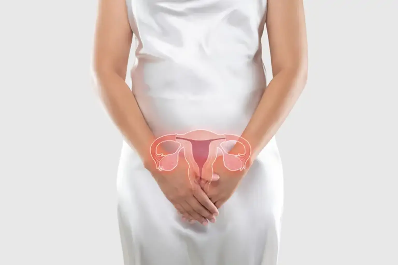
Following a consultation with a gynecologist, some women may require a treatment to preserve their health. Gynecologic surgery is surgery on the reproductive system of a woman. Excessive bleeding, finding fibroids, removing the uterus, and other concerns can be addressed with these operations.


Why a woman might need to undergo gynecology surgery?

Gynecological disorder

Gynecological disorders have an influence on more than just a woman's health. They also have an impact on her quality of life. A woman may require gynecological surgery for a variety of reasons. She may require therapy for one of the following conditions:

  • Endometriosis
  • Fibroids (benign tumors) 
  • Ovarian cysts
  • Cancer 
  • Chronic pelvic pain
  • Pelvic inflammatory illness 
  • Uterine prolapse 
  • Abnormal bleeding
  • Gynecology surgery can also be utilized as a long-term birth control method


What are the common gynecological surgeries?

gynecological surgery

Every year, millions of people have a gynecological treatment or surgery (procedures that deal with uterus, ovaries, cervix, or vagina). Some gynecologic procedures are straightforward and may be performed in the gynecologist's office, while others must be performed in a hospital. Gynecologic operations that are common include:


Therapeutic procedures:

  • Tubal ligation (tying of the tubes)
  • Removal of ovarian cysts (non-cancerous growths on the ovary)
  • Removal of cysts or fibroids (non-cancerous growths) in the uterus 
  • Removal of cervix growths (the lower part of the uterus that connects to the vagina)
  • Uterine artery embolization, also known as uterine fibroid embolization (UFE), which reduces the size of a uterine fibroid by cutting off the blood supply to it.
  • Removal of the uterus (hysterectomy), ovaries, or other reproductive system components of a woman 
  • Surgical treatment of gynecological (cervical, uterine, and ovarian) malignancies


Diagnostic procedures:

  • Colposcopy: a test that looks for symptoms of illness in your cervix, vagina, and vulva.
  • Cystoscopy: this is a technique that allows the doctor to examine the urinary tract for growths, bladder stones, or other issues
  • Urodynamics: a diagnostic assessment of bladder pressure that can be used to explain and cure incontinence
  • Hysteroscopy: this is a technique in which a tiny tube is inserted into the vagina and cervix to let the doctor to see inside the uterus


What is Hysterectomy?

hysterectomy

A hysterectomy is a surgical surgery that involves the removal of the womb (uterus). After the procedure, you will be unable to conceive. You will no longer get periods if you have not previously gone through menopause, regardless of your age. A hysterectomy is performed on a large number of women. It is more frequent in women between the ages of 40 and 50.

A hysterectomy is a surgical procedure that removes the uterus. Noncancerous hysterectomy is often considered only when all other therapeutic options have been exhausted. This operation may be performed for a variety of reasons, including:

  • Uterine fibroids causing discomfort, bleeding, or other issues
  • Uterine prolapse, defined as the uterus moving from its usual position into the vaginal canal.
  • Uterine, cervix, or ovarian cancer 
  • Endometriosis
  • Abnormal vaginal bleeding 
  • Chronic pelvic pain
  • Adenomyosis, or uterine thickening


Types of hysterectomy:

There are several kinds of hysterectomy. The kind you have is determined by the reason for the procedure and how much of your womb and reproductive system can be safely kept in situ.

The following are the most common forms of hysterectomy:

  • Total hysterectomy - the womb and cervix (womb's neck) are removed; this is the most frequent procedure.
  • Subtotal hysterectomy - the womb's main body is removed while the cervix remains in place.
  • Total hysterectomy with bilateral salpingo-oophorectomy - removal of the womb, cervix, fallopian tubes (salpingectomy), and ovaries (oophorectomy)


Complications of hysterectomy:

The majority of persons who get a hysterectomy have no substantial difficulties or consequences as a result of the procedure. Nonetheless, a hysterectomy is substantial operation with significant risks. Among the complications are:

  • Urinary incontinence 
  • Vaginal prolapse (a section of the vagina protrudes from the body)
  • Vaginal fistula creation (an abnormal connection that forms between the vagina and bladder or rectum)
  • Chronic pain

Wound infections, blood clots, bleeding, and harm to neighboring organs are further dangers of hysterectomy, albeit these are infrequent.


What is tubal ligation surgery?

Tubal sterilization

Tubal ligation, often known as tubal sterilization or having your tubes tied, is a kind of permanent birth control. Tubal ligation permanently prevents conception by cutting, tying, or blocking the fallopian tubes.

Tubal ligation stops an egg from passing from the ovaries to the fallopian tubes and also inhibits sperm from passing from the ovaries to the egg. Your menstrual cycle is unaffected by the surgery. Tubal ligation can be performed at any time, including after childbirth or as part of another abdominal surgery, such as a C-section.


Procedure:

If you have an outpatient interval tubal ligation, a needle or an incision through your belly button is performed so that your abdomen may be filled with gas (carbon dioxide or nitrous oxide). A laparoscope is then put into your abdomen. In most situations, your doctor will make a second tiny incision into the abdominal wall to introduce specific equipment. These gadgets are used by your doctor to close the fallopian tubes by damaging sections of the tubes or obstructing them with plastic rings or clips.

After vaginal childbirth, your doctor will most likely make a tiny incision under your belly button, allowing simple access to your uterus and fallopian tubes. If you have a tubal ligation during a C-section, your doctor will utilize the incision used to deliver the baby.


Complications of tubal ligation:

Tubal ligation is a surgical procedure that requires incisions in your abdomen. It is done under general anesthesia. Tubal ligation has the following risks:

  • Damage to the colon, bladder, or major blood arteries 
  • Anesthesia reaction 
  • Improper wound healing or infection
  • Persistent pelvic or abdominal pain
  • Failure of the operation, leading in an undesired pregnancy in the future


What is cervical cryosurgery:

Cervical cryosurgery chart

Occasionally, abnormal cervical cells are detected after a regular Pap smear (screening for cervical cancer). Although abnormal cells may not necessarily signify cancer, your healthcare professional will most likely want to do some more tests or treatments. He or she may advise cervical cryosurgery, often known as cryotherapy. This is a highly successful gynecological therapy that involves freezing a portion of the cervix.


Procedure:

The technique only takes a few minutes. The healthcare practitioner will do the following while you are laying on an exam table:

  • Insert a metal or plastic instrument known as a speculum into the vagina to open it, as with a Pap test.
  • Insert a cryoprobe, a hollow metal instrument, into the vagina. The chemical is contained and circulated via the probe (which is as low as –50 degrees Celsius).
  • Apply the cryoprobe tip to the cells or tissue that require therapy. The probe's tip will be covered with ice crystals and cold enough to freeze the tissue.
  • Leave the probe in place for a few minutes, or until the tissue freezes at around -20 degrees Celsius.
  • After a few minutes, repeat the ablation if necessary.


Complications of cervical cryosurgery:

Most patients can resume their daily habits immediately. For a few days to a few weeks, your provider may advise you to avoid vaginal intercourse, tampons, and douching. This may aid in your healing and infection prevention.

  • Pain or cramps
  • Vaginal spotting
  • Fainting.
  • Infection.
  • Cervical scarring, is a condition known as cervical stenosis.


What is Colposcopy?

Colposcopy diagnose

A colposcopy is a non-surgical diagnostic technique used to look more carefully at the cervix, vagina, and vulva. It is occasionally utilized when a Pap smear is abnormal. If a smear test (cervical screening) reveals abnormal cells produced by the human papillomavirus (HPV), a colposcopy is usually performed. These cells are not cancerous, but if left untreated, they may develop into cervical cancer.

Following the findings of your cervical screening (smear test), you may be given a colposcopy if:

  • Your smear test findings revealed abnormalities in the cells of your cervix
  • Several screening tests failed to yield a definite result

A colposcopy can also be done to determine the source of other symptoms such unexplained vaginal bleeding (for example bleeding after sex).


Procedure:

A colposcopy is often performed in a hospital or clinic. Your colposcopy should take around 15 to 20 minutes and you should be able to go home afterwards. A colposcope, a magnification tool, is used by a healthcare practitioner during the process. If they see anything suspicious during the operation, they may take a sample and submit it to the laboratory for examination.

LLETZ (large loop excision of the transformation zone)

The most frequent method of removing aberrant cells is by a process known as big loop excision of the transformation zone (LLETZ). LLETZ is frequently performed along with a colposcopy. You will not need to stay in the hospital and will be able to go home the same day.

You will initially be given a local anaesthetic injection to numb your cervix so that you do not feel any pain. The tissue is then removed using a heated loop of wire. A section of tissue the size of a fingernail is removed. LLETZ takes about 15 minutes to complete.

If you have a coil (IUD or Mirena coil), the doctor or nurse will make every effort to avoid cutting the threads. However, if this occurs, the coil may still be securely removed without the threads as necessary.

Cone biopsy

A cone biopsy may be recommended if the doctor or nurse has to remove a significant amount of tissue. A cone-shaped portion of tissue from your cervix is removed. For this procedure, you will be requested to schedule a follow-up visit. Because you will be put to sleep (general anesthesia) for the cone biopsy, you may need to spend the night in the hospital afterwards.


Complications of colposcopy:

You may see very little spotting if you have a colposcopy without a biopsy. If you underwent a colposcopy with a biopsy, you may experience some vaginal bleeding and minor discomfort over the next 24 to 48 hours.

You may notice a black discharge from the solution used by your healthcare practitioner to aid view your cervix in addition to some minor vaginal bleeding. For bleeding and discharge, you should use a sanitary pad rather than a tampon. Over-the-counter pain relievers such as acetaminophen or ibuprofen are commonly used to manage pain.

Following the surgery, your healthcare professional may advise you to avoid putting anything into your vagina for 48 hours. This involves refraining from sexual relations.


What is Dilation and Curettage (D&C)?

Dilation and Curettage

One of the most frequent gynecological treatments is dilation and curettage (D&C). During this treatment, a healthcare practitioner uses a suction device or a sharp curette to remove a part of your uterine lining (a surgical instrument used for scraping).

The method can be used to identify uterine problems such as:

D&Cs are also often utilized for abortion procedures, miscarriage, molar pregnancy, and retained placenta (when a placenta does not come out after childbirth).


Procedure:

Once your healthcare specialists have established that it is safe to proceed, you will be escorted to the operating theatre; as previously said, while this work can be done in a hospital's surgical ward, it can also be done in clinical and office settings.

  • Medication and monitoring: You'll be hooked up to monitoring equipment and an IV that will give drugs and fluids as needed. You may be offered anti-anxiety medication.
  • Catheterization and cleaning: A urinary catheter, which is a type of tube, will be inserted to your urethra. The healthcare practitioner will insert a speculum into the vagina to view the cervix and clean it.
  • Anesthesia: A facemask will be used whether you are receiving general anesthesia or regional anesthesia (in which just the bottom half of your body is numb). If you choose local anaesthetic, you will be given a numbing injection near the cervix.
  • Measuring the uterus: Before the uterus can be accessed, its length must be measured. This procedure entails inserting a thin, tube-shaped device known as a uterine sound, which may cause cramping if you have localized anaesthetic. This is then taken away.
  • Assessing and expanding the cervix: In some circumstances, pieces of cervical tissue may be scraped using a tiny curette. The cervix is then opened up by introducing a succession of progressively bigger tubes.
  • Scraping and/or suction: Once the cervix is opened, the healthcare professional enters the uterus using a curette and scrapes away tissue from the lining. If suction is utilized, the tube is positioned and used here.
  • Testing: Once sufficient tissue has been extracted and the region appears stable, the instruments are withdrawn, and the samples are collected and transported to the laboratory for clinical examination.


Complications of Dilation and Curettage:

D&C surgery, while considered minor surgery, is not without risks:

  • Heavy bleeding as a result of the surgery, unexpected perforation of the uterine lining might occur.
  • Infection can happen at the operation site or if the uterine lining has been pierced
  • Asherman’s syndrome Scar tissue grows on the uterine lining following D&C after a miscarriage, which is an uncommon consequence. While curable, this can have an impact on fertility and menstrual blood flow. 


What is Hysteroscopy? 

hysteroscopy procedure

Your healthcare professional may use hysteroscopy to identify or treat uterine abnormalities. This method might be used to:

  • Get rid of adhesions (scar tissue)
  • Find an intrauterine device
  • Identify the reason of recurring miscarriages.


Procedure:

During this process, a healthcare expert inserts a hysteroscope, a narrow, illuminated, telescope-like equipment, via the vagina into your uterus. The images of your uterus are then sent to a screen for additional evaluation.

  1. Your cervix's opening may need to be dilated or made wider with special equipment.
  2. The hysteroscope is introduced into your uterus through your vagina and cervix.
  3. A liquid or gas is then generally introduced via the hysteroscope to enlarge your uterus so your healthcare professional can see inside better.
  4. A light source shined through the hysteroscope allows your healthcare professional to observe the interior of the uterus and the fallopian tube openings into the uterine cavity.
  5. Small tools are put through the hysteroscope if surgery is necessary.


Complications of hysteroscopy:

Although general anesthesia is occasionally utilized, it is not always essential. Hysteroscopy lets your doctor to view within your uterus, which assists in the proper identification of certain medical disorders. Typically, the operation and recuperation period are brief.

Hysteroscopy is a relatively risk-free technique. Problems that can arise occur in fewer than 1% of instances, although they include:

  • Cervical or uterine injury 
  • Infection 
  • Heavy bleeding 
  • Anesthesia side effects 
  • Fluid overload (when too much fluid is administered during the procedure)


What is uterine fibroid embolization?

uterine fibroid embolization

Uterine fibroid embolization is a technique used to decrease noncancerous uterine tumors known as uterine fibroids. Because it does not require significant surgery, you may recover quicker. You may not even need to remain in the hospital.

Uterine fibroid embolization causes fibroids to shrink by cutting off their blood supply. The doctor injects sand-sized particles into the arteries that feed the fibroids. Particles adhere to the vessel wall. This causes a clot to form, cutting off the blood flow. The fibroids shrink when the blood supply is cut off. Your symptoms will normally subside or disappear with time.


Procedure:

You might get uterine fibroid embolization as an outpatient procedure or stay in the hospital overnight. The technique may differ based on your situation and the procedures of your healthcare professional. This is usually followed by uterine fibroid embolization:

  • You will be placed on the treatment table on your back, and an IV line will be placed in your arm or hand.
  • To drain urine, the doctor will insert a long, thin tube (catheter) into your bladder.
  • During the process, medical personnel will monitor your heart rate, blood pressure, respiration, and blood oxygen level.
  • The doctor will use an antiseptic solution to clean the groyne region.
  • Your doctor will insert a little tube (sheath) into your groyne. This will be used as a guide to place the catheter in the blocked-off location (embolized).
  • The doctor will inject contrast dye into the catheter. The contrast dye will help the doctor find the artery to be blocked off. The doctor will use X-rays to help find the blood vessels that supply blood to each fibroid.
  • The doctor will put a tiny catheter into the groin (femoral) artery. He or she will inject very small particles into the blood vessels.
  • Some doctors will use one groin site to treat both the left and right uterine arteries if needed. Other doctors may use two groin sites.
  • The sheath and catheter will be removed after the embolization is done.
  • The doctor will insert a contrast dye catheter. The contrast dye will assist the doctor in locating the obstructed artery. X-rays will be used to assist the doctor locate the blood arteries that provide blood to each fibroid.
  • A small catheter will be inserted into the groyne (femoral) artery by the doctor. He or she will inject very fine particles into the blood vessels.
  • If necessary, some surgeons will utilize a single groyne site to treat both the left and right uterine arteries. Other physicians may employ two groyne locations.
  • After the embolization, the sheath and catheter will be withdrawn.


Complications of uterine fibroid embolization:

Any operation can result in problems. This technique may result in the following complications:

  • Unusual bleeding (hemorrhage)
  • Injury to the uterus 
  • Infection of the uterus or the groyne puncture site
  • Collection of blood beneath the skin (hematoma) at the groyne puncture site
  • Injury to the artery being utilized 
  • Blood clots 
  • Infertility
  • Menstrual period loss (amenorrhea)

Postembolization syndrome affects certain women. This Syndrome might last anywhere from 2 and 7 days. It is treated with pain medicines and anti-inflammatory drugs. Anti-nausea drugs may be used as well. Among the signs and symptoms are:

  • Pelvic pain and cramps 
  • Nausea and vomiting
  • Fever
  • Fatigue and discomfort


Conclusion

Gynecology

Gynecology is a medical specialty concerned with female health issues, such as the development, diagnosis, prevention, and treatment of illnesses and diseases specific to the female reproductive system. Obstetrics is the medical treatment of a woman and her child before, during, and after childbirth.

Women go through a number of reproductive development events during their lives, including menarche, menstruation, pregnancy, motherhood, and menopause. These developmental processes in female reproduction cause more significant physiological changes than developmental events in male reproduction (e.g., menstrual bleeding, physiological changes in pregnancy, breastfeeding, and menopausal hormone fluctuations).

When medicines and non-invasive methods fail to ease symptoms, surgery is the only acceptable and successful treatment option for a variety of gynecologic problems. Cervical and uterine cancer, uterine fibroids, endometriosis, uterine prolapse, and menorrhagia (excessive bleeding) are examples.