Malignant Brain Gliomas
Overview
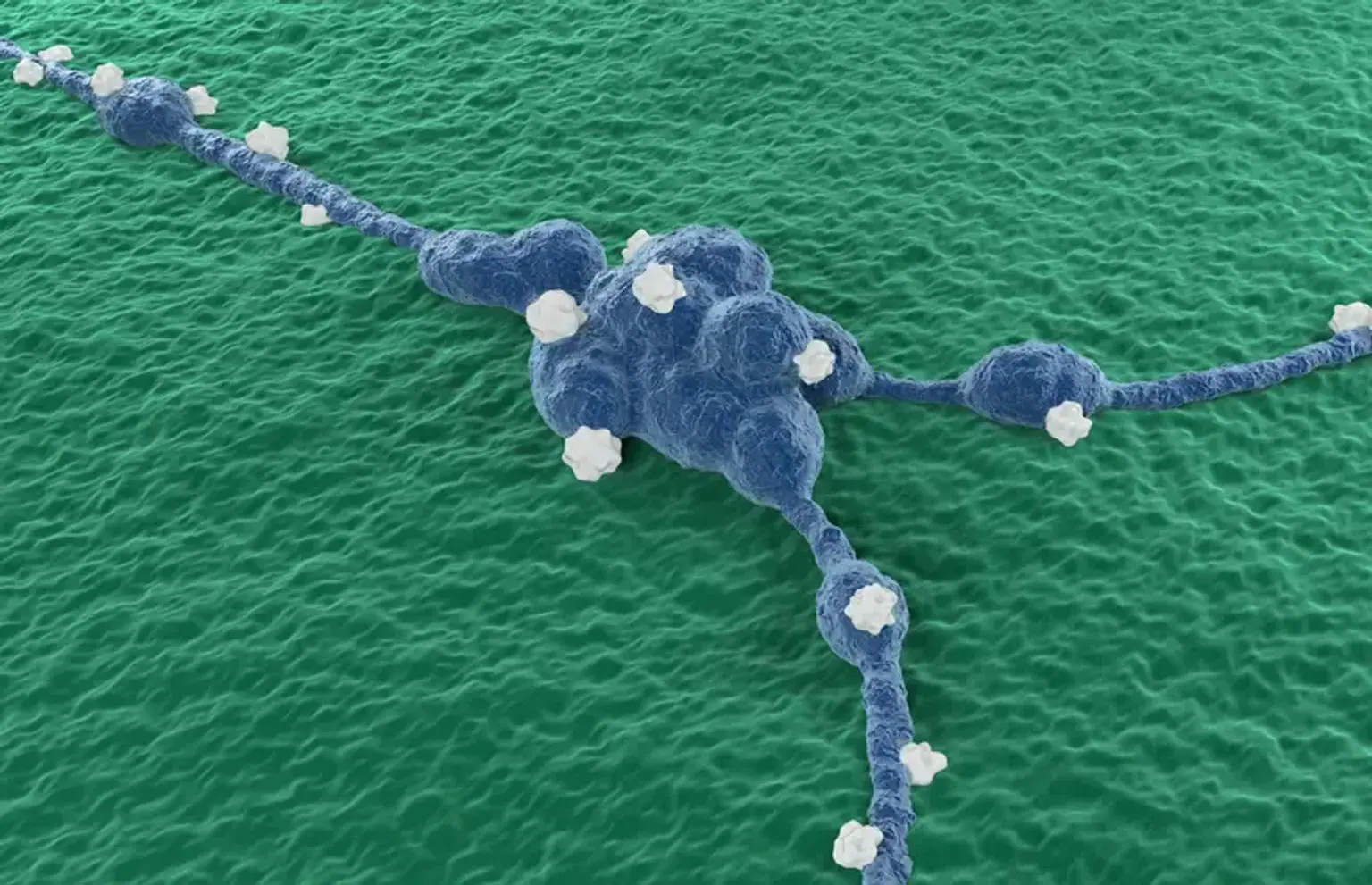
Glioma is a malignant tumor that develops in the brain and spinal cord. Gliomas arise in the gluey supporting cells (glial cells or neuroglia) that surround and support nerve cells. Glioma is a form of brain cancer that is quite frequent. Gliomas account for around 33% of all brain cancers and arise from the glial cells that surround and support neurons in the brain, such as astrocytes, oligodendrocytes, and ependymal cells.
Gliomas can occur in both children and adults. Some expand really fast. The majority of persons with gliomas require a combination of therapies, including surgery, radiation therapy, and chemotherapy.
What are the types of glial cells?
Astrocytes:
- Are the most abundant form of glial cells, accounting for around half of all cells in the brain.
- They control the quantities of extracellular neurotransmitters via unique uptake processes.
- They play a vital role in the creation of synapses (space where two nerve cells attach to each other) by secreting chemicals that encourage synaptic formation.
- Astrocytes establish a physical barrier that is essential for controlling the passage of tiny chemicals into the brain.
- Furthermore, certain astrocytes produce substances that support the survival of specific neurons.
Oligodendrocytes:
- The primary role of oligodendrocytes is to create a coating layer (myelin sheath) that envelopes each axon, allowing for an increase in the speed of transmission of electrical impulses.
Ependymal cells (Also known as, Ependymocytes):
- Create a simple, cuboidal epithelium that coats the brain's inner surfaces.
- Cilia on the apical surface of ependymal cells are considered to be vital in the circulation of cerebrospinal fluid (the fluid that surrounds and protects the brain and spinal cord) around the ventricles and central canal of the spinal cord.
How malignant gliomas are classified?
Gliomas are categorized based on the kind of glial cell involved in the tumor, as well as genetic factors that can help predict how the tumor will behave over time and the therapies that are most likely to succeed.
Gliomas can comprise a variety of cell types. These are referred to as mixed gliomas by specialists. They classify gliomas as low-, mid-, or high-grade depending on how quickly they develop and other characteristics.
Types of gliomas include:
Astrocytomas, (including glioblastomas and diffuse intrinsic pontine gliomas (DIPGs):
- These cancers originate from the astrocytes.
- Glioblastomas are aggressive astrocytomas that develop quickly. They are the most frequent kind of adult malignant brain tumor.
- Astrocytomas are prevalent gliomas in youngsters. DIPG is an uncommon but severe type of brain cancer in children. It originates in the brain stem and primarily affects youngsters.
Ependymomas (including anaplastic ependymoma, myxopapillary ependymoma, and sub-ependymoma):
- These cancers begin in the ependymocytes.
- Ependymomas often develop in the ventricles of the brain or the spinal cord.
- They spread through the cerebrospinal fluid but not beyond the brain or spine.
- Ependymomas account for around 2% of all brain tumors. They affect youngsters more than adults.
Oligodendrogliomas (including oligodendroglioma, anaplastic oligodendroglioma and anaplastic oligoastrocytoma):
- These cancers originate from the oligodendrocytes.
- Oligodendrogliomas develop slowly at first but can become aggressive with time.
- They, like ependymomas, seldom spread outside of the brain or spine.
- They affect adults more than youngsters. Oligodendrogliomas occur for approximately 1% to 2% of all brain cancers.
Mixed gliomas (also called oligo-astrocytomas)
- They are composed of several types of glial cells.
- Their classification as a separate tumor type is debatable, although it may be addressed by genetic testing of tumor samples.
- These tumors are more common in adult men and are frequently discovered in the cerebrum (upper part of the brain).
How prevalent are gliomas?
Each year, an estimated 80,000 new instances of primary brain tumor are detected in the United States. Gliomas account for around one-fourth of these (20,000). Each year, roughly 12,000 cases of glioblastoma are diagnosed (approximately 15 percent of total newly diagnosed brain tumors).
What is the cause of brain glioma?
Gliomas, like most primary brain tumors, have an unknown etiology. According to research, alterations in DNA cause the development of brain and spinal cord malignancies such as gliomas. DNA is found in our genes. They offer cells instructions on how to develop and reproduce. Mutations, or alterations to the DNA in our genes, can cause cells to proliferate uncontrollably. You can inherit genetic mutations from your parents. They might also happen unexpectedly during your lifetime.
What are the risk factors for developing malignant brain glioma?
Gliomas, like most primary brain tumors, have an unknown etiology. However, there are several variables that may raise your chances of developing a brain tumor. These are some examples:
Age:
- Malignant brain gliomas are more frequent in particular age groups, including adults aged 45 to 65 and children under the age of 12.
- A brain tumor, on the other hand, can arise at any age. Children and young adults are more likely to develop certain types of gliomas such as ependymomas and pilocytic astrocytomas.
Ethnic group: White people may be more susceptible than other races to acquire gliomas.
Gender: Gliomas affect men somewhat more than women.
Radiation exposure:
- People who have been exposed to ionizing radiation have an increased chance of developing a brain tumor.
- Examples of ionizing radiation include cancer radiation treatment and atomic bomb radiation exposure.
- Common kinds of radiation, such as electromagnetic fields emitted by power lines and radiofrequency radiation emitted by microwave ovens, have not been demonstrated to raise the risk of glioma.
- It's unclear if smartphone use raises the chance of developing brain cancer. Some studies have discovered a link between smartphone use and a kind of brain cancer known as acoustic neuroma. Many other studies have found no link. Because smartphones are a newer component, additional long-term study is required to fully understand their possible influence on cancer risk. For the time being, if you're concerned about the probable link between smartphones and cancer, doctors advise reducing your exposure by utilizing a speaker or hands-free device, which keeps the telephone away from your head.
Family history of glioma:
- Glioma does not usually run in families. However, having a family history of a glioma increases the likelihood of acquiring it.
- While several genes have been linked to glioma, additional research is needed to demonstrate a relationship between these genetic variants and brain malignancies.
What are the symptoms of brain glioma?
The most common initial presenting symptom of glioma patients is headache. The pathophysiology of headaches is thought to be caused by tumor growth, which causes a mass impact on surrounding tissue. The mass effect, in turn, causes pressure in the microvasculature, which causes edema (accumulation of fluids).
The mass effect causes symptoms of a brain tumor depending on where the tumor is located in the brain. Frontal lobe tumors, for example, might cause behavioral disorders, whereas dominant temporal lobe tumors can cause receptive speech impairments. Other symptoms of mass effects include nausea, vomiting, and visual changes.
Seizures are the second most prevalent presenting symptom. Seizures are caused by tumor irritation of the cerebral cortex, which results in localized or widespread seizures. Other symptoms of gliomas include tingling feelings, weakness, difficulties ambulating, and, in rare cases, a comatose condition owing to bleeding inside the tumor, which causes brain herniation.
Complications of brain glioma include:
- Increased intracranial pressure
- Seizures
- Hydrocephalus (CSF accumulation inside the brain)
- Brain herniation
- Hemorrhage into the tumor
- Local spread
- Spinal metastases
- Death
How are malignant gliomas diagnosed?
Your symptoms are evaluated and your medical history is reviewed by your healthcare practitioner. The doctor will also do a thorough physical and neurological examination.
Following that, he or she will request certain imaging investigations to aid in the diagnosis. Brain abscess, demyelinating disorders, brain infarction, and brain metastasis are all possible alternative diagnoses for malignant brain glioma.
History and physical examination:
Headaches, seizures, nausea, vomiting, seizures, and, in more advanced instances, weakness or altered mental state are the most prevalent symptoms of brain gliomas.
The neurological examination of these individuals may be normal or may reveal various degrees of focal weakness, sensory impairments, or, in a severe case, a changed mental state as a result of an abrupt mass effect caused by tumor enlargement.
Imaging studies:
In the context of a history and physical examination indicative of brain tumors, the following imaging diagnostics are appropriate:
Computed tomogram (CT) head:
- A thorough study should be conducted to assess acute intracranial abnormalities such as bleeding, edema, and asymmetry in brain tissue.
Magnetic resonance imaging (MRI) Brain:
- This is a detailed investigation of the features of brain tumors.
- Low-grade tumors often do not enhance (take up the contrast), but high-grade tumors exhibit varying degrees of enhancement.
- Furthermore, MRI is an excellent technique for determining the extent of cerebral edema.
- A brain MRI will aid in the treatment plan and determining the response to surgery, radiation, and chemotherapy.
Chest X-Ray:
- Patients newly diagnosed with a brain tumor require an adequate metastatic workup (tumor distant spread) based on their history and physical examination.
- Patients with a long history of cigarette use, for example, may require a chest x-ray. If a mass is discovered on the chest x-ray, it must be studied further using a CT scan of the body or positron emission tomography (PET) scan to determine the likely main site of metastatic illness.
Brain biopsy:
Tissue diagnosis can be established during surgical resection or by stereotactic biopsy. A stereotactic core biopsy locates a tumor using a 3D scanning machine (ultrasound, CT scan, or MRI).
Biopsy alone is done when the lesion is not amenable to resection, when a significant quantity of tumor tissue cannot be removed, or when the patient's general clinical status precludes invasive surgery.
Only a biopsy can definitively confirm a brain tumor and provide a prognosis to guide treatment options. A pathologist (a doctor who specializes in identifying cancer and other tissue abnormalities) can assess the grade or stage of a brain tumor based on this information.
Your biopsy sample's physical appearance and growth rate will also be examined by the pathologist (molecular diagnosis). Your doctor will inform you of the pathologist's results. This information aids in making treatment-related decisions.
How malignant brain gliomas are treated?
Patients with gliomas require multidisciplinary therapy. Neurosurgeons, neuro-oncologists, radiation oncologists, and other health professionals will have a role in improving patient care and achieving the best possible outcome. Treatment for glioma is determined by the tumor's origin, size, grade, and location, as well as your age, overall health, and preferences.
Treatment for glioma may include administering medications to minimize the signs and symptoms of your tumor in addition to steps to eliminate the tumor itself.
Steroids may be prescribed by your doctor to decrease swelling and alleviate pressure on affected parts of the brain. To manage seizures, anti-epileptic medicines may be administered.
For the majority of patients, surgery is the first line of therapy for glioma. If the tumor is easily accessible, a surgeon may be able to remove it altogether. However, gliomas can be difficult to entirely eradicate, especially in difficult-to-reach or fragile parts of the brain.
Following surgery, further therapies such as chemotherapy and radiation therapy may be administered. These are adjuvant therapy, which means they eliminate any residual cancer cells or tumor pieces following surgery. However, if your tumor is inoperable, your doctor may recommend chemotherapy or radiation therapy as your primary therapies.
Surgery:
The most frequent form of surgery used to eliminate gliomas is a craniotomy (open brain surgery). You may be a candidate for laser ablation depending on the size and location of the tumor. This minimally invasive procedure employs laser heat to eliminate all or parts of a brain tumor.
A surgeon may employ specialized procedures to guide the surgery, such as imaging or brain mapping. Brain mapping reveals which parts of your brain regulate important activities. This information assists your surgeon in avoiding the removal or damage of healthy brain tissue.
Radiotherapy:
To kill cancers, radiation treatment uses high doses of radiation. Radiation therapy for gliomas may be recommended by your doctor. Radiation treatment addresses the tumor's particular shape, reducing the chance of harm to nearby tissues.
You may also be given brachytherapy, a type of radiation therapy. To treat a tumor, a healthcare professional uses radiation sources near to the tumor. The sources emit radiation without hurting adjacent tissues.
Options for radiation treatment include:
- Using computers to direct radiation therapy to the precise site of the brain tumor. Intensity-modulated radiation treatment and 3D conformal radiation therapy are two techniques.
- Using protons, the positive portion of atoms, as the source of radiation rather than X-rays. Conformal proton beam treatment provides radiation only when proton beams reach the tumor, causing less harm to surrounding tissue than X-rays.
- Using numerous radiation beams to provide a highly targeted kind of radiation therapy. While this approach is known as stereotactic radiation treatment (radiosurgery), it does not entail traditional surgery. Each radiation beam isn't especially powerful, but the place where all the beams come together — at the brain tumor — receives a very high dosage of radiation, killing tumor cells in a relatively tiny area.
Chemotherapy:
Chemotherapy refers to the use of medications to kill cancer cells. It is used to treat a wide range of cancers. This therapy can be either oral or intravenous. Temozolomide is a typical chemotherapy medication used to boost the effectiveness of radiation treatment.
Treatment for recurrence:
Re-operation with chemotherapy and targeted therapy such as angiogenesis (creation of new blood vessels for the tumor) inhibitors or immunotherapy are options for recurrent gliomas. All of these adjuvant treatments are being studied for their efficacy.
Other treatments:
Patients with high-grade glioma are more likely to have seizures, malignant edema, and immobility-related complications. These patients require antiepileptic medicines, DVT prophylaxis, and steroids before, during, and after therapy to prevent cerebral edema.
How can brain gliomas be prevented?
Most glioma risk factors, such as age and ethnicity, are uncontrollable. Early identification and treatment of low-grade gliomas, on the other hand, may limit or prevent the development to high-grade gliomas.
If brain tumors run in your family, you should think about genetic testing. Discuss the risks and advantages of genetic testing with your healthcare physician or a genetic counselor.
It is also a good idea to:
- Limit your head's exposure to radiation.
- Keeping a healthy lifestyle.
What is the prognosis of malignant brain glioma?
Glioma prognosis is determined by various variables, including:
- Patient's age.
- Current comorbidities.
- Grade and location of the tumor.
- Presence of hydrocephalus.
- Response to adjuvant therapy.
- The extent of surgical resection.
Glioma survival rates differ depending on tumor type, tumor grade, and age. Certain mutations can also have an impact on the prognosis. The older a person is when they are diagnosed and treated, the worse their prognosis. Low-grade ependymomas, oligodendrogliomas, and astrocytomas had the greatest five-year survival rates in adults and children. It is the lowest (between 6% and 20%) for glioblastomas.
Malignant gliomas are associated with a significant morbidity and death rate. Even with maximal therapy, the median survival for glioblastomas is just 12-15 months, and 2-5 years for anaplastic gliomas.
Low-grade glioma is a disease that kills young individuals (mean age 41 years), with a 7-year survival rate. Although low-grade glioma patients have a higher survival rate than high-grade (WHO grade III/IV) glioma patients, all low-grade gliomas develop into high-grade glioma and death.
Gliomas in general, and glioblastomas in particular, are extremely difficult to cure. Despite breakthroughs in understanding the molecular biology and genetics of gliomas, there has been little progress in avoiding the mortality of high-grade gliomas. As a result, there is a continual need for clinical and fundamental scientific research to enhance the treatment of this fatal disease.
What happens after treatment of malignant brain glioma?
Maintain constant contact with your healthcare provider following therapy. Regular imaging scans will be required to determine whether cancer has returned.
Brain cancer therapy can harm healthy brain tissue. Rehabilitation with physical or occupational therapists can assist you in regaining abilities such as walking, speaking, and remembering.
Patients should be informed on the value of regular follow-up visits, taking prescriptions on schedule, and adhering to driving restrictions. Support groups for you and your family can help you cope with the physical and emotional difficulties of having a brain tumor.
Rehabilitation after treatment:
Because brain tumors can form in areas of the brain that regulate motor abilities, speech, vision, and thought, rehabilitation may be required as part of the recovery process.
Your doctor may recommend you to appropriate services, such as:
- Physical therapy can aid in the restoration of lost motor skills or muscular strength.
- Occupational therapy: After a brain tumor or other sickness, occupational therapy can help you return to your usual daily activities, including job.
- Speech therapy: with speech pathologists (specialists in speech issues), which can assist if you have trouble speaking.
- Tutoring for school-age children: can assist youngsters in coping with changes in memory and thinking following a brain tumor.
Conclusion
Gliomas are tumors that form in the glial cells of the brain and spinal cord. They are usually cancerous. Gliomas do not usually spread to other regions of the body. They can, however, spread quickly through the brain and spine and can be deadly.
The most common symptoms of brain gliomas include headaches, convulsions, nausea, vomiting, seizures, and, in more advanced cases, paralysis or changed mental state. A comprehensive physical examination, imaging investigations, and a brain tissue sample are all required for the diagnosis of malignant brain glioma.
The majority of glioma patients require a mix of treatments. Surgery, radiation therapy, or chemotherapy may be used in the management. Survival rates for gliomas vary based on tumor type, tumor grade, and age. Certain mutations can also affect the prognosis. When a person is diagnosed and treated, the older they are, the worse their prognosis. The highest likelihood of survival is for young patients with low-grade gliomas.
After undergoing treatment for malignant brain glioma, psychosocial support and rehabilitation therapy are required to improve the final outcome of the recovery process and to improve physical activity, speech, and other abilities.