Microsurgical hepatic artery reconstruction
Last updated date: 10-Jun-2023
Originally Written in English
Microsurgical Hepatic Artery Reconstruction
After providing supportive care to the patient, liver transplantation is the only effective treatment for acute or chronic liver failure. Shorter survival time (which is regarded as less than a year) and unsatisfactory life quality owing to liver disease are the two indications for liver transplantation that are widely accepted by the medical world. Starzl carried out the first successful liver transplant in the 1960s. The importance and popularity of liver transplants are growing daily and are now conducted in two categories: cadaveric donor liver transplantation (CDLT) and living donor liver transplantation (LDLT). Strong kinship ties, insufficient liver donors, and liver procurement issues have all contributed to higher LDLT rates than CDLT. Since it was first published in the 1980s, thousands of LDLT procedures have been carried out in numerous hospitals all over the world.
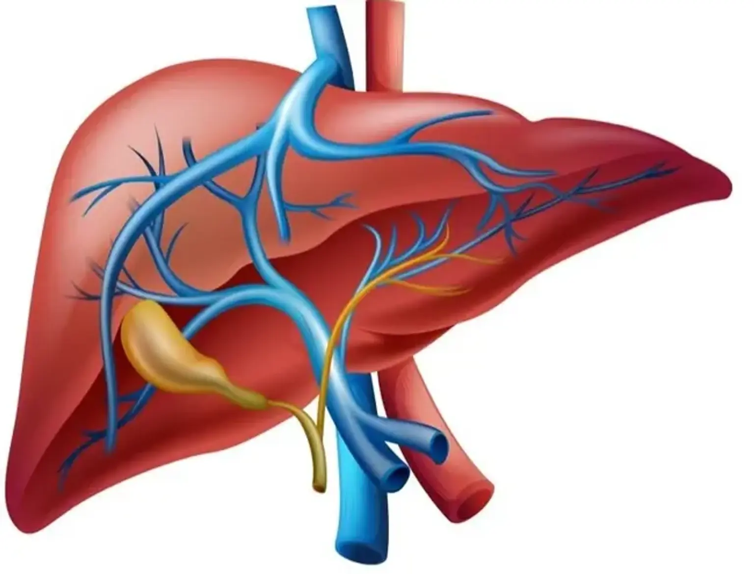
In CDLT, the recipient's hepatic artery system is anastomosed to the hepatic artery system that was removed at the aortic level. Therefore, since this is a gross vascular process, no microsurgical methods are needed. However, in LDLT, the hepatic lobule and its associated hepatic artery are transferred. Due to the procedure's small diameter, microsurgical reconstruction methods and tools are required. The hepatic artery and the portal vein each give blood to the liver separately. As a result, blood enters the parenchyma through both of them, but the biliary tract has no connection to portal circulation. Because of this, the outcome of the surgery and the patient's survival is directly impacted by the reconstruction success. Otherwise, death is the consequence of the situation. While other medical specialties with skills in microsurgery have conducted hepatic artery reconstruction, it is normal practice for plastic surgeons to perform it in many hospitals across the world.
Vascular Microsurgery
Vascular microsurgery or microvascular surgery is carried out using an operative microscope, specialized surgical tools, and tiny needles with ultrafine sutures on blood vessels that are typically 3 to 5 millimeters in diameter. Reattaching severed fingers, hands, arms, and other amputated body parts to the body through microvascular surgery. By reattaching the small blood vessels, circulation is restored before the damaged tissue starts to deteriorate. Reconstructive surgery also employs microvascular technologies. A free flap is a transplant of healthy tissue from a different location. This healthy tissue is sent to the wound site, where it is used to reestablish blood flow.
Microsurgical Hepatic Artery Reconstruction Indications
Microsurgical hepatic artery reconstruction is done in the context of liver transplantation; hence it has the same indications as liver transplantation. Without a functioning liver, you cannot survive. You may require a liver transplant if your liver no longer functions properly. If your liver disease has reached its terminal stages (chronic liver failure), a liver transplant may be advised. This liver condition is severe and potentially fatal. Numerous liver disorders might cause it.
A frequent cause of the advanced liver disease is cirrhosis. It affects the liver and is chronic. It takes place when scar tissue takes the place of healthy liver tissue. This prevents the liver from operating normally. The following illnesses also have a chance of causing end-stage liver disease:
- Acute hepatic necrosis. At this point, liver tissue starts to deteriorate. Acute infections and adverse drug, medication, or toxin reactions are among the causes that could arise.
- Biliary atresia. A newborn is affected with a rare liver and bile duct disorder.
- Viral hepatitis. Most often, hepatitis B or C is to blame.
- Metabolic conditions. disorders that alter the chemical activity of liver-related cells.
- Primary liver cancers. These tumors are malignant and originate in the liver.
- Auto-immune hepatitis. Liver edema or redness (inflammation). It takes place when the immune system in your body attacks your liver.
Microsurgical Hepatic Artery Reconstruction Preparation
The technique will be explained to you by your healthcare provider. Ask him or her about any concerns you may have regarding the procedure. A consent form confirming the surgery may be given to you for signing. If anything on the form is unclear, thoroughly read it and then ask questions. You shouldn't eat for eight hours before the procedure if you are having a scheduled liver transplant. This frequently entails abstaining from eating or drinking after midnight. Once you know a liver is available, you shouldn't eat or drink if it comes from a donor who just passed away. Before the procedure, you can be given a sedative to help you relax. Depending on your needs, your healthcare practitioner might give you different advice.
Microsurgical Hepatic Artery Reconstruction Procedure
The superior mesenteric artery is used to first identify the anatomical right hepatic artery. The superior mesenteric artery branch to the liver (which often has a tiny diameter) is then carefully preserved as the liver arteries are dissected. To further expose the hepatic artery, the tiny R/A-RHA branches that go to the pancreas are knotted and sectioned. To accomplish a reconstruction with a patch from the superior mesenteric artery, only a small portion of the splenic artery trunk is left. The R/A-RHA length is always sufficient for this reconstruction; hence a short splenic artery length is adequate. As an added benefit, since there is no need for a longer mesenteric or splenic artery stump, simultaneous pancreas procurement is possible with such a procedure.
The superior mesenteric artery is then cut into sections (5 mm long on either side of the hepatic branch) and the other side of the cylinder is longitudinally opened to create a Carrel patch that extends 5 mm radially from the artery ostium. By trimming the excess of the superior mesenteric artery around the branch, the Carrel patch is subsequently modified to accommodate the ostium of the splenic artery.
The superior mesenteric artery Carrel patch is then positioned on the splenic artery, with its length and three-dimensional orientation being checked to ensure tension-free anastomosis. The best flow must be preserved under these circumstances. By injecting preservation liquid into the anastomosis, polypropylene sutures are used to perform the flow test and allow for permeability and integrity checks.
To maintain the vertical orientation of the hepatic branches and guarantee their regular flow, the locations of the superior mesenteric artery Carrel-patch and the splenic artery are also confirmed when performing arterial reconstruction on the patient. On days 1 and 5 following surgery, Doppler ultrasound (Doppler USG) is used to measure hepatic blood flow.
The same surgical team at the Department of Liver Transplantation, Hospital das Clnicas, Faculty of Medicine, University of So Paulo, Brazil, successfully used this technique in 4 consecutive situations of R/A-RHA in liver transplantation among a total of 120 deceased donor transplants during the year 2010. It was noted that when the procedures were carried out as instructed, the recipient patients' future vascular anastomosis was made easier. After back-table reconstruction, it was discovered that the method provided for good preservation solution flow.
Microsurgical Hepatic Artery Reconstruction Recovery
You might spend a few hours in the recovery area following surgery before being transferred to the intensive care unit (ICU). For several days, you will be closely monitored in the ICU. Monitors will be connected to you. Your heart rate, blood pressure, other pressure readings, respiratory rate, and oxygen level will all be displayed. You'll have to stay in the hospital for at least a week.
Most probably, a tube will be in your throat. This is done so that until you can breathe on your own, you can breathe with the aid of a machine (a ventilator). Depending on your circumstance, you can need the breathing tube for a few hours or a few days. To get rid of ingested air, a little plastic tube may be put through your nose and into your stomach. When your bowels begin to function regularly once more, the tube will be removed. Until the tube is taken out, you won't be able to eat or drink. Your transplanted liver will be monitored frequently using blood samples. Additionally, your kidneys, lungs, and systemic circulation will be checked.
You can receive IV drips to boost your heart and blood pressure as well as to manage any bleeding issues. These drips will gradually be reduced and stopped as your condition improves. Antibiotics may be given to you.
You can begin drinking liquids once the breathing and stomach tubes have been taken out and you are stable. You can gradually start eating solid foods as instructed. To ensure you are receiving the proper dose and medication combination, your anti-rejection medications will be carefully monitored.
You will be transferred from the ICU to a separate room when your healthcare professional determines you are ready. As you get out of bed and go around for long period, you will gradually be able to move around more. More solid foods will gradually become accessible to you. When you return home, your surgical team will educate you on how to take care of yourself.
When you get home, you need to keep the surgery site dry and clean. You will receive particular bathing instructions from your physician. If they were not taken out before leaving the hospital, any sutures or surgical staples will be removed during a subsequent office visit. Until your doctor instructs you to, you should not drive. Other restrictions on your activity may apply. If you experience any of the following symptoms, contact your doctor right away: fever, redness, swelling, bleeding, or another drainage from the wound area; increased discomfort at the incision site; vomiting or diarrhea; bleeding; or jaundice.
Microsurgical Hepatic Artery Reconstruction Risks
It is possible to see intimal dissection, an unfavorable intraoperative consequence. However, the transplant surgeon may be required to traumatically interrupt the hepatic artery during recipient liver resection as long as this entity has hepatic arterial stasis and hypertension-related cirrhosis. To reduce the possibility of intimal dissection, the recipient arteries must be appropriately cut with specialized atraumatic scissors.
There are two types of hepatic artery thrombosis: early and late. Although there is debate on this matter, early thrombosis is defined as occurring within the first week to 10 days, and late thrombosis is defined as occurring after that. The definition of risk factors is in light of early HAT. Long ischemia times, arterial variations, longer surgical times, low body weight, particularly in pediatric recipients, the use of arterial conduits, and donors who have the cytomegalovirus can be used to categorize these. Twisting and stenosis, two side effects of surgery, can both increase the risk of thrombosis with or without these risk factors. Routine Doppler ultrasonography evaluation is crucial because early hepatic artery thrombosis may not present with aberrant laboratory results or symptoms at baseline. In the literature, it is advised that reconstruction be followed by a 12-hour Doppler USG for 14 days postoperatively. Hepatic artery thrombosis is mostly treated in a three-step manner. They are observation, replantation, and revascularization. Through thrombolysis, thrombectomy, or anastomosis renewal, revascularization can be achieved. When thrombosis develops in cadaveric liver transplants after one month, it is known as late HAT. Late-stage HAT development following collateral development necessitates modifying the treatment strategy to the severity of clinical findings.
Another significant problem that could develop following hepatic artery reconstruction is hepatic artery stenosis. Stenosis might become completely blocked and result in death. Hepatic artery stenosis may be brought on by insufficient surgical equipment, clamp injury, or rejection. Hepatic artery stenosis affects between 3% and 5% of patients. Depending on the location and length of the stenotic segment, stenosis diagnosed by Doppler ultrasound requires a different therapeutic strategy. Short stenotic segments can be treated well using percutaneous transluminal angioplasty (PTA), which also serves as a diagnostic and therapeutic procedure. Surgery is needed for revision if PTA intervention is inadequate.
Aneurysm or pseudoaneurysm, an uncommon consequence following hepatic artery reconstruction, is another potential issue. They come on later. This complication could be brought on by the surgical procedure or by bacterial and fungal infections. Aneurysm or pseudoaneurysm is indicated by an immediate intraabdominal or gastrointestinal hemorrhage that occurs following a liver transplant. Doppler ultrasonography is less useful than angiography. After the removal of the pseudoaneurysm, the treatment involves hepatic artery ligation. If an aneurysm develops late, collaterals might be enough to provide vascularization. If not, retransplantation or revascularization may be required.
Conclusion
One of the most crucial and difficult stages of liver transplantation is hepatic artery reconstruction. The patient undergoing a liver transplant typically has a poorer nutritional condition and an unfavorable coagulation profile compared to patients undergoing the majority of free flap reconstructive surgeries. The autologous musculocutaneous free flap taken from the patient is probably not as effective as the liver graft. The recipient vessel quality could be less favorable to subsequent reduction and worse than those seen in crush injuries. The technical problem of executing reconstruction in a deep recess during respiratory movement is an additional obstacle. To ensure the best results, it is therefore desirable to recruit a group of skilled microsurgeons to plan the harvest of an interpositional graft, if necessary, and carry out the critical reconstruction. This is known as getting one bite at the cherry.