Neonatal Laparoscopic Surgery

Last updated date: 16-Jun-2023

Originally Written in English

Neonatal Laparoscopic Surgery

Laparoscopic and thoracoscopic treatments are both included in minimally invasive surgery (MIS). MIS has gradually gained acceptance as a neonatal surgical disease treatment option. When performed by skilled surgeons, MIS also has the added benefits of lessening tissue trauma, requiring less pain medication, shortening hospital stays, and providing superior clinical data. However, these financial and risk-based expenses of MIS do not come without trade-offs, and these risks increase disproportionately as patient size decreases (naturally), the most vulnerable are the newborns. To treat neonatal surgical conditions, surgeons must face a difficult mechanical and physiological challenge. Pediatric surgeons have historically been reluctant to accept MIS as the gold standard of surgical care. The causes are simultaneous technological, logistical, anatomical, physiological, and anesthesiologic issues. Pediatric surgeons from all over the world participated in a recent study that revealed their concerns about doing MIS for newborn diseases. Only one in ten suggests laparoscopic pyloromyotomy for pyloric stenosis, and only around one in three performs the procedure. Additionally, less than 25% support laparoscopic Ladd's surgery for malrotation.


Neonatal Surgery

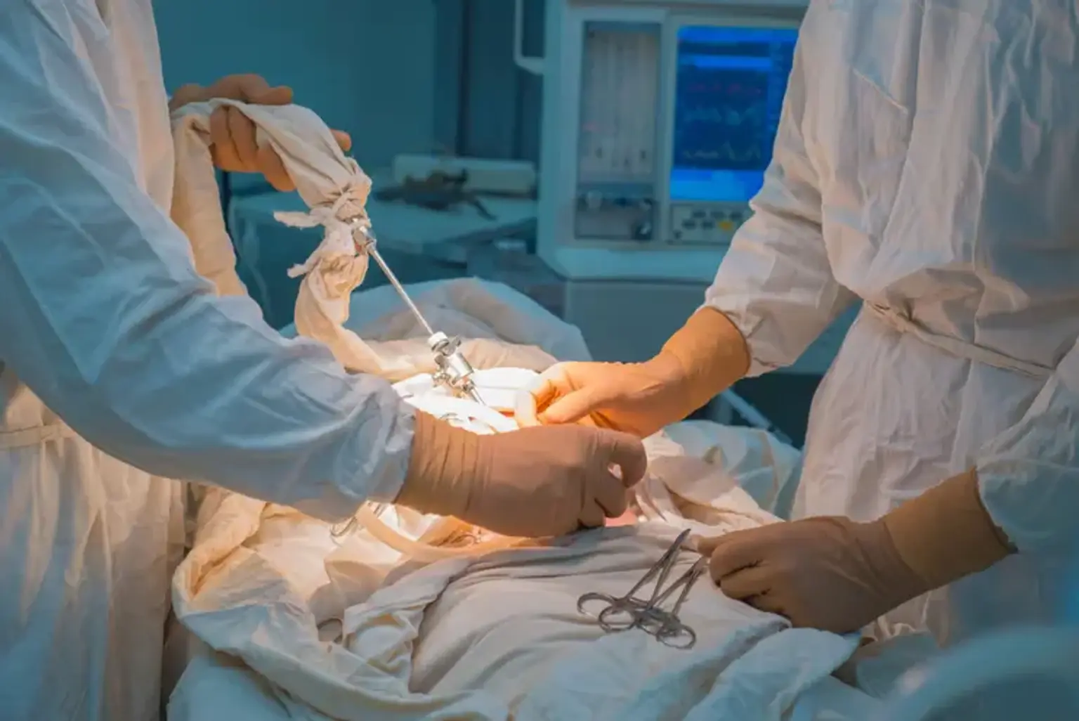
Immediately after birth, newborns undergo neonatal surgery. It is primarily intended to heal diseases that cannot be identified or treated while the baby is still in the uterus. It might also be used to treat diseases that appear soon after birth. Neonatal surgery is frequently needed to treat developmental issues in premature infants. Neonatal surgery is used to treat a wide range of disorders, including the ones listed below:
  • Anorectal malformations
  • Annular pancreas
  • Congenital diaphragmatic hernia
  • Esophageal atresia
  • Gastroschisis
  • Heart defects
  • Hirschsprung’s disease
  • Intestinal blockages
  • Lung lesions and tumors
  • Necrotizing enterocolitis
  • Omphalocele
  • Short bowel syndrome
  • Tracheoesophageal fistula


Neonatal Laparoscopic Surgery Advantages

Neonatal Laparoscopic Surgery Advantages

Even for the neonatal age range, there are obvious benefits of neonatal laparoscopic surgery supported by research. Neonatal laparoscopic surgery may lessen surgical trauma and fluid shifts, which eventually leads to reduced postoperative analgesia; a smaller wound also helps. Additionally, there is less bleeding. Reduced wound size also decreases the risk of dehiscence and incisional hernias, as well as wound infections. Heat loss should be reduced with good instrumentation, which is a crucial component of the effectiveness of newborn surgical treatments. With the development of digital cameras and excellent lighting, visualization and precision have significantly improved with multiple times the magnification; visibility in body cavities' corners with angled telescopes has increased significantly compared to open operations. Less frequently occur postoperative adhesions brought on by handling, talc, rubber, or polyisoprene. Early return to full feeds, early hospital discharge, and lower postoperative morbidity are all aspects that rightfully contribute to the praise that neonatal laparoscopic surgery is given. After neonatal laparoscopic surgery, there is a decreased incidence of postoperative ileus, thrombosis, and nerve entrapment. And last but not least, another benefit of laparoscopic surgery is its power of exploration. Malrotation can be accurately diagnosed by laparoscopic exploration, which can also often reveal information that contrast often cannot. The visibility of the opposite PPV with the same surgical trauma makes neonatal laparoscopic herniotomy preferable to open herniotomy in inguinal hernias.


Laparoscopic Fundoplication

Laparoscopic Fundoplication

One of the most frequent pediatric surgical procedures carried out in the US is this one. The laparoscopic method has demonstrated decreased recurrence rates, enhanced vagus nerve protection, and a quicker recovery time. Laparoscopic fundoplication has been proved in numerous studies to be both safe and effective, even in individuals who have had prior abdominal surgeries, prior open fundoplication, or even those who have had their umbilical abnormalities previously corrected. A laparoscopic Nissen is performed in a manner that is quite similar to an open fundoplication but with far better vision. Even though this surgery is often performed, the laparoscopic version has a challenging learning curve due to the utilization of suturing and other laparoscopic methods.

Four to five ports are typically used. For this treatment, two 5-mm and two 3-mm ports are used, totaling four ports. One 3-mm port is for a liver retractor positioned on the right side just below the xiphisternum, and the other 3-mm port is on the left side, 5 cm away from the umbilical port. One 5-mm umbilical port is for the camera, and the other 5-mm port is on the right side, about 5 cm lateral to the umbilical port for a harmonic scalpel. the size 20-22 bougie and a 2-cm floppy 360° bougie are put across the hiatal suture. By employing Ethibond for intracorporeal suturing, the Nissen's wrap is formed.

The neonatal liver is delicate and susceptible to damage. The already limited working area becomes even more compromised once there is blood on the ground, making the repair even more difficult. For delicate liver retraction, a 3-mm Pincer diamond flex liver retractor is advantageous.

Neonatal tissue is exceedingly thin and friable, making it simple to dissect. It is important to take precautions to protect the vagus nerve and the fragile newborn tissues. Skill and a lot of practice are needed to suture in a small limited area.


Laparoscopic Inguinal Hernia Repair

Laparoscopic Inguinal Hernia Repair

Open inguinal herniorrhaphy, which involves herniotomy and high ligation of the hernia sac, is currently the gold standard for neonatal inguinal hernia repair. Laparoscopic procedures have recently become very popular for treating infant inguinal hernias. The ability to see the contralateral inguinal canal and check for a contralateral patent processus vaginalis (PPV) is one of the main advantages of laparoscopic inguinal hernia repair and is likely the biggest benefit.

The method for treating hernias.  To establish a one-step expandable port and get entry to the peritoneal cavity, a modified Hassan approach was used. A 2.7-mm, 30° telescope was placed through the incision after the CO2 was insufflated at a flow rate of 2 l/minute to a level of 10 mm of mercury to establish the necessary pneumoperitoneum. The surgical approach was changed to include laparoscopic bilateral inguinal hernia repair in the case that a PPV was found on the opposite side. Six centimeters laterally from the umbilicus, two 2-mm incisions were made on the abdominal wall's left and right sides. Without using ports, instruments were inserted straight through the abdominal wall. First, if a hernia sac was present, the contents were reduced using atraumatic graspers. The herniotomy was then completed by circumferentially incising the peritoneum surrounding the deep inguinal ring using laparoscopic scissors or hook cauterization. The defect in the proximal peritoneum was subsequently closed with a circumferential purse-string suture using a vicryl intracorporeal suture, simulating a high ligation utilized in an open hernia repair.

The hernia can be successfully treated without affecting the spermatic cord components or changing the morphology of the inguinal canal by separating the hernia sac and proximally stitching the sac (peritoneum) closed.


Laparoscopic Gastrostomy Tube Placement

Gastrostomy Tube Placement

The insertion of a gastrostomy tube is a routine procedure with low risks. Numerous factors might lead to the necessity for gastrostomy placement, which can begin as early as the neonatal stage if long-term use is anticipated. There are other options, but the one that produces the best outcomes with the fewest difficulties should be chosen.

The few known complications of other gastrostomy tube placement techniques, such as entrapment/fistulization of bowel and colon, insufficient fixation of the stomach to the abdominal wall, and inflation of the balloon outside of the gastric cavity, are reduced by a new technique called Laparoscopic Endoscopic Gastrostomy Tube (LEGT), which involves visualization through endoscopy in addition to laparoscopy.

gastrostomy tube with laparoscopy and endoscopy Into the stomach is advanced a neonate gastroscope. Following that, a tiny port is inserted through the umbilicus to form a pneumoperitoneum. Following insufflation, the stomach is fixed to the abdominal wall using four T fasteners that are inserted under laparoscopic and gastroscopy guidance. The Mickey button is then inserted into the stomach using a Mickey button introducer kit or the guide wire peel-away procedure.


Laparoscopic Pyloromyotomy

Laparoscopic Pyloromyotomy

The ability to view the entire operative area without delivering the pylorus is another advantage of laparoscopic surgery. A 3-mm, 30° telescope is utilized along with a 3-mm port that is inserted into the umbilicus. A pylorus is recognized. The duodenal end of the pylorus is then grasped using a 3-mm grasper that is directly inserted without the use of a port on the right side, about 5 cm above and lateral to the umbilicus. After that, a seromuscular incision is formed on the pylorus using an arthrotomy banana knife (Covidien) from another stab cut made directly from the left side, about 5 cm above and lateral to the umbilicus. The pyloric muscle is spread using a Tan pyloromyotomy spreader. To make sure there isn't a mucosal injury, some surgeons use a gas leak test.

Two key advantages of open versus laparoscopic pyloromyotomy were revealed in a significant study. One benefit is a shorter hospital stay due to the use of less anesthesia, and the other is the cosmetic advantage of using very unnoticeable stab incisions.


Laparoscopic Duodenal Atresia Repair

Laparoscopic surgery has been used to successfully treat duodenal atresia. Three 3-mm ports are used: two functional ports and one 3-mm, 30-degree camera port through the umbilicus. An interrupted end-to-end or end-to-side anastomosis is performed. The liver can be retracted via a fourth port or by placing a suture through the falciform ligament.


Laparoscopic Hirschsprung Disease Repair

Laparoscopic Hirschsprung Disease Repair

Although all methods and multistage procedures are acceptable, a single-stage, laparoscopy-assisted Soave endorectal pull-through is the most often used treatment.

Assisted single-stage laparoscopy Soave anorectal pull-through During the surgery, three 3-mm ports are inserted abdominally. Two ports are placed, one via the umbilicus and the other two around 5 cm laterally to either side. From the rectum to the descending colon, numerous seromuscular mapping biopsies are performed using graspers and a pair of scissors. The laparoscopic dissection starts once the degree of aganglionosis is established. Using hook diathermy or a harmonic scalpel, the mesentery of the rectum and sigmoid colon is removed up to the point of normal bowel. The process is started from the anal/perineal end after this is done. A centimeter or so above the dentate line, submucosal dissection is initiated. A 2-3 cm seromuscular cuff is left behind after a 2-3 cm submucosal dissection. Through the perianal technique, the aganglionic bowel is subsequently removed and sent for histopathology. Transecting the aganglionic bowel is followed by the anastomosis of the ganglionic bowel at the dentate line. To avoid stenosis, the seromuscular sleeve's posterior edge is divided. Excellent visibility, mapping biopsies, precise localization of the level of aganglionosis, avoidance of accidental torsion/twist on the draw through the colon, and avoidance of small bowel trapping are all benefits of laparoscopic intervention. Long-segment Hirschsprung and complete colonic aganglionosis are two current limitations of these procedures.


Laparoscopic Malrotation Ladd’s Procedure

One port is reserved for the camera, and the other two are working ports. Ladd's process is finished. Endoloops can also be used during an appendectomy. The laparoscopic method has less postoperative ileus (paralytic immobile bowel) and allows for early oral feedings. One of the reasons why the rate of recurrence is higher following laparoscopy than after open surgery may be the reduced adhesion development.


Laparoscopic Intestinal Atresia and Bowel Resection

Laparoscopic Intestinal Atresia

Less intestinal manipulation is possible thanks to laparoscopic visualization, reducing the risk of postoperative ileus. The most challenging aspect of these laparoscopic procedures has been anastomosis. Numerous suturing and knot-tying techniques have been tried; however, inadequate anastomosis is frequently prevented by the small bowel's lack of tactile feedback. There are a few cases of successful laparoscopically sutured bowel anastomoses, but the anastomotic leak rates are higher than in open cases. Nitinol clips, which have recently been produced, can approximate the tissue without causing the system to leak much. These haven't been employed by everyone. Despite the lack of small-sized devices, robotic repair may prove to be a more effective technique in the future.


Laparoscopic Neonatal Necrotizing Enterocolitis Repair

Neonatal necrotizing enterocolitis Laparoscopy has been utilized as a diagnostic method in the neonatal critical care unit for patients who have perforation but continue to deteriorate despite receiving the best medical care. The value of experience is controversial and it is limited. Premature babies frequently have substantially enlarged abdomen and dilated bowel loops, which make it challenging to create a pneumoperitoneum.


Laparoscopic Liver biopsy

Babies with coagulation issues can have a surgical biopsy for biliary atresia and newborn hepatitis. Through the umbilicus, a biopsy needle is inserted into the liver while being seen through a 3-mm, 30-degree scope. A core biopsy needle could be used to take two to three biopsies. A diathermy or argon laser could be utilized if there is clotting. If necessary, a wedge biopsy of the liver could be performed with a harmonic scalpel.


Laparoscopic Choledochal Cyst Excision and Biliary Atresia

Laparoscopic surgery can be used for Roux-en-Y hepaticojejunostomy and cyst removal. Patients with biliary atresia receive similar treatment. The learning curve for this process is considerable. The huge size of the cyst frequently makes it difficult to remove it. To avoid future cancer and the development of adhesions to other structures, such as the portal vein, it is removed as much as allowed. Even in the hands of experts, this procedure is quite challenging. The hepaticojejunostomy is typically performed laparoscopically, and the jejunojejunostomy is then carried out extracorporeally.


Laparoscopic Repair of Anorectal Malformations

Laparoscopic Repair of Anorectal Malformations

The recto-bladder neck fistula, however, is where this technique shines. The posterior sagittal anorectoplasty (PSARP) approach is still preferable for the majority of the additional common ones. Three 3-mm ports will be inserted during the surgery, one at the umbilicus and the other two around 5 cm apart on either side of the umbilicus. Laparoscopic dissection of the fistula from the bladder neck is followed by suture tying. Some doctors insert a clip. The muscle complex is then delineated internally and externally, and a one-step expandable port is positioned precisely in its center. To create a neoanus, the rectum is then pushed through the port and stitched to the anal verge. This method works well for recto-bladder neck fistulas or long channel cloaca because it is challenging to locate recto-urethral and recto-vaginal fistulas. It would be challenging to evaluate continence in this group of patients, and the long-term outcomes are not yet known, as the muscle complex is either nonexistent or reduced in the high anomalies.


Conclusion

Neonatal laparoscopic surgery must go through phases of excitement and despair, just like every other field of science and technology. Appropriate infrastructures, logistics that are readily available, neonatal and pediatric surgeons who have received the proper training, careful patient selection, and the ability of the surgeons to spot problems early on by incorporating uncomfortable or foreign techniques into their toolbox are all necessary. It is necessary to provide finer and shorter equipment for use in neonatal laparoscopic surgery, and manufacturing businesses should work to increase profits by improving the health of little infants. Its development and widespread adoption require international cooperation. Last but not least, all neonates should have access to neonatal laparoscopic surgery advantages for their development.