Saline Sonogram(SIS)

    Last updated date: 29-Jan-2023

    Originally Written in English

    Saline Sonogram (SIS)

    Saline Sonogram (SIS)

    Overview

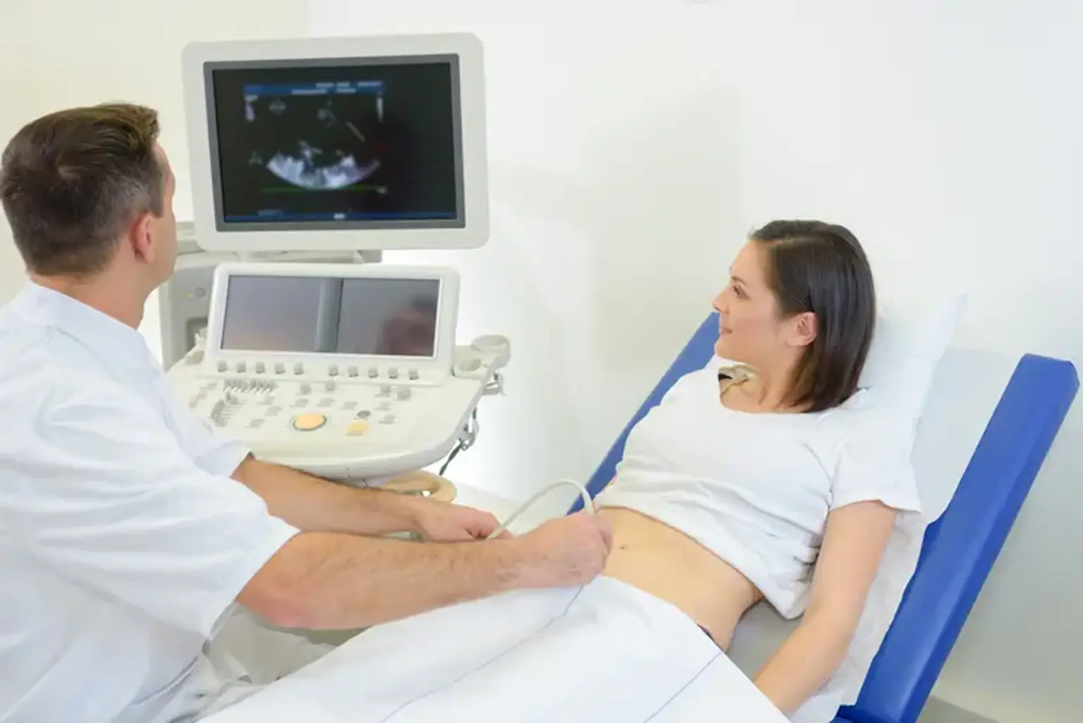
    Hysterosonography, also known as sonohysterography, uses sound waves to create images of the inside of a woman's uterus, which can assist detect a variety of issues such as unexplained vaginal bleeding, infertility, and recurring miscarriages. Hysterosonography is conducted in the same manner as a gynecologic exam. Your doctor will introduce a speculum into your vagina and a catheter into the uterine cavity. Your doctor will use a tiny tube placed into the vagina to inject a small quantity of sterile saline into the uterine cavity and an ultrasound transducer to investigate the uterine lining. Ultrasound does not use ionizing radiation, has no known side effects, and offers a good view of soft tissues that x-rays do not.

    To reduce the risk of infection, hysterosonography should be performed one week after menstruation. This process requires little to no prior preparation. Inform your doctor if you suspect you are pregnant. Wear loose, comfy clothes and leave your jewels at home. You could be required to wear a gown.


    What is Saline Infusion Sonography?

    Saline Infusion Sonography

    The uterus and the morphology of the uterine cavity are evaluated by saline infusion sonohysterography (SIS or SHG). SHG visualizes the uterus and endometrial (uterine lining) cavity using ultrasonography and sterile fluid. The ovaries are also visible during SHG. The goal is to discover any anomalies.


    What are the Benefits vs. Risks?

    Saline Sonogram(SIS) benefits

    Benefits

    • Most ultrasound scanning is noninvasive (no needles or injections).
    • Occasionally, an ultrasound exam may be temporarily uncomfortable, but it should not be painful.
    • Ultrasound is widely available, easy to use, and less expensive than most other imaging methods.
    • Ultrasound imaging is extremely safe and does not use radiation.
    • Ultrasound scanning gives a clear picture of soft tissues that do not show up well on x-ray images.
    • Sonohysterography is a simple, minimally invasive procedure that patients usually tolerate well. It has very few complications.
    • Sonohysterography is a relatively short procedure that provides an excellent view of the uterus and endometrial lining.
    • Many uterine abnormalities that may not be seen adequately with routine transvaginal ultrasound may be viewed in detail with sonohysterography.
    • Sonohysterography can prevent unnecessary surgery. It can also help surgeons remove all polyps and fibroids.


    Risks

    Standard diagnostic ultrasound has no known harmful effects on humans.


    How to Prepare for Saline Sonography?

    Prepare for Saline Sonography

    • It’s best to have the procedure in the week after your period ends. This will help the test be more accurate. It will also lower your risk of infection.
    • You can eat and drink as normal on the day of your procedure. You will likely be able to take all of your medicines as normal.
    • Your healthcare provider may advise taking an over-the-counter pain medicine ahead of time. This is to help prevent discomfort.
    • You may be told to take an antibiotic before the test. This is to help prevent infection.
    • You may want to wear an absorbent pad in your underwear on the day of the procedure. This is because saline fluid will drain from the uterus after the procedure.
    • Your healthcare provider may give you more instructions about how to prepare.


    How Does the Procedure Work?

    Ultrasound for Saline Sonography

    Ultrasound imaging employs the same concepts as sonar used by bats, ships, and fisherman. A sound waves bounce back or echoes when it strikes an item. It is possible to establish how far away an item is by analyzing its echo waves, as well as its size, shape, and consistency. This includes whether or if the thing is solid or fluid.

    Ultrasound is used by doctors to identify changes in the appearance of organs, tissues, and arteries, as well as abnormal structures such as tumors.

    A transducer delivers sound waves and captures the echoing (returning) waves during an ultrasound test. When rubbed against the skin, the transducer emits small pulses of inaudible, high-frequency sound waves into the body. As sound waves bounce off internal organs, fluids, and tissues, the transducer's sensitive receiver detects minute changes in pitch and direction. These characteristic waves are quickly measured by a computer and shown as real-time images on a monitor. Typically, the technician records one or more frames of the moving visuals as still photos. They may also preserve photos in brief video loops.

    Transrectal and transvaginal ultrasound methods follow the same concepts. These checks need the doctor inserting a specific transducer into the patient's body.

    To widen the uterine cavity for sonohysterography, the clinician injects sterile saline into it. The saline outline the endometrium (uterine cavity lining) and provides for simple visibility and measuring. It also detects any polyps or masses that may be present within the cavity. In addition, the doctor may inject saline and air into the uterus to search for air bubbles moving via the fallopian tubes. This aids in determining the patency of the fallopian tubes.

    Doppler ultrasound, a special ultrasound technique, measures the direction and speed of blood cells as they move through vessels. The movement of blood cells causes a change in pitch of the reflected sound waves (called the Doppler effect). A computer collects and processes the sounds and creates graphs or color pictures that represent the flow of blood through the blood vessels.


    When Saline Sonography is Needed?

    Sonohysterogram

    A sonohysterogram is performed in the office of your physician and is less intrusive than other surgical procedures that allow your provider to view into your uterus, such as a hysteroscopy. A normal pelvic ultrasound performs a better job of providing precise pictures of structures inside your uterus that may be causing your problems.

    A sonohysterogram can reveal structures within your uterus that are producing symptoms such as pelvic discomfort, unexplained bleeding, or infertility. Your doctor may order a sonohysterogram if you:

    • Your periods are heavier or longer-lasting than is typical.
    • You have vaginal bleeding in between periods.
    • You’ve got pelvic pain that won’t go away.
    • You’ve been unable to get pregnant.
    • You’ve had two or more miscarriages.

    A sonohysterogram can reveal structures inside your uterus that may be causing these symptoms. A sonohysterogram can show:

         1. Polyps:

    Uterine polyps are growths that connect to the uterine inner wall and expand into the uterine cavity. The overgrowth of cells in the uterine lining (endometrium) results in the production of uterine polyps, also known as endometrial polyps. These polyps are normally noncancerous (benign), although some might be cancerous or progress to malignancy (precancerous polyps).

    Uterine polyps range in size from a few millimeters (about the size of a sesame seed) to several centimeters (about the size of a golf ball). They have a big base or a slender stalk that attaches to the uterine wall.

    You can have one or many uterine polyps. They usually stay contained within your uterus, but occasionally, they slip down through the opening of the uterus (cervix) into your vagina. Uterine polyps most commonly occur in women who are going through or have completed menopause, although younger women can get them, too.

         2. Fibroids

    Uterine fibroids are noncancerous uterine growths that commonly arise during reproductive years. Uterine fibroids, also known as leiomyomas or myomas, are not connected with an elevated risk of uterine cancer and usually never develop into malignancy.

    Fibroids range in size from invisible seedlings to bulky masses that can deform and expand the uterus. You might have a single fibroid or many. Multiple fibroids can cause the uterus to enlarge so much that it approaches the rib cage and adds weight in severe situations.

    Many women get uterine fibroids at some point in their lives. However, because uterine fibroids seldom cause symptoms, you may be unaware that you have them. During a pelvic exam or prenatal ultrasound, your doctor may uncover fibroids by chance.

          3. Atypical uterus shape

    Some women have wombs (uteruses) that are unusual in shape or size. This is known as womb abnormalities or congenital uterine anomaly. It indicates your uterus (womb) developed in an uncommon way before you were born.

    Many women have an abnormally shaped womb but are unaware since there are usually no symptoms. You may only discover through an ultrasound scan or if you have had a miscarriage, bleeding, or difficulty conceiving. Knowing about a womb anomaly has aided some women in planning and preparing for pregnancy.

    Although these anomalies are very prevalent, the impact on pregnancy is not always evident. A uterine anomaly will not always impede your ability to become pregnant, but it may make carrying your baby to full term more challenging. Miscarriage or premature delivery may be more likely depending on the form of the womb (giving birth before 37 weeks). Womb abnormalities can also be associated with a weaker cervix (also known as cervical incompetence), which can result in premature delivery. During pregnancy, you may require additional monitoring to keep you and your baby safe.

    • Bicornuate womb: The top of a bicornuate womb has a deep depression. Women with a bicornuate womb face no additional challenges during conception or early pregnancy, but they are at a slightly increased risk of miscarriage and premature delivery. It may also impact the baby's position later in the pregnancy, necessitating a c-section (caesarean).
    • Unicornuate womb: It is unusual to have a unicornate womb. Because one side of your womb did not develop, it is half the size of a typical womb. Ectopic pregnancy (an abnormal pregnancy that implants and grows outside the womb), late miscarriage, or premature birth are all elevated risks. In later pregnancy, the baby may be in a difficult position, necessitating a c-section (caesarean). Women with unicornate wombs can conceive, although unicornate wombs are more prevalent in infertile women.
    • Didelphic (double) womb: The didelphic womb is divided into two cavities, one on each side. The womb and cervix are commonly affected, however the vulva, bladder, urethra, and vagina can also be affected. Women with a didelphic womb have no additional difficulty conceiving, and it is only associated with a little increase in the risk of premature delivery. 
    • Septate/subseptate womb: A septate womb has a muscular wall running along the center, dividing the area in half. Sometimes the wall only extends halfway down the womb (subseptate), and other times it extends the entire length (septate). Women who have subseptate or septate wombs are more prone to struggle with conception. There is also a higher chance of miscarriage and premature delivery. Because the baby may not be laying in a head-down (cephalic) position later in pregnancy, you may be advised to have a c-section.

          4. Signs of endometriosis.

    Endometriosis is a painful disorder in which tissue comparable to the endometrium, which typically lines the lining of your uterus, develops outside your uterus. Endometriosis most usually affects the ovaries, fallopian tubes, and pelvic tissue. Endometrial-like tissue can occasionally be detected outside of the pelvic organs.

    Endometrial-like tissue behaves similarly to endometrial tissue, thickening, breaking down, and bleeding with each menstrual cycle. However, because this tissue cannot leave your body, it remains trapped. Endometriomas can occur when endometriosis affects the ovaries. Surrounding tissue can become inflamed, leading to the formation of scar tissue and adhesions – bands of fibrous tissue that can cause pelvic tissues and organs to stick together.

    Endometriosis can cause pain — sometimes severe — especially during menstrual periods. Fertility problems also may develop. Fortunately, effective treatments are available.

          5. Signs of endometrial cancer.

    Endometrial cancer is a cancer that develops in the uterus. The uterus is a hollow, pear-shaped pelvic organ in which fetal development takes place.

    Endometrial cancer originates in the layer of cells that forms the uterine lining (endometrium). Endometrial carcinoma is also known as uterine cancer. Other forms of cancer, such as uterine sarcoma, can develop in the uterus, although they are far less prevalent than endometrial cancer.

    Endometrial cancer is commonly discovered at an early stage due to irregular vaginal bleeding. When endometrial cancer is diagnosed early, surgically removing the uterus frequently cures it.


    What Happens During Saline Sonography?

    During Saline Sonography

    The test is performed at the office of a healthcare professional or in a hospital. The operation may be performed by a technician or a radiologist. A radiologist is a physician who specializes in the use of imaging techniques to diagnose and treat diseases. The examination typically lasts less than 30 minutes. In general, you may anticipate the following:

    1. You'll need to empty your bladder before the treatment begins. After that, you'll strip from the waist down and lie on an exam table. For the exam, you will be awake and aware. Mild cramping may occur during and after the treatment.
    2. To rule out any discomfort, your healthcare professional may do a pelvic exam first.
    3. A thin sheath and a specific gel will be applied on the transducer wand by the healthcare provider. After that, the wand will be inserted into your vagina.
    4. To get distinct views of your uterus, the healthcare professional may shift the wand slightly. These visuals are shown on a video screen. This is a routine vaginal ultrasound.
    5. The transducer will be removed by the healthcare professional once these photos have been taken.
    6. The speculum will then be inserted into your vagina. This is the same equipment that is used in a routine vaginal examination. This helps to keep the vagina open so the doctor can view and reach your cervix.
    7. Your healthcare practitioner may clean and wipe your cervix with a swab.
    8. A tiny catheter will then be inserted into the opening of your cervix. As the catheter is implanted, you may feel a minor pinching or cramping. After that, the speculum will be withdrawn from your vagina.
    9. The wand is then reinstalled into your vagina by the healthcare practitioner. Saline will be injected through the catheter into the cervix and uterus. This may result in cramps. Images of the interior of your uterus are taken once enough saline solution has filled it.
    10. The wand and catheter are removed by the healthcare professional once the pictures are taken. Over the following few hours, the saline fluid will drain from your uterus.


    What Happens After a Saline Sonography?

    Result of Saline Sonography

    You will likely be able to go home right after your procedure. In most cases, you can resume your normal activities right away. The saline fluid will drain out of your uterus over the next few hours.

    For a little while after the procedure, you might have symptoms like:

    • Cramping.
    • Light spotting.
    • Fluid leaking from your vagina.

    All of this is typical. The majority of women have no discomfort following the operation. If so, you can use over-the-counter pain relievers.

    Inform your provider straight once if you experience significant discomfort or fever in the coming days. Also, notify your clinician if the kind or volume of your vaginal discharge changes. These might be symptoms of an infection. More instructions may be given to you by your provider.

    Following the exam, a radiologist will review the photos and submit a report to your primary care physician. Discuss these findings with your provider. The results might reveal a medical issue that requires treatment. Another test, like as a hysteroscopy, may be required. 


    Conclusion

    Saline infusion sonogram

    A saline infusion sonogram is performed routinely to ensure that the endometrial cavity (within the uterus) is normal. Benign uterine growths, such as polyps or uterine fibroids, can form in the cavity and produce an environment unfavorable to embryo implantation. The saline infusion sonogram can also detect scar tissue.

    This procedure may be performed as part of a recurrent pregnancy loss examination or prior to an IVF cycle. It may also be used to determine the reasons of irregular or extremely heavy menstrual cycles, as well as some kinds of persistent pelvic discomfort. The saline infusion ultrasonography is often done early in a menstrual cycle, immediately after your menstruation has ended but before ovulation - typically between days 5 and 10 of the cycle. The technique is straightforward and typically takes 10 to 15 minutes to perform. The cervix is washed with an antiseptic solution after a speculum is introduced into the vagina.

    A 1mm diameter catheter (roughly the size of a single strand of spaghetti) is placed through the cervical canal. After that, the ultrasound probe is inserted into the vagina. Sterile saline is injected into the uterine cavity through the catheter. This dilates the endometrial cavity, allowing the doctor to observe the whole cavity on the ultrasound monitor. At this moment, the patient may suffer minor cramping. Several uterine pictures will be captured throughout the process. The ovaries may also be examined. The patient may have slight cramping for a few hours after the treatment.

    The patient may also experience light spotting or watery discharge for as long as 24 hours after the procedure. The patient should refrain from intercourse for approximately 48 hours.