Sports traumatology

Last updated date: 07-Jul-2023

Originally Written in English

Sports Traumatology

Overview

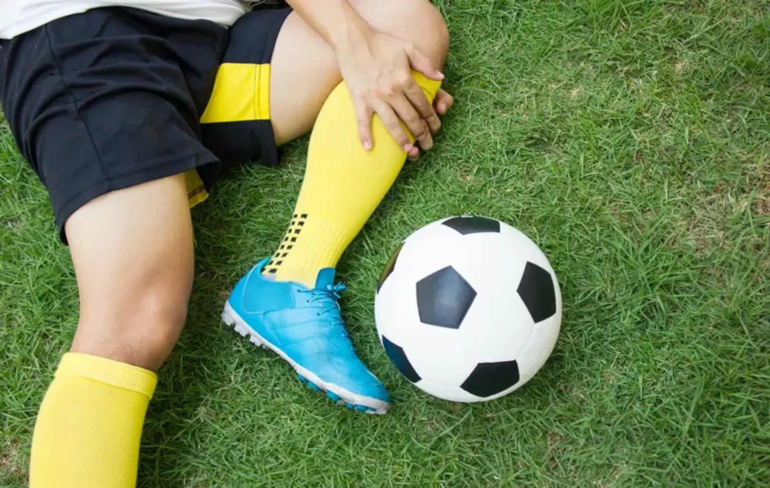
Sport is a life-enriching activity for people of all ages. It maintains us strong and healthy and, at best, slows the aging process. Unfortunately, sports injuries are always a possibility in every sport. This does not necessarily have to be in the form of bruises, abrasions, or broken bones. The body is frequently subjected to inappropriate tension on tendons, muscles, and ligaments, which can lead to more severe physical discomfort and pain over time. Sports medicine is a medical specialty concerned with the prevention, diagnosis, and treatment of sports-related injuries.

Sports injuries can occur unexpectedly as a result of overtraining, improper training, bad technique, incorrect attire, or a lack of fitness, among other factors. The therapies employed enable the individual to resume their usual training program as soon as feasible and in the best possible conditions.

Sprained ankles, muscle tears, tendonitis and tendinopathies (tennis elbow, golfers elbow, Achilles tendon rupture), knee injuries (meniscus fracture or damage to the anterior or posterior cruciate ligament), cartilage injuries, and shoulder injuries (dislocation and tendinopathy), among others, are the most common injuries in sports medicine. 
 

Diagnosis of Sports Injuries

Diagnosis of Sports Injuries

A sports physician or orthopedist can diagnose acute and chronic injuries, but non-physician specialists trained to diagnose and manage these injuries, such as athletic trainers and physical therapists, can also do so.

You will be asked to submit a medical history as well as details on how the accident happened, and you will be subjected to a physical examination.

Your healthcare expert will palpate the region and inquire about the level of discomfort or sensitivity during the physical examination. You will also be asked to move the damaged region in order to assess its range of motion.

Your healthcare professional may order X-rays to rule out any fractured bones, depending on the suspected injury and severity of pain or disability. While some broken bones are visible on an initial X-ray, others (for example, a minor fracture of the wrist or a hairline fracture in the foot) may not be seen until a few days later, once the damage has begun to recover.

Soft-tissue injury may necessitate further diagnostic imaging testing. These, which may be ordered during the initial appointment or after a period of unsuccessful treatment, include the following:

  1. Magnetic resonance imaging (MRI): It is frequently utilized for diagnostic imaging of sports-related muscle injuries, joint damage, sprains, fractures, and head traumas. MRIs examine musculoskeletal structures such as bones, tendons, muscles, ligaments, and nerves by using radio waves in a strong magnetic field.
  2. Ultrasound: An ultrasound takes real-time pictures of superficial soft tissues and is useful for diagnosing tendon injury. The radiologist may ask you to manipulate the joint during the ultrasound to evaluate how motion affects the tendon.
  3. CT scans: A CT scan gives a more comprehensive view of bones and soft tissues. Hairline fractures and minor abnormalities inside complicated joints can be detected with this technique. 


Most Common Sport Injuries

Common Sport Injuries

Injuries to the joints are the most common form of sports injury in the field of sports traumatology. Excessive joint deterioration (arthrosis) is also common. Injuries to the legs are the most common, accounting for 70% of all injuries. However, every fifth patient experiences shoulder and arm discomfort. Accidents to the spine or head are, fortunately, considerably less common.

These are the most common sites of injury in sports traumatology, organized by body region:

In the knee area:

  • Ligament tears, with the cruciate ligament tear as a classic.
  • Meniscus damage.
  • Tendon injuries in the area of the knee joint.
  • Jumping out of the kneecap (patellar luxation).
  • Arthrosis of the knee joints.

In the shoulder:

In the ankle joint:

  • Torn ligament.
  • Achilles tendon rupture or tearing off.


Acromioclavicular Joint (AC) Injuries

Acromioclavicular Joint (AC) Injuries

The Acromioclavicular (AC) Joint is a common site of injury, especially in contact and impact sports like Australian football and rugby (league and union), as well as throwing sports like shotput.

Signs and Symptoms:

  • The end of the collarbone hurts.
  • Until the first pain subsides, it may seem diffuse across the shoulder; after that, it is more likely to be a relatively precise spot of pain over the joint itself.
  • Swelling is common.
  • A step-deformity may be seen depending on the amount of the damage. On more serious injuries, this is a noticeable bump where the joint has been damaged.
  • Pain when moving the shoulder, particularly when raising the arms above shoulder height.


Prevention:

  • Wearing precautionary strapping to support an AC Joint that has already been damaged, especially in contact sports or sports where full arm elevation is not required. Rugby, for example, makes use of protective padding.
  • Warming up, stretching, and cooling down are all important steps.
  • Participating in exercise programs to improve strength, balance, coordination, and flexibility.
  • Training before to competition to guarantee ready to compete.
  • Gradually increasing exercise intensity and duration.
  • Allowing enough time to recover between workouts or training sessions.
  • Wearing the proper protective gear, including footwear.
  • Water consumption before, during, and after play.
  • Prior to training and match play, inspect the athletic environment for risks.
  • Avoiding actions that cause discomfort. If pain does arise, stop the activity immediately and begin RICER.


Management:

The RICER protocol - rest, ice, compression, elevation, and referral – is used to treat any soft tissue injury immediately. For 48-72 hours, the RICE protocol should be followed. The goal is to stop the bleeding and damage inside the joint. Every two hours, the shoulder should be raised and an ice pack administered for 20 minutes (never apply ice directly to the skin). In addition, the arm should be immobilized in a sling. This might be for as little as two days in the event of a minor injury or up to six weeks in the case of a major injury.

The No HARM strategy should also be followed, which includes no heat, no alcohol, no running or exercise, and no massage. This will guarantee that swelling and bleeding in the affected region are reduced.

A sports medicine specialist should be visited as soon as possible to assess the degree of the injury and give treatment recommendations. A sports medicine expert may do a physical examination and take shoulder x-rays.


Rehabilitation and Return to Play:

Following the immobilisation period, most AC Joint injuries are managed conservatively with different combinations of strengthening activities, as pain allows. Surgery is normally reserved for situations when the AC Joint is completely dislocated (Grade 3), or when a less severe injury fails to respond well to conservative care. 


Muscle Contusion (Bruise)

Muscle Contusion

Athletes who participate in contact sports are more likely to experience a muscle contusion (bruise). Contusions are the second most common cause of sports injuries, after only muscle strains.

Contusions develop when a blunt object strikes a region of the body, crushing underlying muscle fibers and connective tissue but not breaking the skin. A contusion can occur as a result of falling or collapsing against a hard surface.

Most contusions are mild and heal rapidly, allowing the player to return to the game. Severe contusions, on the other hand, can cause profound tissue injury and problems that may keep an athlete from playing sports for months.


Symptoms:

Contusions cause swelling and pain and can limit joint range of motion near the injury. Torn blood vessels may cause bluish discoloration. The injured muscle may feel weak and stiff.

Sometimes a pool of blood collects within damaged tissue, forming a lump over the injury (hematoma). If tissue damage is extensive, you may also have a broken bone, dislocated joint, sprain, torn muscle, or other injuries. Contusions to the abdomen may damage internal organs.


Treatment:

Keep the muscle in a gentle stretch posture and follow the RICE procedure to decrease pain, bleeding, and inflammation:

  • Rest. Stop play to protect the damaged region from additional harm. You could also utilize a safety device (i.e., crutches, sling).
  • Ice. Several times a day, apply cold packs for 20 minutes at a time. Ice should not be applied straight to the skin.
  • Compression. Wrap the damaged area lightly with a soft bandage or ace wrap.
  • Elevation. Elevate the afflicted location over the heart.

Most athletes who suffer from contusions recover rapidly with modest therapy. For pain treatment, your doctor may prescribe nonsteroidal anti-inflammatory medicines (NSAIDs) such as ibuprofen or other medications. Avoid massaging the damaged region.

Rest, ice, compression bandages, and elevation of the wounded region are likely to be necessary within the first 24 to 48 hours following injury (acute phase). While the damaged muscle recovers, continue to exercise the uninjured sections of your body to maintain your total fitness.

If a big hematoma does not disappear within a few days, your doctor may drain it surgically to speed healing.


Return to Play:

When you regain full strength, mobility, and endurance, you may be able to resume contact sports. When your doctor and sports trainer agree that you are ready to return to play, they may want you to wear a specialized protective device to avoid additional harm to the area that had a contusion. 


Ligament Injuries

Ligament Injuries

Common joints involved in Ligaments injuries:

  • Knee Ligament injuries: 

Depending upon your sport, you may get special padding made of firm or semi-firm materials. The padding spreads out the force of impact when direct blows from blunt objects strike your body.

  1. Anterior Cruciate Ligament (ACL) Tear: Common in soccer, football and basketball, an ACL injury is damage to the ligaments on the inside of the knee. The severity can range from a mild sprain to a full tear of the ligament(s). ACL tears frequently require surgery.
  2. Medial Collateral Ligament (MCL) or Lateral Collateral Ligament (LCL) Tear: A Medial Collateral Ligament (MCL) or Lateral Collateral Ligament (LCL) injury is a sprain or tear of the inner or outer knee ligaments. It is usually a result of an injury that occurs while participating in an activity that puts a heavy strain on the knees, such as skiing.  Most MCL and LCL injuries will recover without surgery
  3. Posterior Cruciate Ligament (PCL) Tear: Injury to the PCL is the least common type of ligament injury in the knee and frequently occurs from direct trauma or fall on the knee.  Most PCL injuries will heal without surgery.


The Ulnar Collateral Ligament (UCL) is the most often injured ligament in the elbow. This is also known as the Tommy John injury. Baseball injuries are most typically caused by excessive pitching and can be acute or chronic in nature. Traumatic tears can also develop as a result of falls or activities such as wrestling. The UCL is located on the inside of the elbow and can be injured with a slight sprain, partial tear, or total tear. Learn more about the elbow.


Shoulder ligaments are most typically damaged in ball and socket joint dislocations. These are often severe injuries that necessitate a trip to the emergency department to have the joint reduced (put back into place). A shoulder separation includes tearing of the ligaments that hold the Acromioclavicular Joint (ACJ), which is located on the outside of the collar bone. 


  • Thumb ligament injuries: 

The most common type of ligament injury is called a gamekeeper's thumb. Gamekeepers thumb is a tear of the ligament between the base of the thumb and the wrist bones. These commonly occur during a fall when you’re holding something in your hand, such as a ski pole or the handlebars of a bike. Learn more about the hand.


What Are the Symptoms of a Ligament Injury?

Torn ligament symptoms will vary depending on the severity of an injury to a tendon, but may include:

  • Pain to the area, often sudden and severe.
  • A loud pop or snapping sound at the time of the injury.
  • Swelling to the area of the injury.
  • Looseness of the joint.
  • Inability to move or put weight on the joint without pain.
  • Spasms in the muscles surrounding the injury.

Ligament injuries can be as painful as fractures and should be treated as soon as possible. One of our healthcare experts can assess you and recommend the best treatment option for your injury.


Ligament injuries treatment:

Mild ligament injuries can be treated non-surgically with the R.I.C.E. technique (Rest, Ice, Compression, and Elevation) and anti-inflammatory medicine. For comfort and protection, a brace or splint may be required. Physical therapy may also be recommended by your doctor as part of your treatment plan.

A surgical technique to repair a ligament may be necessary for severe strains or tears. Ligament surgery may involve mending or replacing the ligament. A tendon transplant is sometimes required to hold the damaged joint together. The tendon utilized may be autografted from the wounded individual or allografted from an organ donor. Physical therapy is usually often needed after surgical repair.


Tendonitis

Tendonitis

Tendonitis, sometimes called tendinitis, is an inflammation of the tendons that occurs when they become irritated and inflamed. Tendinitis can create deep, nagging discomfort that hinders simple, pleasant motions. 

Some areas of the body that commonly develop tendon injuries include:

  • Tennis Elbow (Lateral Epicondylitis).
  • Golfer’s Elbow (Medial Epicondylitis).
  • Achilles Tendinitis.
  • Wrist Tendinitis.
  • Carpal Tunnel Syndrome.


Causes of Tendinitis:

Tendinitis or tendinopathy can develop as a result of poor sports technique or biomechanical difficulties, in which case working with a coach or trainer is the best approach to avoid a persistent condition. Making careful to warm up properly and include appropriate cross-training is also beneficial in preventing tendon overuse issues.

Overuse injuries are caused by repeated use, stress, and damage to the body's soft tissues (muscles, tendons, bones, and joints) without enough rest. They are also known as cumulative trauma injuries or repeated stress injuries.

An acute injury that pushes a tendon to stretch beyond its usual range of motion and produces pain, swelling, and inflammation is the most prevalent cause of tendonitis in sports.


Tendinitis treatment:

If you experience a sudden soreness or pain in a tendon and suspect tendonitis, the first thing you should do is halt all activity and rest. Tendonitis will be helped by the R.I.C.E. approach (rest, ice, compression, and elevation). This approach reduces inflammation and swelling while also providing short pain relief. In most cases, conservative therapy is all that is required to heal from true tendonitis. Tendinitis normally goes away within a few days to a few weeks.

Unfortunately, long-term tendinopathy might take two to six months to recover. Many tendon injuries progress into chronic difficulties when the athlete continues to participate despite the persistent discomfort.

If your tendon discomfort persists after a few days of rest and conservative therapy, you should consult a sports medicine professional and work with a physical therapist to rehab the tendon.

Ultrasound or other modalities may be used by physical therapists to aid in the healing of tendinopathy. Splints or braces may be used in some circumstances to relieve pressure on the tendon as it recovers. Ultrasound, medicines, massage, bracing or splinting are all common ways of rehabilitation.

The last step of tendinopathy rehabilitation comprises strengthening and flexibility exercises. Your therapist will assist you in determining the best rehab path for you, but it's critical to remember that starting any workouts before the tendon has healed may aggravate the condition, so it's critical to follow your therapist's or physician's recommendations. 


Clavicular fractures

Clavicular fractures

Younger people are more likely to incur these injuries as a result of moderate to high-energy mechanisms such as car accidents or sports injuries, whereas the elderly are more likely to sustain injuries as a result of a low-energy fall.

Although a fall into an outstretched hand was often thought to be the most common mechanism, it has been shown that the clavicle most frequently fails under direct compression from a force delivered directly to the shoulder. A clavicle fracture is caused by a direct fall onto the lateral shoulder in around 87 percent of reported cases.


Signs & Symptoms:

The patient may appear with the following signs and symptoms:

  • The intact arm can be used to cradle the wounded extremity.
  • When an injury occurs, a patient may describe hearing a snapping or cracking sound.
  • The shoulder may look shorter and droopy in comparison to the opposing side.
  • Over the clavicle, swelling, ecchymosis, and discomfort may be seen.
  • Abrasion across the clavicle is possible, indicating that the fracture was caused by a direct process.
  • Gentle manipulation may reveal crepitus caused by the fracture ends pressing against one other.
  • Breathing difficulties or decreased breath sounds on the afflicted side may suggest a pulmonary injury, such as pneumothorax or even pneumonia.
  • A concurrent injury may be revealed by palpating the scapula and ribs.
  • Tenting and blanching of the skin at the fracture site may suggest an imminent open fracture that will most likely necessitate surgical stabilization.
  • A brachial plexus injury is indicated by associated distal nerve dysfunction.
  • A subclavian artery damage may be indicated by decreased pulses.
  • A subclavian venous injury is indicated by venous stasis, discolouration, and edema.


Management:

Clavicle fractures are addressed surgically or conservatively depending on a variety of parameters such as location (mid-shaft, distal, proximal), nature (displaced, undisplaced, comminuted), open VS closed damage, age, and neurovascular constraints.

Traditionally, clavicle fractures have been treated conservatively with sling immobilization and subsequent rehabilitation. Conservative therapy of displaced mid-shaft clavicle fractures resulted in higher rates of re-injury, longer return periods to sport, and poor shoulder function due to clavicular mal-union and shortening, with resulting thoracoscapular dyskinesia. Similarly, conservative therapy of displaced lateral fractures in the athletic patient has been demonstrated to result in high rates of nonunion and eventual shoulder function impairment.

The chief goal of this treatment is to achieve a healed clavicular strut in a normal anatomical position as possible.

The following are the indications for clavicular fracture surgery: 

  • Severe displacement caused by comminution, with resulting angulation and tenting of the skin severe enough to jeopardize its integrity and that does not respond to closed reduction.
  • Neurovascular impairment due to symptomatic non-union of the shoulder girdle.
  • Neurovascular damage or compromise that is progressive or fails to revert after the fracture has been repaired.
  • An open fracture
  • Distal clavicular fracture of type II (displaced).
  • Multiple traumas, where patient movement is desired and closed techniques of immobilization are impractical or impossible.
  • Inability to withstand closed immobilization, such as Parkinsonism or seizure disorders.
  • For cosmetic reasons.
  • Relative indications include shortening of more than 15 to 20mm and displacement more than the width of the clavicle.

Surgical procedure includes:

  • Internal fixation with plates and screws. (most common)
  • Intramedullary (IM) fixation.


Physical Therapy/Rehabilitation:

The major objective of therapy is to enhance and restore shoulder function for daily living, occupational, and athletic activities. The rehabilitation plan may differ slightly in the first few weeks depending on the primary treatment technique (conservative vs. surgical). 


Achilles tendon rupture

Achilles tendon rupture

Achilles tendon rupture is a lower-leg ailment that affects the rear of the leg. It mostly affects persons who participate in leisure activities, although it may happen to anybody.

The Achilles tendon is a strong fibrous cord that links your calf muscles to your heel bone. When you overstretch your Achilles tendon, it can totally or partially tear (rupture).

If your Achilles tendon ruptures, you may hear a pop followed by an instant acute pain in the back of your ankle and lower leg, which may impair your ability to walk normally. Surgery is frequently used to heal the rupture. Nonsurgical therapy, on the other hand, works just as effectively for many people.


Symptoms:

Although it's possible to have no signs or symptoms with an Achilles tendon rupture, most people have:

  • The feeling of having been kicked in the calf.
  • Pain, possibly severe, and swelling near the heel.
  • An inability to bend the foot downward or "push off" the injured leg when walking.
  • An inability to stand on the toes on the injured leg.
  • A popping or snapping sound when the injury occurs.


Achilles tendon rupture treatment:

  • Surgical Treatment:

Some people may benefit from surgery to repair a totally torn Achilles tendon. The surgery's purpose is to reattach the tendon. However, in rare circumstances, the injured part (or the entire tendon) may need to be removed and replaced with tissues from another area of your foot.

  • Nonsurgical Options:

Other patients may benefit from avoiding surgery. Nonsurgical Achilles tendon rupture therapy relies on letting the tendon to heal spontaneously while immobilized in a boot. Early functional weight-bearing is an important element of Achilles tendon recovery, both surgical and nonsurgical.


Conclusion

Sports traumatology

If you play sports, whether you're a top athlete or a weekend warrior, you've undoubtedly been injured at some point. Sprains, strains, inflamed muscles, shin splints, rotator cuff injuries, knee injuries, fractures, and dislocations are all common sports injuries.

Some sports injuries are the consequence of a sudden occurrence that generates extremely apparent symptoms. Others are chronic, overuse illnesses with more modest symptoms, either at initially or over time.

An accident, impact, bad training habits, incorrect equipment, a lack of fitness, or insufficient warm-up and stretching can all result in a sports injury. Sprains and strains of muscles, ligament and tendon rips, dislocated joints, broken bones, and head traumas are all prevalent.

While joints are particularly sensitive to sports injuries, any component of the body can be injured on the court or field. Here's a deeper look at some of the most prevalent injuries for various sections of the body.