Total gastric resection

Last updated date: 03-Nov-2023

Originally Written in English

Total Gastric Resection

For many benign and malignant stomach conditions, gastric resection represents the recommended surgical treatment choice. Stomach cancer is the world's fifth most prevalent cancer, with gastric resection or total gastrectomy remaining the primary treatment option for long-term survival and cure. A subtotal gastrectomy removes 75-80% of the distal stomach, whereas a total gastrectomy removes the whole stomach, including the pylorus.

Despite a continuous drop in the incidence and mortality of gastric carcinoma over the previous century, the absolute number of cases continues to rise each year as the population becomes older. Early detection of stomach cancer is extremely rare, and nodal metastases are common. Because lymphatic dissemination is the most important prognostic marker in gastric cancer, proper lymphadenectomy is necessary for both curative and staging resection.

Stomach cancers are classified as either intestinal or diffuse. Helicobacter pylori infection, which can lead to atrophic gastritis and intestinal metaplasia, is the most common underlying cause of intestinal-type cancer. Marked fibrosis and early penetration into the submucosa indicate diffuse-type cancer. Genetics (CDH1 gene), H. pylori infection, stomach ulcers, gastroesophageal reflux disease (GERD), cigarette or alcohol use, nutrition, and chemical exposure are all risk factors for gastric cancer.
 

What is Gastric Resection?

Gastric Resection Definition

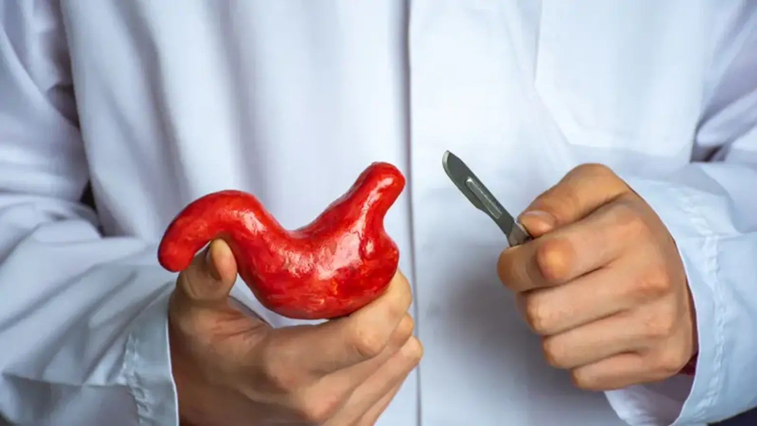
A Gastric resection (gastrectomy) is a surgical procedure that removes all or part of the stomach. This technique is used to remove adenocarcinoma and lymphoma of the stomach, as well as other benign and malignant neoplasms (tumors) of the stomach. Partial gastrectomy can also be used to treat a variety of less common benign tumors of the stomach or stomach wall. Furthermore, the procedure is occasionally used to treat peptic ulcers because it removes the gastric-acid-secreting parietal cells in the stomach lining and stops the release of the acid-stimulating hormone gastrin, removing the underlying cellular components that produce an ulcer. Gastrectomy, which was once a common treatment for people with painful ulcers, is now only used as a last resort if all other options have failed or if the ulcer is perforated or bleeding.


Total Gastric Resection Indications

Gastric Resection Indications

The following are the most frequent reasons for gastric resection:

  • Malignancy. Adenocarcinoma, gastrointestinal stromal tumors (GIST), neuroendocrine neoplasms, signet ring cell gastric cancer, and hereditary diffuse gastric cancer (CDH1 mutation carriers) are all cancers that can occur in the stomach.
  • Peptic ulcer disease. Refractory stomach ulcer disease can lead to bleeding, perforation, or stricture.
  • Bariatric procedures. Roux-en-Y gastric bypass surgery, sleeve gastrectomy, or gastric banding are all bariatric operations.

For malignancies of the gastric body or antrum, subtotal distal gastrectomy is the gold standard if a 4-6 cm proximal border can be obtained while keeping an adequately sized remnant pouch. For malignancies affecting the entire or proximal stomach, signet ring cell gastric cancer (diffuse submucosal invasion), or hereditary diffuse gastric cancer (multifocal pattern), total gastrectomy is recommended.


Total Gastric Resection Contraindications

Total Gastric Resection Contraindications

Any patient who is unsuited for general anesthesia is an absolute contraindication to gastric resection. Patients who are elderly, have a significant cardiopulmonary failure, or have a reduced life expectancy due to other chronic diseases are relative contraindications to gastric resection.

When wide negative margins (4 to 6 cm) can be accomplished with partial gastrectomy, total gastrectomy is contraindicated, because partial gastrectomy has significantly better safety and long-term functional outcome profile, particularly in patients with older age, malnutrition, and extensive comorbidities.


Equipment

The surgeon's ability to perform this treatment successfully requires a typical open or laparoscopic abdominal operative tray. Surgical drapes, scalpels, and electrocautery should all be included in the kit. A self-retaining table-mounted retractor, such as the Book-Walter retractor system, is useful for open operations. CO2 gas insufflation tube, trocars, a 30-degree laparoscope with a light source, and a monitor are all necessary for laparoscopic surgeries.

Suction irrigators, bowel graspers, liver retractors, scissors, ultrasonic dissector (harmonic scalpel), and vessel sealing devices are all often used devices for both open and laparoscopic surgeries. Depending on the type of gastric resection and reconstruction method employed, a linear GIA or circular EEA stapler may be used.


Personnel

A surgeon with sufficient competence in foregut and oncological surgery should be able to conduct a gastric resection or total gastrectomy. The operating surgeon, first assistant, anesthesiologist, scrub nurse, and circulating nurse are all essential members of the surgical team.


Total Gastric Resection Preparation

Total Gastric Resection Preparation

The majority of individuals with stomach cancer are identified at an advanced stage, which is unfortunate. Weight loss, cachexia, anorexia, early satiety, indigestion, gastric outlet obstruction, and malnutrition are all possible symptoms. A range of diagnostic techniques is used to determine which people are candidates for stomach resection during a gastric cancer screening. Routine laboratory tests, such as baseline hemoglobin, platelet count, and complete metabolic profile, should be done. Serum biomarkers albumin and prealbumin can be used to determine nutritional status.

If a stomach tumor is suspected, the diagnostic process begins with an esophagogastroduodenoscopy (EGD). EGD offers a histopathologic diagnosis, as well as the tumors' location and size. Endoscopic ultrasonography (EUS) is utilized to determine the depth of the tumor (T stage) and the presence of any nodal involvement. To rule out the occurrence of distant metastatic disease or bulky lymphadenopathy, a CT scan of the chest, abdomen, and pelvis with oral and intravenous contrast should be done. A positron emission tomography (PET) scan is an additional imaging technique that can be used in conjunction with other imaging modalities to help with staging.

To establish surgical resectability, preoperative plan, and the role of neoadjuvant chemotherapy or radiotherapy, a multidisciplinary approach should be used. For medical optimization, a comprehensive examination of the patient's comorbidities, as well as an evaluation of the patient's performance status and capacity to withstand surgery, is essential. Furthermore, any modifiable risk factors, such as preoperative smoking cessation, which has been shown to enhance outcomes after gastrectomy for malignancy, must be managed before surgical intervention.


Total Gastric Resection Procedure

Total Gastric Resection Procedure

An open, laparoscopic, or robotic-assisted technique for gastric resection is possible. Oncologic equivalency and favorable postoperative recovery outcomes with minimally invasive techniques have been proven in randomized controlled trials comparing laparoscopic and open gastrectomy. In comparison to open operations, minimally invasive gastrectomy has been demonstrated to have a shorter hospital stay, fewer perioperative problems, and less intraoperative bleeding. Surgeon preference, higher surgical complexity, port site recurrence, and insufficient lymph node dissection are all factors to consider while having an open gastrectomy.

The patient is put in the supine position on the operating room table and prepped and draped in the usual sterile manner for all methods. Antibiotics are given before surgery, and a nasogastric tube is inserted for gastric decompression. The key processes for gastric resection surgery are outlined below.

  • Laparoscopic staging and general inspection

To exclude possible occult metastatic illness of the liver or peritoneum, a diagnostic laparoscopy should be performed first. The surgeon can proceed with gastric resection if there is no sign of grossly metastatic illness. The left lobe of the liver may be mobilized and retracted using a hepatic retractor.

  • Mobilization of the Stomach greater curvature and entry into the lesser sac

The patient is in a partly reverse Trendelenburg position, and the gastrocolic ligament is divided, allowing access to the lesser sac. Injury to the transverse colon mesentery and vasculature should be avoided at all costs. The left gastroepiploic artery and short gastric arteries are dissected, as well as the splenic flexure. The splenic hilum lymph nodes are left intact to be collected with the samples. The right gastroepiploic vessels are separated and omentectomy is performed laterally down the transverse colon towards the liver. Dissection is carried out until the pylorus is about 2 cm away.

  • Mobilization of the stomach and duodenum lesser curvature

The stomach's lesser curvature is moved toward the esophagus and the diaphragm right crus. The gastrohepatic ligament is divided, and an accessory right hepatic artery should be avoided if possible. The gastric vessels on the left and right sides are separated. Following the common and proper hepatic arteries, lymphatic tissue is mobilized and collected together with the specimen. Dissection of the intraabdominal esophagus is performed during total gastrectomy. The specimen is dissected together with the pericardial lymph nodes (stations 1 and 2). At the level of the esophageal hiatus, the gastrohepatic omentum must be divided.

  • Lymphadenectomy

In the case of stomach cancer, D2 nodal dissection is the gold standard. The perigastric nodes are collected en bloc with the gastric samples at Stations 1-7. Lymphadenectomy continues through the proper hepatic artery to the common hepatic artery (station 8) and then to the celiac axis (station 9) and splenic artery (station 11p and 11d). After that, the dissection is carried down to the hepatoduodenal ligament, where the accompanying lymph nodes (12a station) are harvested.

  • Gastric transaction

After the nasogastric tube is withdrawn, the duodenum is divided with a linear GIA stapler. The inferior and superior pyloric nodes (stations 5 and 6) are taken together with the duodenum. Once the tumor has been identified, a linear GIA stapler is used to transect the stomach or distal esophagus to establish acceptable oncologic proximal margins of 4-6 cm.

  • Reconstruction

For gastric resection, a variety of reconstruction procedures have been described. Reconstruction with Billroth 1 gastroduodenostomy, Billroth 2 gastrojejunostomy, or Roux-en-Y gastrojejunostomy is an option for total gastrectomy. A Roux-en-Y esophagojejunostomy or a Hunt-Lawrence jejunal pouch may be used to rebuild a total gastrectomy. Roux-en-Y reconstruction has been found in randomized controlled trials to have a lower rate of long-term postoperative outcomes after gastric resection.

Roux-en-Y Reconstruction. The jejunum is separated 30 to 50 cm from the Treitz ligament, with the Roux limb proximally and the biliopancreatic limb distally, using a linear GIA stapler. 60-70 cm along the Roux limb, a jejunojejunostomy is performed. The biliopancreatic and Roux limbs' antimesenteric margins are aligned, and enterotomies are established for the linear GIA stapler. A TA stapler is utilized to establish the anastomosis. To prevent internal hernias, the mesenteric defect is sealed with an absorbable suture. In an antecolic or retrocolic way, the Roux limb is carried up to the proximal stomach or esophagus. The end-to-end esophagojejunostomy or gastrojejunostomy anastomosis is stapled or hand-sewn. To rule out a leak, run a water-bubble test. On surgical day 5, a water-soluble upper GI contrast study can be done to ensure the anastomosis' integrity before starting oral intake.

  • Distal feeding tube placement

Patients with preoperative weight loss or risk factors for a problematic or delayed postoperative recovery should be given special attention. In such circumstances, a jejunostomy feeding tube, inserted in the distal jejunum in a Stamm or Witzel way, may be recommended. A feeding jejunostomy tube is not typically advised, although it may be necessary for patients with severe preoperative nutritional deficits or if postoperative nutrition is a concern.


Total Gastric Resection Recovery

After gastrectomy

After a gastrectomy, a nasogastric tube is sometimes temporarily put through the nose and connected to the stomach area to allow for the drainage of intestinal fluid and gas, as well as to reduce nausea and vomiting. However, in recent years, the necessity of employing a nasogastric tube has become a source of debate.

The patient is nourished intravenously in the days following surgery. After about a week, the patient usually returns to a light diet, eating smaller meals more frequently than before.

Over the course of a few months, the patient gradually regains the ability to consume a normal diet and feels comfortable consuming a typical amount of food in one sitting. After a gastric resection surgery, the patient will likely need to make some dietary modifications, such as eating less fiber and taking certain vitamin and mineral supplements.


Total Gastric Resection Risks

Gastric Resection Risks

There is a danger of bleeding, infection, and injury to adjacent structures with any surgical procedure. High-risk patients may be more susceptible to complications, which can lead to substantial morbidity or even mortality. Tobacco usage, preoperative malnutrition, total gastrectomy, resection for non-malignant reasons, and blood transfusions have all been associated with an increased risk of morbidity.

Early complications (days to weeks)

  • Anastomotic Leak
  • Postoperative Ileus of bowel obstruction
  • Duodenal stump blowout
  • Delayed gastric emptying

Late complications (after 6 weeks)

  • Bile reflux gastritis
  • Dumping syndrome
  • Afferent and efferent limb syndrome
  • Malnutrition and nutritional deficiencies
  • Internal hernia or Peterson hernia
  • Anastomotic stricture
  • Marginal ulcerations
  • Cancer recurrence

Nutritional deficits, dumping syndrome, small gastric remnant, post-vagotomy diarrhea, delayed gastric emptying, afferent or efferent loop syndrome, and bile reflux gastritis are the most prevalent post-gastrectomy problems.

  • Internal hernia. Internal hernias are a documented cause of acute abdominal pain in gastric resection and Roux-en-Y reconstruction patients. In these patients, three forms of trans-mesenteric hernias are frequent. Herniation through the surgical gap in the transverse mesocolon, through which the alimentary limb descends, is known as a transmesocolic hernia. Peterson's hernia is a hernia that arises behind the alimentary limb, in the potential space between the Roux limb mesentery and the mesocolon. Finally, the colon can herniate through the mesentery of the small bowel, especially at the jejunostomy site.
  • Dumping Syndrome. It is a group of gastrointestinal and vasomotor symptoms caused by the fast emptying of hyperosmolar contents from the stomach into the proximal intestine. Abdominal pain, diarrhea, bloating, nausea, flushing, palpitations, excessive sweating, tachycardia, syncope, and hypertension are all symptoms of early dumping, which occurs within 30 minutes of eating. Late dumping happens 2 to 4 hours after a meal and is predominantly caused by hypoglycemia-related vasomotor symptoms. Dietary changes, medical treatment with somatostatin analogs, and surgical intervention for refractory cases are the most common treatments for dumping syndrome.
  • Bile reflux gastritis. This is produced by the loss of the pylorus, which exposes the gastric remnant to biliopancreatic secretions over time. Epigastric discomfort, nausea with vomiting, and pain that is only partially related to meals are all symptoms. In extreme situations, endoscopic findings of bile and inflammation in the distal stomach or HIDA scan observations of bile pooling in the stomach or esophagus might be used to make the diagnosis. The majority of treatment is surgical correction, which entails converting to a Roux-en-Y gastrojejunostomy with at least a 60 cm Roux limb to divert biliopancreatic materials away from the gastric remnant.
  • Afferent and efferent loop syndromes. gastric resection problems such as afferent and efferent loop syndromes are well-known. Internal hernia, marginal ulcers, adhesions, recurrent malignancy, or intussusception can all cause afferent loop syndrome in patients who have had a Billroth II gastrectomy. Symptoms include acute postprandial pain and cramping, which is followed by vomiting, which relieves symptoms completely. Acute afferent loop syndrome, which develops 1 to 2 weeks after surgery and might lead to a duodenal stump leak, must be detected. Anastomotic stricture, marginal ulcers, recurrent malignancy, or adhesions can cause efferent loop syndrome, which is a mechanical blockage at the gastrojejunostomy. Bilious emesis and delayed stomach emptying are common symptoms.


Conclusion

For many stomach disorders, such as cancer, peptic ulcer disease, and bariatric surgeries, gastric resection remains the recommended surgical therapeutic choice. In comparison to open operations, minimally invasive gastrectomy has been demonstrated to have a shorter hospital stay, fewer perioperative problems, and less intraoperative bleeding. Post-gastrectomy syndromes such as internal hernias, dumping syndrome, bile reflux gastritis, and afferent or efferent loop syndromes are common complications following gastric resection.