Transaxillary robotic-assisted thyroid surgery
Last updated date: 11-Jul-2023
Originally Written in English
Transaxillary Robotic-assisted Thyroid Surgery
Overview
The surgical removal of all or part of your thyroid gland is known as a thyroidectomy. The thyroid gland is a butterfly-shaped gland near the front of your neck. It produces hormones that regulate every aspect of your metabolism, from your heart rate to the pace at which you burn calories.
Thyroidectomy is a surgical procedure used to treat thyroid problems. These include malignancy, noncancerous thyroid enlargement (goiter), and hyperactive thyroid (hyperthyroidism).
The extent to which your thyroid gland is removed during thyroidectomy is determined on the purpose for the surgery. If only a portion of your thyroid is removed (partial thyroidectomy), your thyroid may function normally following surgery. If your entire thyroid is removed (total thyroidectomy), you will require daily thyroid hormone medication to replace your thyroid's natural function.
What is the Thyroid?
Your thyroid is a tiny, butterfly-shaped gland found under your skin at the front of your neck. It is part of your endocrine system and regulates many of your body's critical activities by creating and releasing specific hormones known as thyroid hormone.
Hormones are molecules that help your body coordinate diverse processes by transporting information through your blood to your organs, skin, muscles, and other tissues. These signals instruct your body on what to do and when.
The primary function of your thyroid is to regulate the rate of your metabolism (metabolic rate), which is the process through which your body converts the food you eat into energy. Every cell in your body need energy to function.
There are two main parts of your thyroid: the two halves (lobes) and the middle of the thyroid that connects the two lobes (thyroid isthmus).
Types of Thyroidectomy
There are two main categories of thyroidectomies: Total and partial.
Types of partial thyroidectomies, which involve removal of part of your thyroid include:
- Hemi-thyroidectomy or thyroid lobectomy: The surgeon removes one lobe (one half) of your thyroid.
- Isthmusectomy: The surgeon removes the thyroid tissue between the two lobes (thyroid isthmus). Surgeons perform this surgery specifically for small tumors that are located in the isthmus.
- Open thyroid biopsy: In this operation, the surgeon removes a thyroid nodule directly. Surgeons rarely perform this surgery.
The surgical removal of all or most of your thyroid tissue is known as a complete or near-total thyroidectomy.
The kind of thyroidectomy required is determined on the purpose for the procedure. If you have a nodule on one side of your thyroid, for example, you may require a hemithyroidectomy to remove the afflicted lobe. A complete thyroidectomy is likely if you have a significant goiter or a huge malignant tumor.
You, your endocrinologist, and your surgeon will work together to identify the optimal surgical plan for you.
Different Thyroid Surgery Methods
1. Open surgery:
In Australia, open thyroid surgery is by far the most common. It is both safe and effective. The scar is located in a natural skin crease on the front of your neck, and the aesthetic outcome is typically great. The incision will be made by your surgeon, and part or all of your thyroid will be removed. The incision is 4 to 7 cm or larger in length, depending on the size of your thyroid and neck.
2. Keyhole (endoscopic) surgery:
This surgery is performed through a series of tiny incisions, which are typically between 2 and 2.5 cm in length. Your surgeon views your thyroid nodule with an endoscope (a tube with a light and camera on the tip) and removes it with instruments placed through the incision.
Keyhole surgery takes longer than conventional surgery and may carry more risks. You'll need to find an endocrine surgeon who is experienced in keyhole surgery.
If you're undergoing keyhole surgery, keep in mind that if your surgeon has trouble with the operation, they may need to switch to open surgery.
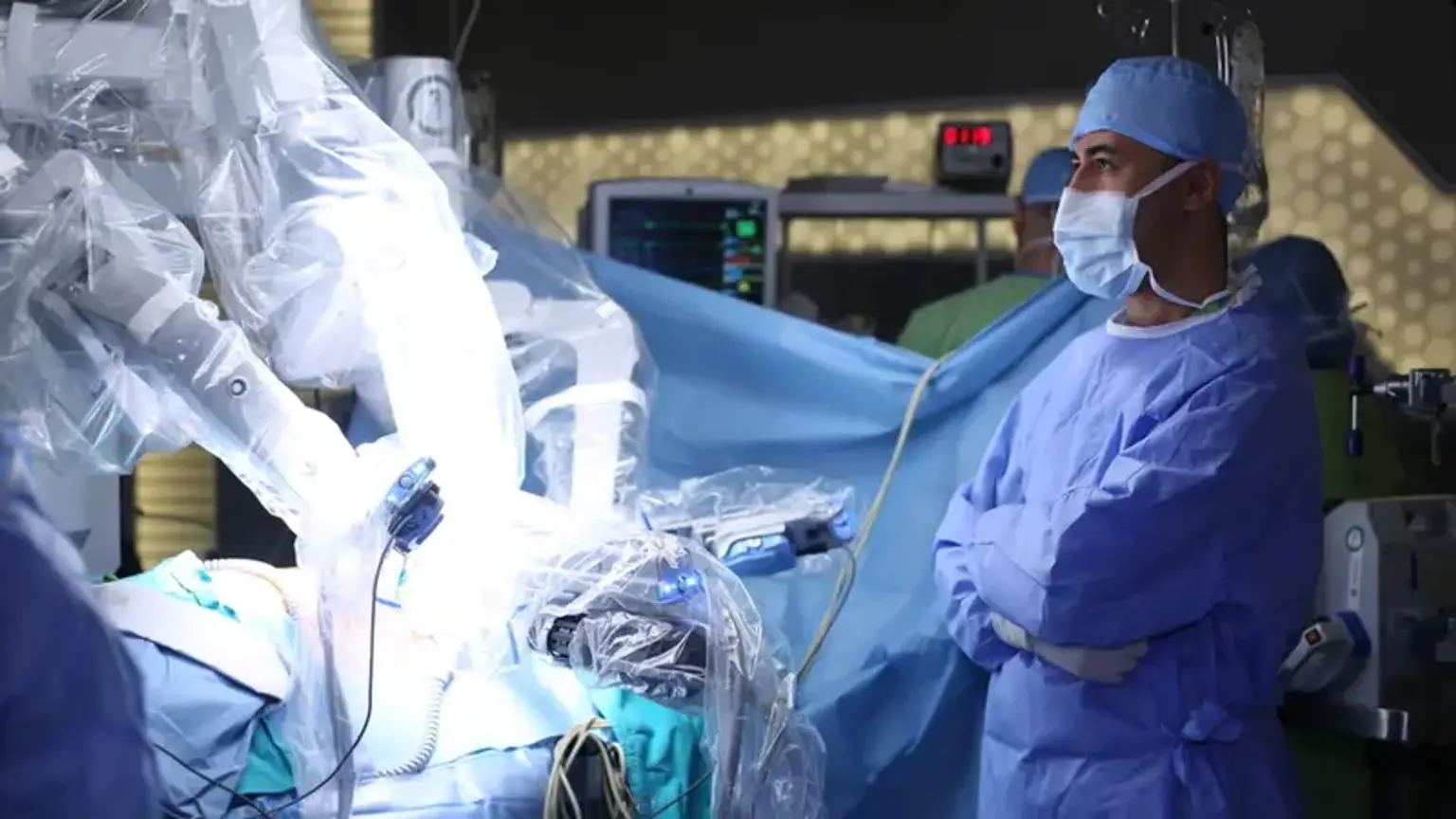
3. Robotic surgery:
At the time, there are just few surgeons that do robotic thyroid surgery, and their experience is limited. This form of thyroid surgery has been stopped in the United States due to safety concerns, although it is still common in Asia.
When Transaxillary Robotic-assisted Thyroid Surgery is Indicated?
Thyroid nodules, hyperthyroidism, obstructive or substernal goiter, differentiated (papillary or follicular) thyroid cancer, medullary thyroid cancer (MTC), anaplastic thyroid cancer, primary thyroid lymphoma (surgery is limited to obtaining tissue biopsy), and metastases to the thyroid from extrathyroidal primary cancer can all be treated with thyroidectomy (most commonly renal cell and lung cancer).
Thyroid nodules are a global phenomena that affects 1% of men and 5% of women clinically. The vast majority of nodules are benign, with about 5% containing malignancy. Thyroid nodules can be found by high-resolution ultrasonography in up to 68% of randomly selected people who have a screening ultrasound, with a preference for women and the elderly.
A goiter is an abnormal development of the thyroid gland that can be diffuse or nodular. Goiter is associated with iodine deficiency and is so endemic in iodine-deficient parts of the world. A goiter can be identified in around a quarter of the population in asymptomatic, iodine-deficient individuals, with an increasing prevalence in elderly populations. The large bulk, however, will not become surgical candidates or acquire thyroid nodules that need surgery.
Goiter can also arise in areas where there is no significant iodine deficiency. Goiter is often multinodular in certain areas and may be due to thyroid autoimmune disorders such as Hashimoto's thyroiditis or Graves' disease.
Thyroidectomy may be justified in both malignant and benign diseases with high selectivity. Thyroid cancer, toxic multinodular goiter, toxic adenomas, goiter with compressive symptoms, Graves disease that is either not responding to medical therapy or for whom medical management is not recommended, such as those seeking to become pregnant, are examples of indications.
The majority of thyroid nodules do not require removal. Fine needle aspiration (FNA) is frequently used to differentiate between benign and malignant nodules in nodules that are at high risk of cancer. When nodules are larger than 1 cm in size, non-functional (referred to as a "cold" nodule), and/or exhibit worrying ultrasonography results, they often fulfill the requirements for biopsy. Several associations have published treatment strategies for thyroid nodules. The American Thyroid Association recommends two parameters: nodule size and sonographic pattern.
Advantages of Transaxillary Robotic-assisted Thyroid Surgery
The most obvious advantage of Robotic-assisted Thyroid Surgery (RATS) over traditional cervical thyroidectomy is that no cervical incision is required. This cosmetic element makes RATS appealing, particularly to young female patients and those who have a proclivity for keloid development.
Over the open and endoscopic procedures, the RATS offers significant technological benefits. First, the robotic system provides three-dimensional magnified visualization, making it easier to identify the RLN and parathyroid glands than the cervical approach; second, it eliminates the natural surgeon tremor; and third, it enables a wider range of motion through the robot's EndoWrist and arm articulations.
All of this resulted in low complication rates, as well as good cancer control and functional outcomes. Furthermore, as compared to open or endoscopic surgery, the better visibility and operative ergonomics result in less musculoskeletal pain for the surgeon.
RATS was reported to improve patient outcomes by reducing pain and increasing aesthetic satisfaction, as well as lowering rates of paresthesia, postoperative voice alteration, and swallowing difficulty.
Disadvantages of Transaxillary Robotic-assisted Thyroid Surgery
RATS, on the other hand, introduces possible additional issues such as tracheal and esophageal damage due to the novel approach to the surrounding anatomy and the lack of tactile sense. Only a few studies took such difficulties into consideration, and then only in a minimal degree, with no need to convert to open thyroidectomy (OT). Furthermore, there is a danger of brachial plexus neuropathy due to the ipsilateral arm posture. This danger can be avoided by flexing the arm overhead 90 degrees, which reduces the possibility of nerve stretching.
Intra-operative monitoring of the ulnar, radial, and median nerves may further reduce the possibility of brachial plexus injury, by identification of any impending damage to these nerves and enabling the patient to be repositioned.
Another disadvantage of RATS is the longer operative time due to the creation of the working space and the robot docking. However, several studies have examined the learning curves of the RATS and have shown that increased experience led to decreased total operative time. RATS involves a steep learning curve, compared to the conventional approach. However, it has been demonstrated that compared to the endoscopic approach which requires 55-60 procedures, the RATS required only 35-40 procedures.
In terms of cost, the RATS is a more expensive procedure compared to the OT, due to the cost of the equipment and the longer operative time. However, some studies have pointed out that RT eliminated the need for an additional surgical assistant, and, combined with the potentially shorter hospital stay and the expected decrease in the maintenance cost of the robot, this may eventually result in an equally cost-effective procedure.
How Transaxillary Robotic-assisted Thyroid Surgery is Performed?
Preparation and positioning
Under general anesthesia, the patient's axilla is exposed by bringing the arm over the head and flexing the elbow 90° to 100°. To reduce the danger of brachial plexus damage, the arm should rest naturally across the forehead. The head is rotated slightly to the contralateral side of the incision, and the neck is extended. Anatomical markers such as the sternal notch, cricoid and thyroid cartilage, mandible angle, and sternocleidomastoid (SCM) muscle are traced on the patient's skin.
The patient's axilla is exposed while under general anesthesia by raising the arm over the head and bending the elbow 90° to 100°. The arm should naturally rest across the forehead to avoid the risk of brachial plexus injuries. The head is slightly moved to the other side of the incision, and the neck is lengthened. On the patient's skin, anatomical markers such as the sternal notch, cricoid and thyroid cartilage, mandible angle, and sternocleidomastoid (SCM) muscle are traced.
The procedure is best performed using a headlamp and surgical loupes, especially to aid precise hemostasis of perforating arteries during muscle dissection. The da Vinci robot endoscope may also be used to increase visibility and for teaching but requires a second assistant to manipulate it. The view from the endoscope may also help the assistant holding the retractors to obtain the best exposure possible by giving direct feedback.
Incision and approach to neck midline
A 4- to 6-cm skin incision is performed immediately posterior to the anterior axillary fold, following the axillary hairline (thus the scar is hidden in the axilla), and the lateral border of the pectoralis major is exposed. Using monopolar electrocautery, dissection is performed above the pectoral fascia toward the clavicle and the SCM. A thin layer of fat tissue may be left on the pectoral muscle fascia so as not to expose perforating arteries, which can retract into the muscle and cause delayed bleeding.
The surgical path is expanded superiorly and inferiorly before opening the SCM muscle to optimize exposure and visibility. By following the lateral boundary of the SCM's clavicular head, the dissection should remain on a subplatysmal plane above the clavicle. Starting at the SCM, the dissection should be carried out laterally until two fingers can be comfortably inserted in the workspace. The skin and muscle of the clavicle are extremely thin, and the external jugular vein is superficial directly under the platysma.
Neck midline and exposure of thyroid gland
Once the sternal and clavicular heads of the SCM have been identified, the neck midline is approached by dissecting between both branches. Electrocautery dissection should remain close to the sternal head of the SCM to prevent any injury of the internal jugular vein (IJV), which lies at the same depth and is immediately medial to the clavicular head.
The sternal head is elevated, exposing the strap muscles. The omohyoid muscle is an important landmark that must be identified, as it is immediately superior to the IJV and usually indicates the upper pole of the thyroid gland. As with the SCM, dissection must remain close to the omohyoid to prevent IJV injury. The omohyoid muscle can be sectioned if a larger workspace is required (lymph node dissection or large gland).
The strap muscles are elevated as the dissection progresses, exposing the thyroid gland. Again, dissection must be careful, and the monopolar electrocautery should remain in direct contact with the inferior border of the strap muscles to keep a safe distance from IJV. In case of IJV injury, direct ligature or vascular clips (3 or 5 mm) may be used.
Dissection must be pursued until the contralateral omohyoid muscle is reached. Thus, the da Vinci Kuppersmith retractor can be put into place, elevating the skin and creating a workspace sufficient for robotic dissection of both thyroid lobes.
Closure After Robotic Surgery
It is of paramount importance to check the entire workspace for bleeding, especially from the muscle tissue, to prevent hematoma. This is especially true during the first cases of TARS for inexperienced surgeons, when hemostasis may have been undermined during the initial approach. Careful inspection of the muscles under the retractor is necessary as injury can be sustained during the robotic sequence.
Ropivacaine is injected in the wound to facilitate postoperative pain management. No drain is required (no risk of hematoma airway compression as any bleeding seeps into the transaxillary surgical workspace).
Wound closure is done as follows: Vicryl 3-0 suture of the muscle at the level of the clavicle limits dead space in the wound. Skin closure combines Vicryl 3-0 interrupted deep suture and Monocryl 3-0 subcutaneous continuous suture.
Transaxillary Robotic-assisted Thyroid Surgery Risks
Thyroidectomy is normally quite safe when performed by a well trained and experienced surgeon.
Complications are infrequent, however the following are the most significant hazards of thyroidectomy:
- Bleeding after surgery that could lead to acute respiratory distress.
- Injury to a recurrent laryngeal nerve, which can result in temporary or permanent hoarseness as well as acute respiratory difficulty if both nerves are affected.
- Parathyroid gland damage, which is located behind your thyroid. By secreting parathyroid hormone, these glands regulate the calcium levels in your blood. If they are injured during surgery, it can result in either temporary or chronic hypoparathyroidism and hypocalcemia (low levels of calcium in your blood).
- While these complications are rare, they’re more likely to happen if: You have an invasive tumor and/or the cancer has spread to nearby lymph nodes in your neck.
- You’re undergoing a second thyroid surgery.
- You have a large goiter that goes below your collarbone into the top of your chest (substernal goiter).
If you’re concerned about possible complications of your surgery, talk to your surgeon.
What is the Recovery Time For Robotic-assisted Thyroid Surgery?
After a few hours of monitoring in the hospital, many patients who undergo a thyroidectomy, particularly a hemithyroidectomy, are able to go home the same day. Some patients must spend the night in the hospital before being released the next morning.
Before you leave, your physician will teach you on how to care for your incision and stitches, as well as what complications and symptoms to watch for.
You should be able to return to work in two to three weeks.
You’ll likely be able to go back to your normal, light activities the first day after your thyroidectomy. Your surgeon will likely recommend that you limit more intense physical activities for a week or two. This is mainly to reduce the risk of a neck hematoma (blood clot) and breaking open your stitches.
You should wait at least 10 days to two weeks before returning to vigorous sports and activities, such as swimming and heavy lifting.
When Should I Call My Healthcare Provider?
It is critical to contact your healthcare practitioner if you have any of the following symptoms or conditions following your thyroidectomy:
- Swelling at the incision site.
- Bleeding at the incision site.
- Redness or warmth at the incision site.
- Fever of 101 degrees or higher.
- Numbness or tingling in your face, hands or lips.
Conclusion
Thyroidectomy through transaxillary robotic surgery (TARS) was created in 2007 with the da Vinci robot. It is utilized in many populations as a safe and efficient alternative to traditional cervicotomy to remove everything from minor thyroid nodules to Graves' disease goiters and cervical lymph nodes. The technique's advantages include cosmetics, a high-definition view of structures, and a lower risk of compressive cervical hematoma (due to the vast surgical space); the primary disadvantages are the expense and time of the treatment.
Another significant barrier is the learning curve; as robotic surgery necessitates the acquisition of new skills; training is necessary before the first operation can be conducted. Furthermore, because it is unusual for head and neck surgeons to reach the midline of the neck laterally, the transaxillary method has its own learning curve.