Transcranial Doppler (TCD) Ultrasound

Transcranial Doppler (TCD)

Overview

TCD ultrasonography is a painless test that uses sound waves to detect medical issues that alter blood flow in your brain. It can identify strokes caused by blood clots, restricted blood channel sections, vasospasm produced by a subarachnoid hemorrhage, micro blood clots, and other conditions. Discover how the operation is carried out.
 

What is transcranial doppler (TCD) ultrasound?

TCD ultrasound

TCD ultrasonography is a non-invasive examination that uses sound waves to monitor blood flow in your brain. This test has been prescribed by your doctor to diagnose a medical problem that affects blood flow to and within the brain. The test is also used to track the effectiveness of some therapies, such as the dissolution of clots inside brain arteries. Sound waves are delivered through the tissues of your skull during TCD. These sound waves reflect off blood cells as they move through your blood arteries, allowing the radiologist or neurologist to determine their speed and direction. The sound waves are captured and shown on a computer screen. Transcranial Doppler (TCD) ultrasonography is a noninvasive, real-time evaluation of blood flow parameters and cerebrovascular hemodynamics inside the brain's basal arteries. The physiologic data received from these measures complement the structural data collected from the many techniques of vascular imaging now accessible. TCD is the most practical technique to evaluate vascular changes in response to therapies at the bedside during acute cerebrovascular episodes. Given the simplicity of use of this instrument as a diagnostic technique, clinical and scientific applications in the numerous illnesses of the cerebral artery will continue to grow.

Transcranial Doppler (TCD) ultrasonography monitors cerebrovascular function in real time and is noninvasive. TCD may be used to analyze relative flow changes in the brain's basal arteries, identify localized vascular stenosis, and detect embolic signals inside these arteries. TCD may also be used to examine the physiologic health of a specific vascular region by evaluating blood flow responses to changes in blood pressure (cerebral autoregulation), end-tidal CO2 fluctuations (cerebral vasoreactivity), or cognitive and motor stimulation (neurovascular coupling or functional hyperemia). TCD has shown useful in the clinical diagnosis of a variety of cerebrovascular illnesses, including acute ischemic stroke, vasospasm, subarachnoid hemorrhage, sickle cell disease, and brain death.


What medical problems may transcranial doppler ultrasound diagnose or monitor?

transcranial doppler ultrasound

Transcranial Doppler ultrasonography is used to diagnose or monitor a variety of blood flow-related medical problems, including:

  • Acute ischemic stroke occurs when a blood clot forms in a blood artery in the brain, shutting off oxygen-rich blood supply to the brain. 

TCD is especially effective in acute ischemic stroke, when repeated TCD tests may be utilized to assess the progression of an arterial blockage before to and after thrombolysis.  TCD has high sensitivity, specificity, and positive and negative predictive values for acute MCA occlusions.  TCD may identify the blockage in the ICA siphon, vertebral, and basilar arteries with acceptable sensitivity and positive predictive value, as well as excellent specificity and negative predictive value 

  • Vasospasm is the construction of a blood vessel portion caused by contraction. It's a response to a brain bleed in this case - a subarachnoid hemorrhage/brain aneurysm rupture.

Two-thirds of patients with aneurysmal SAH experience angiographic cerebral vasospasm (VSP), with half becoming symptomatic. Although anatomic and technical considerations reduce the link for the ICA and ACA, there is a substantial direct correlation between VSP severity after SAH and flow velocities in most cerebral arteries.

 TCD sensitivity and specificity for the detection of VSP following SAH in the proximal parts of each intracranial artery have previously been described. TCD detects proximal vs distal VSP substantially more sensitively.

  • Stenosis in brain arteries: A narrowing or blockage of an artery, most usually caused by atherosclerosis (hardening of the arteries).
  • Cerebral microemboli are minute blood clots that circulate in the circulation and induce a transient ischemic event (mini stroke).

TCD is the first medical instrument that can detect both solid and gaseous circulating cerebral microemboli in real time. The detection of TCD microemboli is based on the backscatter of ultrasound waves from emboli, which results in high-intensity transient signals (HITS) or embolic signals in the Doppler spectrum as they transit through the insonated vascular.

  • Stroke risk in sickle cell anemia adults and children: In sickle cell anemia, the altered shape of blood cells can result in blood clots and blocked blood arteries, increasing the risk of stroke.

Chronic hemolysis causes decreased hemoglobin concentration in children with sickle cell disease (SCD). Angiogenesis and neovascularization are triggered by chronic anemia and hypoxia. Furthermore, the interaction of sickled red cells with endothelial cells induces inflammation and cerebral stenosis. These youngsters are predisposed to both ischemic and hemorrhagic infarcts due to their impaired vascular system.

  • Detection of patent foramen ovale/right-to-left shunt: A solution is injected into a vein in the forearm during this examination. The appearance of bubbles in the brain arteries shows that blood is flowing backwards due to a breach in the wall between the heart's two upper chambers (called a patent foramen ovale [PTO]). A PTO can cause a stroke in youngsters.
  • Flow of Collateral: Understanding the collateral flow patterns of the brain's basal arteries has important therapeutic implications in the care of individuals with the cerebrovascular atherothrombotic illness. A number of clinical investigations have shown that the degree of collateral flow is associated with infarct magnitude and clinical prognosis in ischemic stroke patients. TCD can be utilized to give real-time information on blood flow direction and velocity in known cerebral collateral channels that become active in acute and/or chronic steno-occlusive cerebrovascular disorders.
  • Circulatory Arrest in the Brain: A drop in cerebral perfusion pressure, along with a rise in ICP and PI, causes intracranial artery compression and a halt of supply to the brain, resulting in cerebral circulatory arrest (CCA). TCD can visualize and continually monitor the pattern of cerebral blood flow that leads to CCA and brain death.

What are the future implications of transcranial Doppler ultrasound?

transcranial Doppler ultrasound implications

TCD Intraoperative Monitoring 

Noninvasively monitoring cerebral blood flow velocity during surgery can also offer real-time information on changes in velocity or the emergence of microemboli, which can be rectified quickly to prevent intraoperative cerebral ischemia damage. Several prior investigations looked into cerebrovascular hemodynamics after coronary artery bypass graft and carotid endarterectomy.

TCD in Alzheimer's Disease 

TCD might be useful in dementia studies as well. One recent study found that individuals with Alzheimer's disease and vascular dementia had decreased cerebral blood flow velocity and greater PI when compared to healthy age-matched controls, confirming the link between dementia and hemodynamic abnormalities. Another recent study has linked microemboli to rapid cognitive deterioration in dementia patients. The use of TCD in cognitive studies is fast growing.

TBI (Traumatic Brain Injury)

TBI is one of the top causes of mortality and disability in the United States. Cerebral vascular damage and hemodynamic impairment are major contributors to poor prognosis in TBI patients. We now have a poor understanding of the processes behind cerebrovascular injury in TBI. One putative cause is disruptions in cerebral hemodynamics, with an early period of cerebral hypoperfusion followed by hyperemia and subsequent rises in ICP. 


What are the many types of transcranial Doppler devices?

TCD equipment

TCD equipment is now available in two varieties:

  1. non-duplex (nonimaging) 
  2. duplex (imaging).

The arteries are recognized "blindly" in non-duplex devices based on the auditory Doppler shift and the spectral display. Standard criteria are used to identify specific vessels, which include the cranial window used, probe orientation, depth of sample volume, direction of blood flow, relationship to the terminal internal carotid artery, and response to various maneuvers such as common carotid artery compression and eye-opening and closing.

B-mode transcranial color-coded duplex (TCCD) imaging combines pulsed wave Doppler ultrasonography with a cross-sectional picture of the region of insonation, allowing the detection of arteries in relation to various anatomic sites. While recording blood flow velocities, the color-coded Doppler also indicates the flow direction in respect to the probe (transducer). To reduce the Doppler shift measurement error, the angle of insonation in TCD is considered to be 30 degrees (as near to zero as feasible). The angle of insonation, on the other hand, may be measured and utilized to adjust the flow velocity measurement in TCCD. More recently, a more advanced technique known as power motion-mode TCD (PMD/TCD) has been available, which gives multi-gate flow information in the power M-mode display at the same time. It displays flow signals using numerous overlapping sample volumes. PMD/TCD appears to improve TCD processing by enabling temporal window location and incident signal alignment to allow cerebral blood flow velocity measurements via numerous arteries.

Although these imaging TCD modalities significantly improve TCD reliability with better insonation angle correction, clinical applications of the more recent imaging modalities are still in development, and the majority of currently used clinical applications have been best developed using the non-duplex mode of TCD. As a result, the investigation and clinical uses of the non-duplex TCD will be the focus of this study. Duplex TCD modalities will likely replace nonduplex TCD in clinical practice over time, not simply expanding TCD's therapeutic value.  


What happens during a doppler transcranial ultrasound?

Ultrasound process

How to Get Ready for the Exam?

This test requires no extra preparation. You are not required to wear a medical gown.

Keep in mind to:

  • If you wear contact lenses, remove them before the test.
  • Keep your eyes closed while the gel is being applied to your eyelids to avoid getting it in your eyes.

How the Examination Will Feel?

On your skin, the gel may feel chilly. As the transducer moves around your head and neck, you may feel some pressure. The pressure should not be painful. A "whooshing" sound may also be heard. This is typical.

How the Test is Performed?

  • It is not essential to remove jewelry or change into a hospital gown.
  • A board-certified radiologist or neurologist interprets your ultrasound test, which is conducted by properly trained technicians.
  • During the examination, you will either recline on a cushioned examining table or sit in a chair.
  • On the skin over the region to be inspected, a little quantity of water-soluble gel is placed. Typically, the gel is applied to the back of the neck, above the cheekbone, in front of the ear, or over the eyelid. These are the blood artery sites that feed the brain with oxygen and nutrients. The gel is safe for your skin and will not stain your clothes.
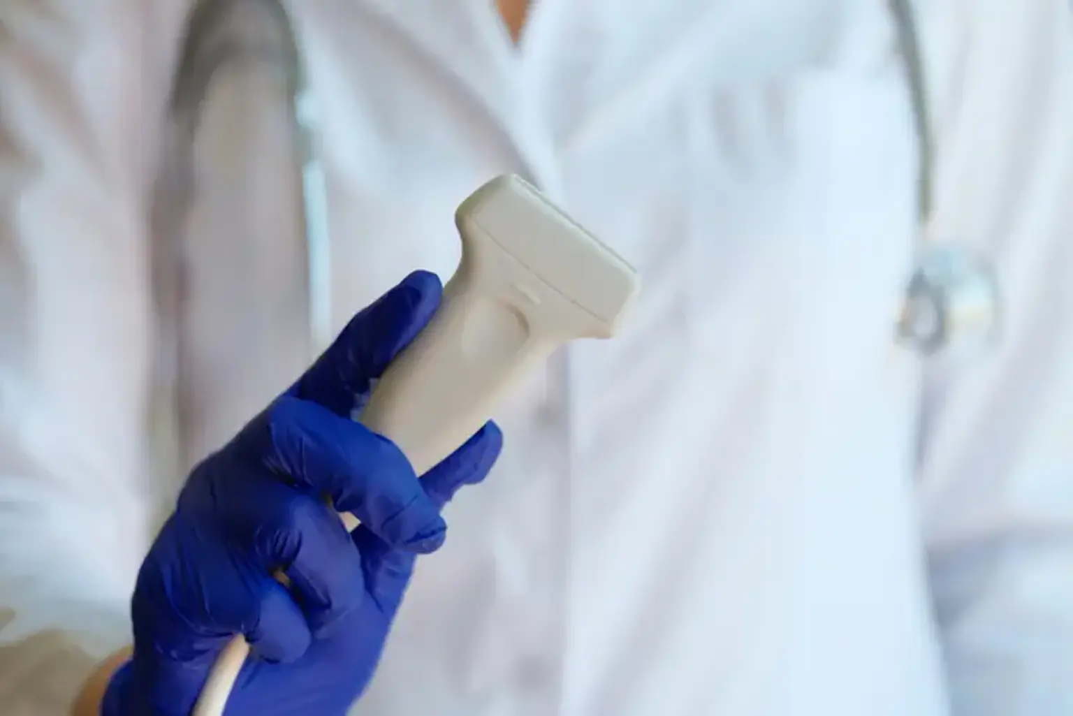
  • A transducer, a tiny microphone-like device, is kept in position on the test region. The transducer transmits high-frequency sound waves into the brain and records the resulting blood flow data. The ultrasound signal is converted into graphs or color images that are displayed on the screen.
  • During the exam, there is hardly any pain. As the transducer is put against your skin, you may feel some small pressure.
  • During the exam, you must keep your head steady and refrain from speaking.
  • The ultrasound procedure takes between 30 and 60 minutes to complete.
  • The gel will be wiped from your skin after the test. 


What are the limitations of doppler transcranial ultrasound? 

TCD limitations

TCD has two key drawbacks that prevent it from becoming more widely used. The portable approach necessitates a thorough three-dimensional understanding of cerebrovascular architecture and its variations, making it extremely operator-dependent. 

TCD utilization is further impeded by the 10 to 15% prevalence of insufficient acoustic windows among Blacks, Asians, and elderly women. This might be due to bone thickness and porosity around the acoustic windows, as well as attenuation of ultrasound energy transfer. TCD measures are likewise restricted to the main basal arteries and can only offer a global rather than local indication of cerebral blood flow velocity.


How will the findings of my transcranial Doppler ultrasonography be sent to me?

Ultrasonography

A radiologist or neurologist will review the photos and submit a report to the doctor who ordered the test. Your doctor will go through the findings of the tests with you. A follow-up test may be required to get further information or to monitor a specific medical condition for which the test was ordered, or to determine whether therapy was effective.


Are there any risks associated with the transcranial Doppler ultrasound test?

Risk of TDU

The use of transcranial doppler ultrasonography is not dangerous. There are no negative side effects. Unlike X-ray testing, ultrasound does not utilize radiation.


Conclusion 

TCD is a low-cost yet important technique that may be used in conjunction with a battery of other tests to help diagnose a variety of cerebrovascular illnesses such as acute ischemic stroke, vasospasm, traumatic brain injury, and cerebral microembolization. TCD can also be used to identify collateral flow and manage cerebrovascular atherosclerosis. Children with SCD who are at risk of having a stroke can be evaluated for TCD and treated with blood transfusions. TCD can also be used to confirm brain death. TCD is also commonly utilized in research settings to investigate cerebral autoregulation, vasoreactivity to CO2, and neurovascular coupling in both healthy and ill populations. Greater knowledge of these physiologic processes may lead to new therapeutic targets in conditions such as acute ischemic stroke, vasospasm, TBI, and dementia, where we have the most restricted therapeutic therapies.