Brain Endoscopic Surgery
Overview
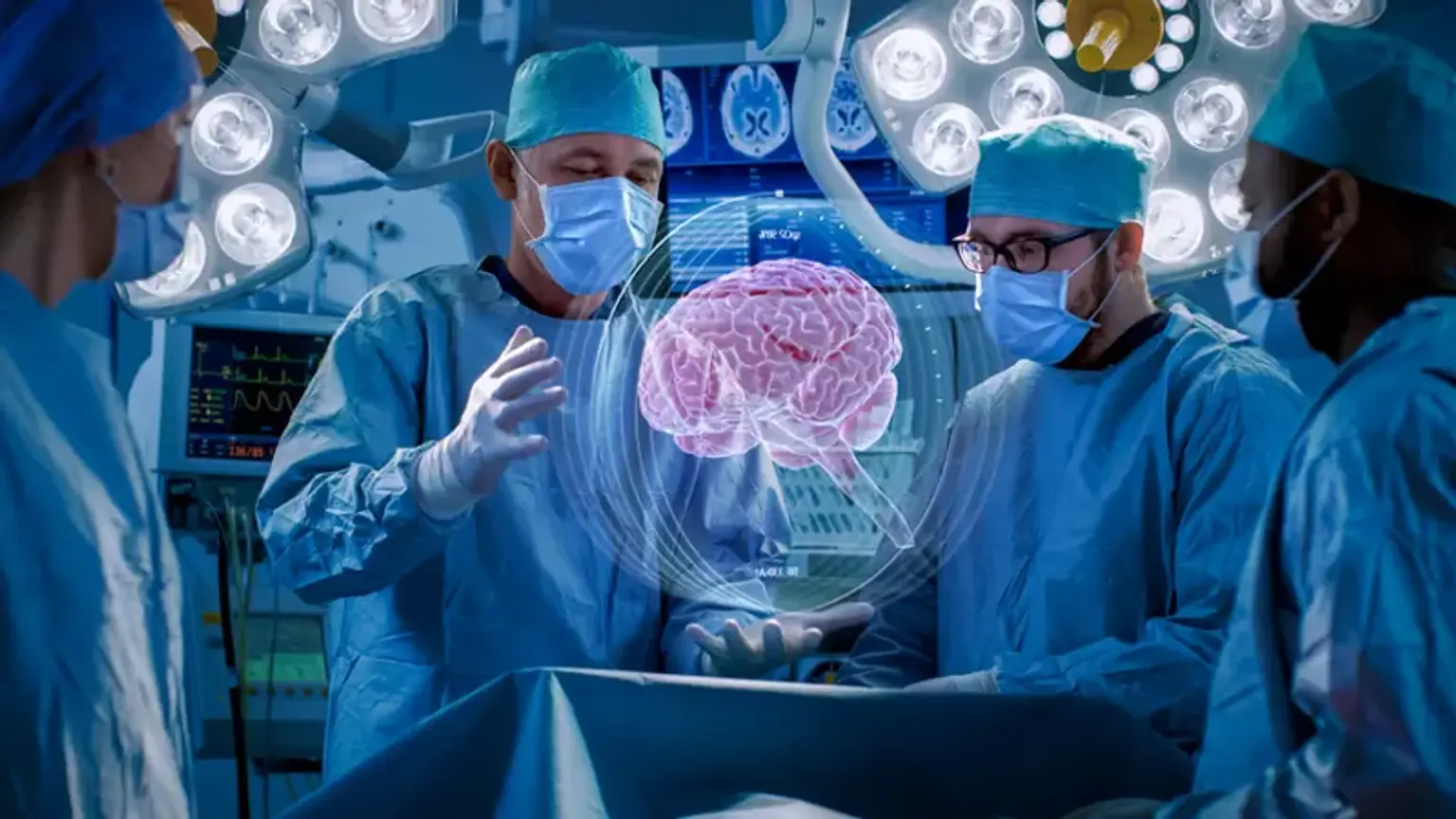
Endoscopic brain surgery is a type of minimally invasive brain surgery that can cure a variety of neurological problems. During this sort of brain surgery, tiny tubes with a camera on the end, similar to a telescope, are introduced into the brain through small incision marks (either in the skull or other regions of the body) and used to produce pictures of the damaged area. The pictures from the endoscope are used as a guide to assist the surgeon in determining the location of the tumor and the precise region that has to be removed. Specialist surgical equipment is used to remove the afflicted regions.
What is Brain Surgery?
Tumors, blood clots, aneurysms, epilepsy, and Parkinson's disease are among the disorders treated by brain surgery. It is conducted by a neurosurgeon with the assistance of a medical anesthesiologist who is well-versed in the forms of anesthetic, monitoring, and post-operative care necessary for these delicate procedures. This medical anesthesiologist may be a neuroanesthesiologist with extensive clinical training in anesthesia for brain and spinal procedures in some situations.
Some examples of brain surgery?
These are some of the most common procedures that involve the brain or regions around it.
- Craniotomy. A section of the skull is removed to provide surgeons access to the brain in order to remove a brain tumor, abnormal tissue, blood, or blood clots, alleviate pressure after an accident or stroke, repair a brain aneurysm or skull fractures, or treat other brain disorders. Following surgery, the portion of the skull is reinserted.
- Biopsy. Biopsies are usually performed when imaging has revealed a possible problem. The surgeon creates an incision in the skull or inserts a needle to extract brain cells or tissue for pathology evaluation.
- Deep neural stimulation (DBS). An implantable pulse generator, a battery-powered medical device, is implanted to administer electrical stimulation to particular parts of the brain. DBS is most typically used to treat movement disorders such as Parkinson's.
- Neuroendoscopy. A narrow tube called an endoscope is threaded via the mouth, nose, or small incisions in the skull to access or remove brain tissue in this minimally invasive method. On the end of the endoscope is a light and a camera, and the operation is conducted using instruments inserted through the endoscope. One kind of neuroendoscopy is endoscopic transsphenoidal surgery (also known as endoscopic pituitary surgery). It entails inserting an endoscope via the nose in order to remove brain tumors and lesions near the pituitary gland, which is located directly beneath the bridge of the nose.
- Decompression of the posterior fossa. This therapy includes cerebellar and brainstem areas of the brain. To cure a Chiari malformation, for example, the surgeon makes an incision at the back of the patient's head and removes a tiny part of the bone near the base of the skull. This removal gives the cerebellum more room and relieves strain on the spinal cord.
- Thrombectomy and repair of a cerebral aneurysm. The surgeon directs surgical devices such as a catheter or small metal wires via a big blood artery in the patient's groin to reach the brain vessels, using contrast dye to locate the troublesome blood vessel without having to open up the patient's skull. Patients with a blood clot in a brain artery, a cerebral aneurysm (a weakened and bulging region in an arterial wall), or a burst aneurysm that causes bleeding into the brain are the most common candidates for the treatment.
Some patients with brain cancer are treated with stereotactic radiosurgery, which may include the use of a Gamma Knife, however this is not surgery in the traditional sense of the word, and the "knife" is not truly a knife. Radiosurgery is a type of external radiation therapy that does not need an incision. Specialized equipment, such as the Gamma Knife brand, carefully administers a high dosage of radiation that targets tumors or other lesions while causing minimal harm to surrounding healthy tissue.
What is Endoscopy?
Endoscopy is the method of seeing into a bodily cavity through a tiny hole using a tube containing a lens and a light source. An endoscope is a little camera that examines the pathology or disease. The endoscope is equipped with a light source and is linked to a monitor. The procedure is carried out while gazing at a screen, and the real target size is amplified more than a hundred times. This method corresponds to the transition from open abdominal surgery to laparoscopic surgery. The goal is to get at the disease or problem while causing as little harm to the surrounding tissues as possible. In neurosurgery, this method can be utilized to treat both brain and spine illness.
Types of Brain Surgery
There are several forms of brain surgery. Your surgeon's choice of brain surgery may be influenced by a number of factors, including the nature and location of the problem being treated.
- Open brain surgery, also known as classical brain surgery, involves making one or more incisions in the scalp in the area that has to be treated. To obtain access to the brain, the surgeon makes a hole in the skull known as a bone flap. Additional treatments, such as the excision of a brain tumor or the clipping of a cerebral aneurysm, are used to address the specific ailment.
- Minimally invasive brain surgery employs cutting-edge technologies that can reduce the size and number of incisions while boosting precision and accuracy. When compared to traditional open operations, this usually results in less discomfort for the patient, shorter hospital stays, and faster recovery times.
Minimally invasive brain surgery may be used to treat conditions such as aneurysms, brain tumors, hydrocephalus and pituitary tumors.
- Endoscopic brain surgery (includes endonasal and neuroendoscopy) is a form of minimally invasive therapy that employs an endoscope, a narrow instrument outfitted with a light and a camera. The endoscope allows the surgeon to see the region being treated as well as get access to the brain through the nose or tiny holes in the skull.
- The Neurosurgery Imaging is used to help with minimally invasive brain surgery. Advanced surgical planning software, tool tracking technology, and intra-operative imaging are all part of the system. The device enables the surgeon to make data-driven decisions while accurately tracking their surgical tools in respect to the anatomy of the patient.
What is Endoscopic brain Surgery?
Endoscopes, which are small, flexible, illuminated tubes, are used in minimally invasive neurosurgery to see various areas of the brain, skull base, or spinal cord through small incisions. Endoscopes function as miniature microscopes, magnifying essential anatomical structures so that the surgeon can examine the numerous sick parts that need to be repaired, removed, or replaced. Endoscopic neurosurgery is referred to as minimally invasive neurosurgery because the use of endoscopes is substantially less intrusive into these anatomical structures than conventional surgery.
Endoscopic neurosurgery, for example, does not require big incisions on the head or skull, nor does it necessitate considerable dissection of brain tissue. As a result, the operation is faster and easier, with fewer post-operative problems, faster recovery, and less pain and scarring. The issue is removed or repaired as efficiently, safely, and completely as with traditional "open" surgery.
Recent technological advances in endoscope design have resulted in equipment that is smaller and more efficient than previous generations, with higher resolution and brighter illumination. Endoscopic neurosurgery has dramatically eased the therapy of numerous intracranial (within the brain) diseases in both adults and children, reducing surgical complications, shortening hospital stays, and minimizing aesthetic problems associated with many important neurosurgical disorders.
- Intracranial Surgery:
Endoscopes are inserted into the brain through extremely small skull holes in intracranial surgery, and tiny tools are then implanted through the same aperture to conduct endoscopic tumor biopsy, colloid cyst excision, and endoscopic cyst fenestration, as well as to treat hydrocephalus. Patients are usually discharged from the hospital the next day after operations that last between 15 minutes and an hour.
What are the Benefits of Endoscopic Brain Surgery?
This form of surgery is less intrusive than other open surgical treatments, allowing the patient to recover faster and with less discomfort. While each patient's medical history, age, condition, and other factors influence the success of endoscopic brain surgery, the following are some of the most typical advantages of the procedure:
- Minimally invasive (results in less pain for the patient)
- Faster recovery time than open brain surgery
- Reduced risk of brain trauma
- Reduced risk of side effects
- Reduced hospital stay
What happens before surgery?
Before surgery, you will meet with a neurosurgeon, an ENT surgeon, and an endocrinologist. If you have visual difficulties, you may need to see an ophthalmologist. The surgeon will describe the operation, its risks and advantages, and answer any questions during the office visit. Following that, you will sign consent documents and fill out paperwork to provide the surgeon with information about your medical history (i.e., allergies, medicines, bleeding history, anesthesia reactions, previous surgeries).
Discuss with your health care provider all drugs you are taking (prescription, over-the-counter, and herbal supplements). Some drugs must be continued or discontinued on the day of surgery. Preoperative testing (e.g., blood tests, electrocardiograms) may be required many days before surgery. Consult your primary care physician about discontinuing certain drugs and ensuring you are surgically cleared.
7 days before surgery, discontinue all nonsteroidal anti-inflammatory medications (ibuprofen, naproxen, etc.) and blood thinners (Coumadin, Plavix, aspirin, etc.). To avoid bleeding and healing issues, stop using nicotine and drinking alcohol one week before and two weeks after surgery. Before surgery, you may be requested to cleanse your skin with Hibiclens (CHG) or Dial soap. It eliminates microorganisms and lowers the risk of surgery site infections. (Do not get CHG in your eyes, ears, nose, or genital regions.)
Morning of surgery
- No food, drink, gum or candy.
- Use antibacterial soap to shower. Dress in clean, loose-fitting attire.
- Wear closed-back flat-heeled shoes.
- If you've been told to take your normal medicine with small sips of water the morning before surgery, do so.
- Remove any make-up, hairpins, contacts, body piercings, nail paint, and other jewelry.
- All valuables and jewelry should be left at home (including wedding bands).
- Bring a list of medications, including dosages and times of day they are generally taken.
- Bring a list of any drug or food sensitivities.
The day of operation, patients are admitted to the hospital. The nurse will go through the preoperative procedure with you and answer any questions you may have. An anesthesiologist will meet with you to discuss the benefits and dangers of anesthesia.
How is it done?
We seek to reach pathologies such as a tumor through natural apertures such as a nostril or tiny incisions with minimum or no brain retraction in minimally invasive endoscopic brain surgery. An endoscope is placed via this incision to visualize and execute the neurosurgical procedure. The procedure is carried out while a monitor displays enlarged real-time footage from the endoscope. The remainder of the procedure is performed using comparable approaches to open surgery, but necessitates the use of specific equipment and allows for less retraction and risk of brain damage. The same concepts apply to spine procedures.
Endoscopic Surgery for Brain Tumors
Traditionally, anterior skull base and brain cancers were removed through the bony skull, necessitating major skull and facial incisions, bone flaps, and brain retraction to access and remove these tumors through a maze of blood arteries and nerves.
A minimally invasive option is now available in which a team of surgeons removes malignant tumors endoscopically through the patient's nose, a natural entrance, avoiding face or skull incisions associated with major surgery. First, an otolaryngologist uses an endoscope, a narrow, illuminated tube with a small video camera on the end, to build a surgical track to the skull base. The otolaryngologist next performs the delicate technique of delicately extracting and removing the tumor through the nose in collaboration with a neurosurgeon.
The endoscope enables doctors to remove the tumor while preserving normal tissues. The surgeons next repair the skull base to close the gap between the brain and the nose and give full closure. Most patients experience fewer issues, have no visible scars, and recover far more quickly.
This novel endoscopic surgical method is most typically employed in individuals with benign tumors like pituitary adenomas. However, surgeons are beginning to utilize it to remove different types of brain tumors. This innovative method may be used to treat the following types of lesions:
- Pituitary tumors
- Meningiomas (tumors arising from the lining of the brain)
- Craniopharyngiomas (a type of tumor derived from pituitary gland tissue)
- Rathke's cleft cysts (benign cystic lesions that affect mainly the pituitary gland)
- Chordomas (rare, slow-growing malignant tumors at the base of the skull)
- Spinal fluid leaks/rhinorrhea
What is “awake” brain surgery?
Awake brain surgery is most commonly used to treat epileptic convulsions and Parkinson's disease, but it is also increasingly utilized to remove brain tumors near key brain processes. Maintaining consciousness allows you to respond to questions that can assist the surgeon in identifying parts of the brain impacting activities such as vision, movement, or speech. That information is used by the surgeon to precisely focus the therapy.
You will still get sedation and pain relief medication from your physician anesthesiologist, who will monitor your blood pressure, heart rate, and oxygenation and will always stay by your side during awake brain surgery. Furthermore, your surgeon may use a local anesthetic to numb your scalp.
You may not be entirely aware or cognizant throughout the operation. During surgery, the physician anesthesiologist can change medicine dosage to wake you just when required. Even so, you may not need to be brought to full consciousness in order to talk. Instead, you could be led to a point where you can answer by squeezing your hand or making another indication. Some patients have no recollection of being awake.
Preoperative consultations with your physician anesthesiologist can help alleviate any concerns you may have about this sort of surgery.
What is Transsphenoidal Pituitary Surgery?
Endoscopic surgery is used to remove tumors from the pituitary gland and skull base through the nose. In this minimally invasive surgery, the surgeon uses a small endoscopic camera and light to remove malignancies using long tools via the nostrils. Pituitary tumors can induce hormonal imbalances as well as eyesight loss. Tumor excision frequently improves eyesight and restores normal hormone balance.
The term "transsphenoidal" literally translates to "through the sphenoid sinus." It is a procedure that removes pituitary tumors through the nose and sphenoid sinus. Endoscopes, microscopes, or both can be used to do transsphenoidal surgery. Neurosurgeons and ear, nose, and throat (ENT) surgeons frequently collaborate.
A conventional microscopy approach involves making a skin incision beneath the upper lip and removing a big part of the nasal septum to allow the surgeon to examine the sphenoid sinus region directly.
Endoscopic endonasal surgery is a minimally invasive method that employs a tiny incision at the rear of the nasal cavity and produces little damage of the nasal tissues. The ENT surgeon uses a small camera and light called an endoscope to operate via the nostrils. To access the pituitary, bone holes are produced in the nasal septum, sphenoid sinus, and sella in both procedures. The neurosurgeon removes the tumor once the pituitary is revealed.
What Risks are Associated with Endoscopic Brain Surgery?
There are dangers associated with every procedure. Rebound surgeons educate their patients about the best treatment choices for their specific disease as well as the hazards involved. Possible hazards include:
- Temporary or permanent interference with sensory and motor functions like speech, vision, memory, balance, coordination, etc.
- Blood clot
- Bleeding
- Swelling
- Infection
- Seizure
- Stroke
- Coma
While these consequences are uncommon, we believe in open patient-doctor communication so that each patient knows their surgery and the risks involved.
Conclusion
Endoscopic cranial surgery involves passing equipment and a tiny camera via small holes in the skull. Once the camera and equipment are in place, the neurosurgeon can utilize them to repair or remove structures in or around the brain.