Carotid Ultrasonography
Overview
Stroke is the third greatest cause of mortality in the United States, with around 400,000 new cases diagnosed each year and over 163,000 fatalities in 2002. Ultrasonography (US) of the carotid arteries is the modality of choice for atheromatous disease triage, diagnosis, and monitoring.
This is an operator-dependent test that necessitates a thorough grasp of Doppler physics and hemodynamic physiology. The accuracy of carotid US is dependent on adhering to established norms and employing thorough scanning procedures. There are various problems that might cause the operator to interpret color and spectral Doppler findings incorrectly.
Carotid ultrasonography is the most often used noninvasive imaging modality for identifying, grading, and monitoring extracranial internal carotid artery (ICA) stenosis, which is a well-known surrogate marker for stroke risk. Furthermore, Doppler US is the predominant imaging modality for monitoring patients after carotid intervention with endarterectomy or stent implantation.
What is Carotid ultrasound?
Ultrasound imaging is a noninvasive medical diagnostic that aids doctors in the diagnosis and treatment of medical disorders. It is risk-free and painless. It uses sound waves to create images of the interior of the body. Sonography is another name for ultrasound imaging. It employs a tiny probe known as a transducer and gel that is applied directly to the skin. High-frequency sound waves flow from the probe into the body via the gel. The noises that bounce back are collected by the probe.
These sound waves are used by a computer to generate a picture. Ultrasound examinations do not involve the use of radiation (x-rays). Because ultrasound pictures are captured in real time, they can reveal the structure and movement of the body's interior organs. Blood can also be seen flowing via blood vessels in the photos.
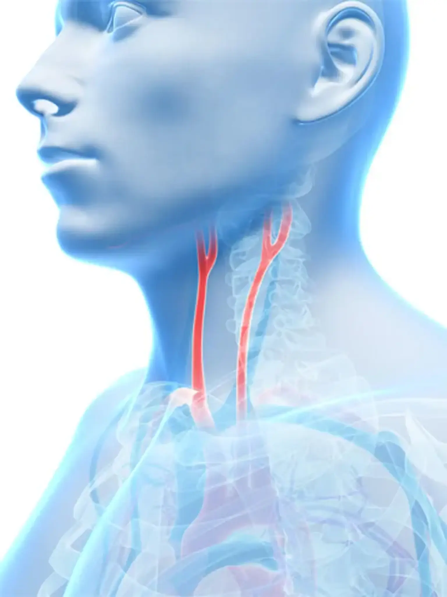
An ultrasound of the body's two carotid arteries, positioned on either side of the neck and carrying blood from the heart to the brain, offers comprehensive images of these blood vessels as well as information about the blood flowing through them.
A Doppler ultrasound examination is typically included as part of a carotid ultrasound evaluation. Doppler ultrasonography is a kind of ultrasound that measures the movement of materials in the body. It lets the clinician to examine and analyze blood flow via the body's arteries and veins.
The carotid artery is "representative" of the body's other arteries (i.e. coronary arteries). As a result, carotid ultrasonography can also be used to determine the total burden of atherosclerosis (and therefore the risk of cardio-cerebrovascular events). Carotid ultrasonography produces high-resolution pictures that allow us to assess intima media thickness, detect plaques, and grade stenosis.
Candidates for Carotid Ultrasound
Carotid ultrasonography examines the carotid arteries for plaque accumulation. Plaque can cause your carotid arteries to narrow or block, preventing oxygen-rich blood from reaching your brain. If you have any of the following symptoms, your doctor may advise you to get a carotid ultrasound:
- Recently experienced a stroke or mini-stroke.
- You may experience some or all of the symptoms of a stroke during a mini-stroke. The symptoms, on the other hand, normally go away on their own within 24 hours.
- Have a carotid bruit, which is an odd sound in your carotid artery. A carotid bruit can be detected by your doctor using a stethoscope placed on your neck over the carotid artery. A bruit might indicate a partial blockage in your carotid artery, which could result in a stroke.
Your doctor may also recommend a carotid ultrasound if he or she suspects you may have:
- Blood clots that can slow blood flow in your carotid artery.
- A split between the layers of your carotid artery wall that weakens the wall or reduces blood flow to your brain.
A carotid ultrasound may also be performed to determine whether your carotid artery surgery, also known as a carotid endarterectomy, has restored normal blood flow via your carotid artery. After a carotid stenting operation, you may undergo a carotid ultrasound to evaluate the location of the stent in your carotid artery. The stent, a tiny mesh tube, aids in preventing the artery from narrowing or being blocked again.
Carotid ultrasonography is often performed as a preventative screening test in persons who have medical problems that enhance their risk of stroke, such as high blood pressure or diabetes. Even if there is no evidence of plaque accumulation, people with certain illnesses may benefit from having their carotid arteries examined on a regular basis.
An odd sound in your carotid artery is another symptom that you may require a carotid ultrasound. A carotid bruit is a symptom that doctors can detect by placing a stethoscope on your neck above the carotid artery. Bruises can indicate a partial blockage in this artery, prompting your doctor to recommend a carotid ultrasound.
Limitations of carotid duplex ultrasound?
- Vessels are small (0.3-1.5cm), good hand eye coordination required
- We only see the extracranial arteries
- The origin of the common carotid arteries difficult to image
- The vertebral arteries difficult to see DD hypoplasia or complete occlusion of the vertebral arteries
- Rely on indirect signs to quantify intracranial stenosis
- Sometimes difficult to differentiate the ECA from the ICA
- Calcifications make grading of stenosis difficult
- Trauma (cervical collar)
- Image quality
How to perform Carotid Duplex ultrasound?
Sonographers or medical physicians do Carotid sonography (internists, angiologists, cardiologists, radiologists, vascular surgeons). There is also a growing interest in carotid duplex sonography among family doctors, general practitioners, and other primary care health care providers.
- Carotid ultrasound is conducted using a linear transducer with a frequency range of 7-12mHz.
- The linear transducer has the benefit of having a large aperture (footprint) and hence providing high-resolution pictures of surface structures (such as the carotid artery)
- The patient is positioned on his back, with his head rotated to the opposite side of the artery being examined.
- The observer is either at the patient's head, to the left, or to the right.
- Scanning can be done with either the left or right hand. Some people may swap the scanning hand depending on which side of the carotid artery is being examined.
- Scanning takes place in both the longitudinal and transverse planes.
How does the procedure work?
An ultrasound examination can be done in a variety of methods. Some of the methods are listed below. Turn the patient's head contralateral to the side being examined, cover the transducer with a towel for protection, generously apply gel to the transducer or neck, and begin the scan transversely from the proximal common carotid artery (CCA) proceeding distally.
Note the carotid bifurcation, look for plaques, try to characterize the nature of the plaque, and switch to the sagittal view; by convention, the patient's head is at the left on the screen, and images at the top of the screen are closest to the transducer; color may be used at this point to identify flow within the artery and potential high velocity areas.
The on-screen probe is inserted into the artery parallel to the vessel walls; excessive angles should be corrected. The "gate" is the breadth of the listening window; the wider the gate, the more probable it is that the signal will be detected. The trade-off, though, is greater noise. Conduct a spectrum analysis to determine the maximum velocity or frequency.
The technique is performed in the CCA, internal carotid artery (ICA), and external carotid artery (ECA), with at least two or three spectral studies acquired for each vessel. Color imaging and power Doppler may be employed, however they do not always offer useful information.
After evaluating the anterior circulation, evaluate the vertebral circulation. The C4-C6 section is usually accessible; to locate the vertebral artery, tilt the transducer laterally and inferiorly and determine the presence and direction of flow; velocity data can also be acquired.
Doppler ultrasound is a type of ultrasound that monitors the direction and speed of blood cells as they travel through vessels. The mobility of blood cells causes the pitch of reflected sound waves to alter (called the Doppler effect). A computer captures and analyses the noises, producing graphs or color images that depict the flow of blood via the blood vessels.
Ultrasound imaging works on the same principles as sonar, which is used by bats, ships, and fisherman. A sound wave bounces back or echoes when it strikes an item. It is possible to establish how far away an item is by analyzing its echo waves, as well as its size, shape, and consistency. This includes whether or if the thing is solid or fluid-filled. Ultrasound is used by doctors to identify changes in the appearance of organs, tissues, and arteries, as well as abnormal structures such as tumors.
A transducer delivers sound waves and captures the echoing (returning) waves during an ultrasound test. When the transducer is rubbed against the skin, it emits inaudible high-frequency sound waves into the body. As sound waves bounce off interior organs, fluids, and tissues, the transducer's sensitive receiver detects minute changes in pitch and direction. These characteristic waves are quickly measured by a computer and shown as real-time images on a monitor. Typically, the technician records one or more frames of the moving visuals as still photos. They may also preserve photos in brief video loops.
Vessel identification
Identify the common carotid artery as follows:
- Pulsatile walls
- Smaller caliber than jugular vein
- Systolic peak and diastolic endpoints in between that of external and internal carotid arteries on spectral analysis
How should I prepare?
Wear loose-fitting, comfy clothes. You may be required to remove all clothes and jewelry from the area being checked. It's best to wear a loose-fitting, open-necked shirt or blouse.
Ultrasound tests are extremely sensitive to motion, and a busy or screaming youngster might cause the examination to take longer. It is generally helpful to explain the procedure to the youngster ahead to the exam to ensure a seamless encounter. Bring books, tiny toys, music, or activities to keep the youngster occupied and the time pass fast. There might be a television in the exam room. Feel free to request your child's preferred channel.
What does the equipment look like?
Ultrasound machines are made out of a computer console, a video display, and a transducer attached to it. The transducer is a tiny, handheld device that looks like a microphone. During a single exam, certain exams may employ many transducers (each with a unique set of capabilities). The transducer emits inaudible high-frequency sound waves into the body and listens for echoes. The same concepts apply to boat and submarine sonar.
The technician puts the transducer in the region under inspection after applying a little quantity of gel to it. The gel permits sound waves to bounce back and forth between the transducer and the region being examined. On a video display, the ultrasound picture is instantly apparent. The picture is generated by the computer using the loudness (amplitude), pitch (frequency), and time it takes for the ultrasound signal to return to the transducer. It also examines the sort of bodily structure and/or tissue through which the sound is traveling.
How is the procedure performed?
Most ultrasound tests will have you lying face-up on an exam table that may be tilted or adjusted. Patients can turn to either side to increase the visual quality.
The technician adds a transparent water-based gel to the part of the body being examined. This allows the transducer to form a secure connection with the body. It also aids in the removal of air pockets between the transducer and the skin, which can prevent sound waves from entering your body. The technician or radiologist moves the transducer over the skin, skimming over the region of interest. They may also slant the sound beam from a different point in order to better see a problem spot.
In children with sickle cell disease, the branches of the carotid arteries inside the brain cannot be viewed directly but can be assessed using Doppler ultrasonography. The transducer is placed over the child's temple and the blood flow in the middle of the skull is recorded. This ultrasound examination normally takes 30 to 45 minutes.
Determining Degree of Stenosis
While some ultrasonographers attempt to quantify the degree of stenosis simply based on visual features (similar to the North American Symptomatic Carotid Endarterectomy Trial technique of angiographic estimate), this criterion is not accurate.
Some laboratories quantify the degree of stenosis in percentages. A range (for example, 50-69 % stenosis) is likely to be more accurate. The ranges and measurements differ from one laboratory to the next. The equipment utilized, the person doing the ultrasonography, and the sites sampled for measurement are all factors that influence measurements (eg, the distal ICA often has higher velocities than the proximal ICA). For quality control, laboratories should make their own correlations using angiographic measures wherever possible.
Doppler ultrasonography and pulse oximetry Carotid artery stenosis has been detected using Doppler ultrasonography. The physical features of Doppler ultrasonography are unique. Its 2-dimensional gray scale may be used to measure intima-media thickness, which is an excellent biomarker for atherosclerosis and can help with plaque characterisation. The shape of a plaque is linked to the likelihood of having a stroke. Plaque ulceration is also regarded as a powerful indicator of future embolic event risk.
A consensus conference of the Society of Radiologists in Ultrasound recommended the following criteria for estimating stenosis:
- Normal: ICA PSV < 125 cm/s and no plaque or intimal thickening is visible.
- < 50% stenosis: ICA PSV < 125 cm/s and plaque or intimal thickening is visible.
- 50-69% stenosis: ICA PSV is 125-230 cm/s and plaque is visible.
- >70% stenosis to near occlusion: ICA PSV >230 cm/s and visible plaque and lumen narrowing are seen.
When stenosis exceeds 90% (near occlusion), velocities may actually decrease as flow-maintenance systems fail. Ratios can be especially useful when cardiovascular variables (such as a low ejection fraction) limit the rise in velocity. In such instances, ICA/CCA ratios greater than 3 may indicate severe stenosis.
What are the risks of carotid (duplex) ultrasound?
In general, ultrasound is a fairly safe and painless process. It does not emit any dangerous radiation. Ultrasound does not injure tissue, and there is no chance that manipulating the transducer would dislodge a plaque and trigger a stroke. It is also unlikely that a scan will cause asystole or clinically severe bradycardia (hypersensitive carotid sinus).
What will I experience during and after the procedure?
The majority of ultrasound tests are painless, quick, and well tolerated. After you lie down on the exam table, the radiologist or sonographer will apply some warm, water-based gel to your skin before pressing the transducer against your body. To capture the necessary photographs, they will move it back and forth over the region of interest. There is typically little discomfort as they press the transducer on the area being examined.
If the transducer is placed over an area of discomfort, you may feel pressure or slight pain. Because the transducer is swept across the whole length of your neck on both sides to capture images of the artery from different viewpoints, you may need to tilt or twist your head for the optimum exposure. It also assists in keeping your arm and shoulder down. To keep your head motionless, it will be supported.
If your doctor does a Doppler ultrasound test, you may hear pulse-like noises that change pitch as they monitor and measure blood flow. The technologist will wipe away the transparent ultrasonic gel from your skin once the imaging is finished. Any remaining parts will dry rapidly. Generally, ultrasound gel does not stain or discolor garments.
You should be able to resume your usual activities soon following an ultrasound test.
Who interprets the results and how do I get them?
The pictures will be analyzed by a radiologist, a doctor who is qualified to monitor and interpret radiology tests. The radiologist will provide the doctor who requested the exam with a signed report. Your doctor will then discuss the findings with you. After the exam, the radiologist may share the results with you in specific instances.
You may require a follow-up examination. If this is the case, your doctor will explain why. A follow-up test may be necessary to further analyze a suspected problem with more views or a particular imaging technology. It may also check to determine whether a problem has changed over time. Follow-up assessments are frequently the most effective approach to determine if therapy is effective or whether an issue requires attention.
Conclusion
Carotid ultrasonography is a technique that employs sound waves to create images of the carotid arteries in the neck, which transport blood from the heart to the brain. A Doppler ultrasound – which measures blood flow via a blood vessel – is generally included in this evaluation. It is most commonly used to test patients for carotid artery blockage or narrowing, a disease known as stenosis, which may raise the risk of stroke.