Endoscopic Spine Surgery
Endoscopic spine surgery is the least invasive spine surgery we are aware of, and it is used to treat herniated, protruded, extruded, bulging discs, and disc ruptures that compress or irritate the spinal nerves, resulting in back or leg pain. Before choosing conventional, open, or minimally invasive spine methods such as laminectomy, microdiscectomy, or spinal fusion, patients with painful spinal disorders should explore all less invasive options such as pain management and endoscopic procedures.
For the endoscopic spine surgeon to locate the cause of the pain and selectively manage the painful condition without causing the patient considerable postoperative pain or recovery, a correct diagnosis is required in addition to therapeutic and diagnostic injections. Endoscopic spine surgery has been shown to be clinically equivalent to open and minimally invasive spine surgery (MIS) with the right indication, diagnosis, and training. Endoscopic patients heal faster, require fewer narcotic drugs, and return to work sooner than MIS surgical patients, despite having similar benefits in alleviating pain for spinal disorders.
Endoscopic spine surgery can be beneficial in the treatment of teenage disc herniations, particularly for active and athletic individuals who participate in competitive sports and require less tissue stress and a faster return to function. Before undergoing spine fusion surgery, athletes and physically active patients should get a second opinion from an experienced endoscopic spine doctor.
What is Endoscopic Spine Surgery?
Endoscopic spine surgery is a minimally invasive spine surgery that removes a herniated disc (and treats other spinal problems) through very small incisions using specialized video cameras and tools. To obtain entry to the spine and perform the surgery, the approach is performed from the back, chest, or abdomen.
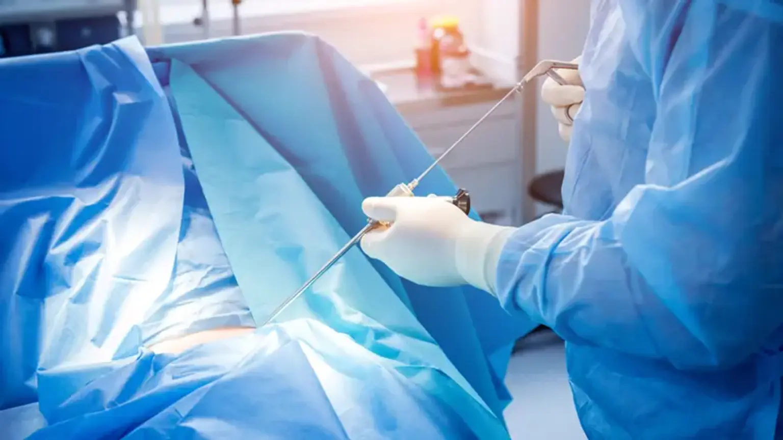
An endoscope is a small tube with a tiny video camera at the end that is used to perform endoscopic spine operations. The camera projects images of the inside of the body onto television sets, making it easier for the doctor to observe what's going on. Fluoroscopes (x-ray scanners) are used to provide a nice view of your spine throughout your surgery. The endoscope is guided to the problematic location after being inserted through a tiny incision. Your doctor will use an x-ray and a camera to locate the piece, as well as specific devices to remove it. Sutures are used to close the incisions, which are then bandaged with surgical tape.
Endoscopic Spine Surgery Benefits
When compared to traditional treatments such as microdiscectomy (open surgery using an operative microscope), doctors highlight the procedure's lower complication risk, faster recovery time, reduced postoperative pain, and faster come back to work and normal activity.
Another significant potential benefit, which has yet to be demonstrated, is a reduced likelihood of subsequent problems at the index treatment level.
Given that more than 1.5 million spinal procedures are conducted in the United States each year, the mainstreaming of endoscopic spine surgery in the United States would be a massive benefit to the treatment options for patients who require surgery for spinal issues.
The operation is most commonly used to treat herniated disks, although it can also be used to treat some types of central and foraminal stenosis, certain types of spinal infections, and certain synovial cysts. Scoliosis, spinal instabilities, and trauma are among the most common indications for spine surgery.
But, for the time being, endoscopic spine surgery is not generally available in the United States, owing to significant equipment costs and the steep learning curve required to understand its technical complexities.
Endoscopic Spine Surgery Indications
Endoscopic procedures started in the lumbar spine and have since grown to cover the cervical, thoracic, and thoracolumbar junction. The transforaminal and interlaminar techniques in the lumbar spine, as well as the anterior and posterior approaches in the cervical region, are famous. Treatment of primary and recurrent disc disorder, failed back surgery syndrome, spinal stenosis, spondylolisthesis, synovial cysts, radiculopathy, infectious diseases, discogenic back pain, spinal tumors, dural tears, and tethered cord syndrome are among the most endoscopic spinal surgery cases that have been published. As more technique publications and case reports are released in the literature, surgical choices and indications are expected to expand.
Diseases Treated with Endoscopic Spine Surgery
There are a variety of spinal diseases that might cause discomfort in a patient. The most common issues observed in a spine surgeon's office are mentioned below. The surgeon can use endoscopic spine surgery to pinpoint and treat the following spinal disorders with the least amount of effect on the patient's muscles and spinal elements.
- Arthritis and Bone Spurs of the Spine
- Bulged Disc
- Discogenic Back Pain
- Herniated Disc
- Failed Back Surgery
- Foraminal Stenosis (Constriction of the spinal canal)
- Sciatica
- Radiculitis or Radiculopathy
- Chronic Facet Disease (Back spasms and pain when leaning back)
Minimally Invasive Spine Surgery Types
Endoscopic Discectomy
Endoscopic Discectomy is the least invasive spine operation we are aware of for treating herniated discs and low back pain. To remove herniated disc material, the technique is conducted as an outpatient surgical spine operation. Because of the lengthy postoperative recovery, significant infection rates, and morbidity associated with open spinal surgery, the term minimally invasive spine surgery (MIS) was developed. Although MIS has alleviated some of the issues associated with open surgery, it still restricts spinal movement and requires lengthy recovery and time off work.
Because there is no traumatic back muscle dissection, no bone extraction, and no major skin cut, endoscopic discectomy differs from open or MIS lumbar microdiscectomy. With this endoscopic disc surgery, the potential of problems such as scar formation, blood loss, infection, and anesthesia that can occur with MIS or conventional surgery is greatly decreased or avoided. Endoscopic discectomy surgery was developed to repair herniated discs effectively while eliminating as many hazards as feasible.
Endoscopic Foraminoplasty
Endoscopic foraminoplasty is a minimally invasive spine operation for treating back pain produced by pressure on the leaving nerve or spinal cord caused by herniated discs, bone spurs, or scar tissue. This constriction of the spine or foramen is caused by degenerative disc disease, foraminal stenosis, and facet ailment. Using an endoscopic method, rongeurs, reamers, and small-motorized burrs are used to selectively remove bone through the endoscope to extend the foramen and decompress the nerves.
Endoscopic Rhizotomy
For patients suffering from persistent low back pain and muscle spasms caused by facet joint pathology, the Endoscopic Rhizotomy is the least invasive outpatient surgery that effectively gives up to 5 years of pain relief. Patients are alright when they lean forward, but when they lean backward or stand for long periods of time, they experience substantial low back pain. Patients with low back discomfort will most likely visit a pain management professional and be treated with facet injections, medial branch blocks, or percutaneous radiofrequency rhizotomies (RFA) to alleviate the pain. The patient's RFA outcomes are usually just transient, lasting between six and twelve months. These individuals are usually suitable candidates for endoscopic rhizotomy treatment if the pain returns or if the RFA does not provide enough long-term pain relief.
Cervical Endoscopic Rhizotomy
A cervical endoscopic rhizotomy is a minimally invasive procedure performed by a spine surgeon that provides direct viewing of the nerves that run through your neck's problematic facet joints. This procedure targets tiny nerves called the dorsal medial branches that transmit pain signals from your neck and shoulders to your brain using a 12-inch cut and an HD camera connected to an endoscope. Through the endoscope, a radiofrequency probe is used to directly ablate and kill these dorsal ramus medial branch facet nerves, interrupting the pain signal and relieving neck pain perception.
The dorsal ramus medial branch facet nerves in your neck and shoulders do not influence any muscle function. As a result, after surgery, the cervical endoscopic rhizotomy has no effect on your capacity to function. This simple procedure may even prevent or postpone the need for major spinal fusion surgery.
Endoscopic Interlaminar Discectomy
Endoscopic Interlaminar Discectomy is a minimally invasive outpatient treatment that treats low back and leg discomfort caused by herniated, bulging, or extruded spinal disc material squeezing the spinal cord or exiting spinal neurons.
A minimally invasive spine (MIS) lumbar microdiscectomy is similar to an endoscopic interlaminar discectomy. There are three main differences between the two techniques. First, in contrast to MIS microdiscectomy, endoscopic surgery uses an 8mm or 0.5-inch surgical operative tube. The endoscopic technique is less stressful on the muscles and supporting structures of the spine as a result of the smaller tube. Second, unlike the MIS microdiscectomy, endoscopic surgery does not need any bone excision, which results in more scar tissue formation. Third, endoscopic surgery makes use of a 30-degree HD optical scope, which allows the surgeon to see the disc and surrounding tissue more clearly. As a result, the discectomy is more precise. Endoscopic treatment reduces or eliminates the possibility of problems from scarring, blood loss, infection, and anesthesia that can occur with MIS or conventional surgery.
Endoscopic Spine Surgery Procedure
The patient is first prepared for surgery by receiving a local anesthetic to relieve pain. A tubular trocar (about the width of a pencil) is inserted through a 1-inch or smaller surgical incision. The endoscopic surgeon may enter the spine using one of two ways, depending on the patient's specific diagnosis: intralaminar (from the back of the column between two laminae) or transforaminal (from the back/side of the column into the neuroforamen; a nerve pathway).
Then, through the trocar, a small camera is placed into the targeted location of the spine. The camera captures and displays real-time images of the surgical site onto a monitor in the surgeon's direct vision throughout ESS. During the surgical operation, the endoscopic camera aids and guides the surgeon.
The endoscopic camera and trocar are gently withdrawn after the operation, and the small cut is stitched with a suture and a little dressing (e.g., Band-Aid).
Endoscopic Spine Surgery Recovery
Everyone's situation is unique, and it is mostly determined by your work and daily routines. Many individuals are able to drive just two days after a decompression or discectomy procedure. They will be able to travel for business on day five. Most patients can anticipate resuming their normal daily activities two weeks after surgery. A spinal fusion treatment necessitates a longer recovery period, with patients being restricted for 3 months to allow for adequate bone fusion and healing.
Endoscopic Spine Surgery Complications
The risks of major complications or injury are modest, as they are with arthroscopic knee surgery; in the author's experience, they are around 1% or less. As with any operation, the dangers of infection, nerve injury, dural rupture, hemorrhage, and scar tissue formation are always present. The most frequent postoperative complaint, transient dysesthesia, occurs in about 5% to 15% of cases and is almost invariably temporary. Its etiology is unknown, however, it could be related to nerve healing, operating near the dorsal root ganglion of the leaving nerve, or a tiny hematoma near the ganglion of the leaving nerve, as it can develop days or weeks following surgery. It cannot be totally prevented, and it has happened even when there were no adverse intraoperative incidents and no signs of nerve irritation on continuous electromyography (EMG) or somatosensory evoked potentials (SEP). Most endoscopic specialists do not report it as a complication because the symptoms are sometimes so minimal. The more severe dysesthesia symptoms resemble a form of complex regional pain syndrome; however, they are usually less severe and do not include skin changes. Transforaminal epidurals, sympathetic blocks, and off-label usage of Neurontin titrated to 1800-3200 mg/day are used to treat postoperative dysesthesia. It is FDA-approved for post-herpetic neuralgia, but gabapentin (Neurontin) is also beneficial in the treatment of neuropathic pain.
The capacity to properly visualize normal and patho-anatomy, as well as the use of local anesthetic and conscious sedation rather than general or spinal anesthesia, all help to reduce the risk of problems. The entire process is normally completed with the patient feeling comfortable throughout the surgery, and should not cause considerable pain unless it is anticipated, such as during Evocative Chromo-Discography, annular fenestration, or when tools are maneuvered past the exiting nerve. When .5% lidocaine is used as a local anesthetic, it is possible to use a large amount of this dilute anesthetic for pain control while still allowing the patient to feel discomfort when the nerve root is manipulated.
Endoscopic Spine Surgery vs Traditional Spine Surgery
Consider endoscopic spine surgery for a variety of reasons. After discussing all endoscopic spine surgery options, many patients should consider minimally invasive spine surgery. Endoscopic spine surgery aims to ease pain, avoid spinal fusion, and preserve the patient's spinal structure to reduce the need for future surgery.
- Uses a high-definition camera and an endoscope to provide the spine surgeon with a better picture than traditional surgical approaches.
- The incision is less than 0.5 inches, which reduces the risk of skin scarring.
- There is no tearing of muscle or tissue, thus there is less scar tissue formation and spinal flexibility is preserved.
- Conscious sedation lowers the dangers of general anesthesia.
- Postoperative pain is reduced, and narcotic medications are not required.
- Recovery time is reduced.
- Return to work as quickly as one week after your surgery (under some restrictions for lifting and certain physical activities)
Conclusion
Treatment for spinal problems is always evolving and improving in tandem with technological advancements. Improved endoscopic optics and instruments have permitted enhanced vision of spinal anatomy with the least degree of approach-related trauma utilizing an ultra-minimally invasive technique. To achieve optimal results, careful patient selection is required, taking into account both the location and kind of disc disease as well as approach-related restrictions (position of the iliac wing and retroperitoneal contents). A thorough understanding of the lumbar neuroforaminal architecture, as well as MRI interpretation of disc disease, is also important to attaining operational success. Endoscopic procedures can be used to treat central, paracentral, foraminal, and far lateral lumbar disc herniations. Decompression of the posterior interlaminar lumbar spine, anterior and posterior cervical decompression, and posterior-lateral thoracic decompression are all possible with modern endoscopic techniques. Finally, by rigorous cadaveric training and surgical supervision, the learning curve needed in learning endoscopic procedures can be reduced.