Pneumothorax

Overview
Pneumothorax is an emergency that must be addressed as soon as it is diagnosed. There are two types of pneumothorax: primary and secondary. A primary pneumothorax is one that arises without a known etiology and in the absence of substantial lung disease. Secondary pneumothorax, on the other hand, happens in the context of pre-existing lung disease. A tension pneumothorax occurs when the volume of air in the chest grows significantly and a one-way valve form. Unless effectively treated, this condition has the potential to worsen and lead to death.
Pneumothorax can occur as a result of physical trauma to the chest or as a side effect of medicinal or surgical intervention (biopsy). Typical symptoms include chest discomfort and shortness of breath. A chest X-ray or computed tomography (CT) scan is required to diagnose a pneumothorax. Small spontaneous pneumothoraces usually resolve without therapy and simply require observation.
What is a Pneumothorax?
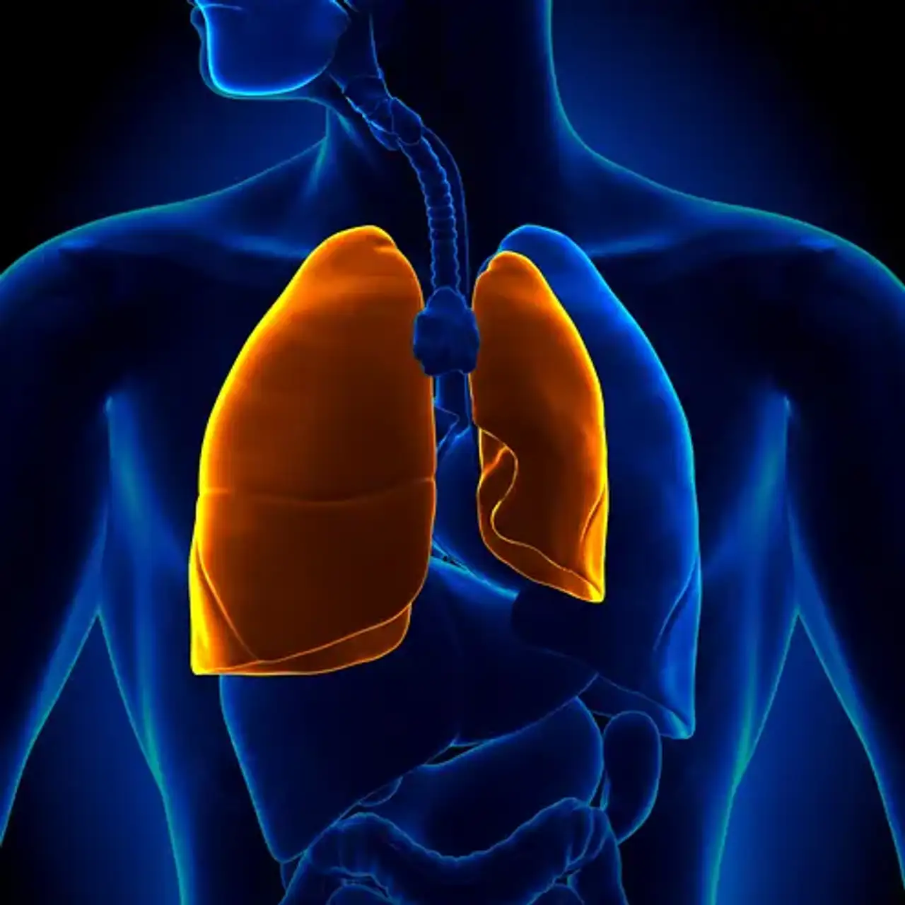
A pneumothorax is a collection of air that exists outside the lung but within the pleural cavity. It happens when air is trapped between the parietal and visceral pleurae of the chest. The buildup of air can put pressure on the lung and cause it to collapse. The clinical manifestation of pneumothorax is determined by the degree of collapse. Air can enter the pleural space via two mechanisms: trauma generating a connection through the chest wall or lung rupture causing a rupture of the visceral pleura.
Pneumothorax is classified into two types: traumatic and atraumatic. Primary and secondary atraumatic pneumothorax are the two subtypes. A primary spontaneous pneumothorax (PSP) happens without any known triggering event, whereas a secondary spontaneous pneumothorax (SSP) happens after an underlying lung illness.
Traumatic pneumothorax can occur as a result of either blunt or penetrating trauma. Pneumothoraces are further subdivided into simple, tension, and open. A simple pneumothorax does not cause mediastinal structures to move, however a tension pneumothorax does. An open pneumothorax is a wound in the chest wall that allows air to enter and exit.
Types of Pneumothorax
- Primary spontaneous
There are two forms of spontaneous pneumothoraces: primary, which arise in the absence of established lung disease, and secondary, which occur in people who have underlying lung disease. Until recently, the etiology of primary spontaneous pneumothorax (PSP) has not been discovered; nevertheless, various risk factors such as smoking, male sex, and a family history of pneumothorax have been reported.
Furthermore, a PSP is more likely to arise in a young adult who does not have any underlying lung abnormalities. Typical symptoms include chest discomfort and, in some cases, slight dyspnea. There are various circumstances where a PSP poses a life-threatening hazard to a patient; yet, some people may wait several days before seeking medical assistance. PSPs seldom induce tension pneumothoraces.
- Secondary spontaneous
Secondary spontaneous pneumothorax happens as a result of underlying chest diseases. They are most typically seen in people with Chronic obstructive pulmonary disease (COPD), which accounts for around 70% of cases. Tuberculosis, necrotizing Pneumonia, Lung cancer, Sarcoma involving the lung, sarcoidosis, Endometriosis, Cystic fibrosis, acute severe Asthma, Idiopathic Pulmonary fibrosis, Rheumatoid arthritis, Ankylosing spondylitis, systemic sclerosis, and Marfan's syndrome are all known lung diseases that may increase the risk of pneumothorax.
Secondary spontaneous pneumothoraces (SSPs) are defined as occurring in people who have considerable underlying lung illness. In more severe instances, symptoms such as hypoxemia and hypercapnia are common. The abrupt onset of dyspnea in individuals with established underlying lung disorders, such as COPD, cystic fibrosis, or other significant lung diseases, should trigger examinations to rule out a pneumothorax.
- Traumatic pneumothorax
When the chest wall is breached, such as by a knife wound or a gunshot wound, air enters the pleural space and causes traumatic pneumothorax. Only rib fractures are more prevalent in this group. Traumatic pneumothorax has been observed to occur in up to half of all instances of chest trauma. In half of these cases, the pneumothorax is occult, but it may enlarge—especially if mechanical ventilation is necessary. This sort of pneumothorax has also been seen in individuals who are already on mechanical ventilation for another cause.
Mechanism of injury
The lungs, heart, and several main blood arteries are all housed in the thoracic cavity. A pleural membrane covers the surface of the lung (visceral pleura) on each side of the cavity and also borders the interior of the chest wall (parietal pleura). A modest quantity of lubricating serous fluid exists between the two layers. Because the pressure inside the airways is greater than the pressure inside the pleural space, the lungs are completely inflated within the cavity. Pneumothorax may only occur if air is allowed to enter the pleural space, either by injury to the chest wall or to the lung itself, or on rare occasions because bacteria in the pleural space create gas.
Pneumothorax Symptoms
The patient is asymptomatic in primary spontaneous pneumothorax because otherwise healthy people endure physiologic effects well. Chest discomfort and shortness of breath are the most prevalent symptoms. The discomfort in the chest is pleuritic, acute, and intense, radiating to the ipsilateral shoulder. Dyspnea is more severe in SSP due to lower underlying lung reserve.
A previous history of pneumothorax is crucial since recurrence occurs in 15-40% of patients. Recurrence on the other side is also possible.
On examination, the following findings are noted
- Respiratory discomfort
- Increased respiratory rate
- Asymmetrical lung expansion
- Decreased tactile fremitus
- Hyperresonant percussion note
- Decreased intensity of breath sounds or absent breath sounds
In tension pneumothorax following additional findings are seen:
- Tachycardia more than 134 beats per minute
- Hypotension
- Jugular venous distension
- Cyanosis
- Respiratory failure
- Cardiac arrest
Subcutaneous emphysema is related with several traumatic pneumothoraces. A physical exam may be difficult to identify pneumothorax, especially in a loud trauma bay. However, a physical exam is required to make the diagnosis of tension pneumothorax.
Pneumothorax Diagnosis
Diagnosis can be made via chest radiography, ultrasonography, or CT, albeit a chest x-ray is most commonly utilized. A 2.5 cm air gap on radiographs is similar to a 30% pneumothorax. CT can detect occult pneumothoraces, however they are generally clinically inconsequential. The extended focused abdominal sonography for trauma (E-FAST) test is a more contemporary pneumothorax diagnosis technique. The lack of lung sliding, the absence of a comet-tails artifact, and the existence of a lung point are commonly used to make an ultrasound diagnosis.
Unfortunately, the sensitivity and specificity of this diagnostic approach are very operator dependent. Ultrasonography offers a sensitivity of up to 94 percent and a specificity of 100 % in competent hands (better than chest x-ray). If a patient is hemodynamically unstable and has a suspected tension pneumothorax, care is not delayed while imaging is being performed. If the patient is hemodynamically unstable and has a compelling history and physical exam confirming tension pneumothorax, needle decompression can be done.
Pneumothorax Management
A pneumothorax is treated by needle decompression in individuals who have concomitant symptoms and exhibit evidence of instability. This is often done using a 14- to 16-gauge, 4.5-cm-long angiocatheter placed just superior to the rib in the second intercostal gap at the midclavicular line. After needle decompression or for stable pneumothoraces, a thoracostomy tube is inserted. This is normally positioned in the fifth intercostal gap, anterior to the midaxillary line, above the rib. The size of the thoracostomy tube is normally determined by the patient's height and weight, as well as the presence of an accompanying hemothorax.
An open "sucking" chest wound is initially treated with a three-sided occlusive bandage. Further therapy may necessitate a tube thoracostomy and/or correction of a chest wall defect.
Asymptomatic small primary spontaneous pneumothorax (depth less than 2cm) patients are frequently discharged with outpatient follow-up in 2-4 weeks. If the patient is symptomatic or the depth/size is greater than 2cm, needle aspiration is performed; if the patient recovers and the remaining depth is less than 2cm, the patient is released; otherwise, tube thoracostomy is performed.
If the size/depth of the pneumothorax is less than 1cm and there is no dyspnea, the patient is hospitalized, high flow oxygen is administered, and the patient is observed for 24 hours. If the size/depth is between 1-2cm, needle aspiration is performed, and the residual size of the pneumothorax is determined; if the depth after needle aspiration is less than 1cm, management is accomplished through oxygen inhalation and observation; and if the depth is greater than 2cm, tube thoracostomy is performed. Tube thoracostomy is performed when the depth is greater than 2cm or when there is a lack of air.
The pleural space may reabsorb air at a rate of 1.5 percent every day. This reabsorption rate can be increased by using supplementary oxygen. The nitrogen in atmospheric air is displaced by raising the proportion of inspired oxygen concentration, affecting the pressure differential between the air in the pleural space and the capillaries. Pneumothorax on chest radiography of 25% or greater typically requires treatment with needle aspiration if symptomatic, and tube thoracostomy if this fails.
Indications for surgical intervention (VATS vs. Thoracotomy)
- Continuous air leak for longer than seven days
- Bilateral pneumothoraces
- The first episode in high-risk profession patient, i.e., Divers, pilots
- Recurrent ipsilateral pneumothorax
- Contralateral pneumothorax
- Patients who have AIDS
Pleurodesis is used to occlude the pleural space in patients undergoing video-assisted thoracic surgery (VATS). Mechanical pleurodesis combined with bleb/bullectomy reduces pneumothorax recurrence to 5%. Mechanical pleurodesis techniques include removing the parietal pleura vs using an abrasive "scratchpad" or dry gauze. In patients who are unable to endure mechanical pleurodesis, chemical pleurodesis is an alternative. Talc, tetracycline, doxycycline, and minocycline are all pleural lining irritants that can be used for chemical pleurodesis.
Pleurodesis and surgery for Pneumothorax
Pleurodesis is regarded as the ultimate treatment. It is a surgical treatment that permanently closes the pleural gap and connects the lung to the chest wall. The most successful treatment is a surgical thoracotomy with detection of any source of air leakage and bleb stapling, followed by pleurectomy of the outer pleural layer and pleural abrasion of the interior layer. The lung attaches to the chest wall throughout the healing process, thereby obliterating the pleural gap. The recurrence rate is roughly 1%. Pain after a thoracotomy is common.
Thoracoscopy, primarily in the form of video-assisted thoracoscopic surgery(VATS), is a less intrusive method. VATS outcomes are less effective than thoracotomy, however they leave less scars in the skin. VATS allows for shorter in-hospital stays, less postoperative pain management, and a lower chance of lung complications after surgery. VATS can also be used to achieve chemical pleurodesis by insufflating talc.
Talc inhalation causes inflammation of the pleura surfaces. If a chest tube is already in place, several chemicals, such as talc, tetracycline, minocycline, or doxycycline, may be infused into the tube to accomplish chemical pleurodesis. Chemical pleurodesis has worse outcomes than surgical pleurodesis, whereas talc pleurodesis produces the best results.
Recovery after a Pneumothorax
Following surgery, a chest tube will be inserted. It will remain in place for a few days to a week, depending on your healing process. We will take x-rays and examine you to confirm that we inserted your tube in the best possible spot for your lung. While the chest tube is in place, you will need to stay in the hospital. Before you leave the hospital, your doctor will certify that your lung has not collapsed again. We train you on breathing activities known as incentive spirometry. This helps to dilate your air sacs and enlarge your lungs. This will aid in the prevention of pneumonia.
You should quit smoking if you smoke. Smoking increases your chances of developing a pneumothorax. Quitting smoking will help your body recuperate more completely. It will also aid in the healing of your wounds.
You should also ask your doctor when you will be able to travel again. You should normally wait at least 2 weeks and up to 12 weeks before using this mode of transportation. It is risky to fly in an airplane or travel to locations with elevations more than 8000 feet. If your lung has not yet recovered, the pressure shift may cause it to collapse again.
Complications of Pneumothorax
The 3 main problems encountered when treating pneumothorax are:
- Air leaks
- Failure of the lung to expand
- Re-expansion pulmonary edema
Air leaks are primarily caused by the main defect (continuous loss of air from the lung into the pleural space), but they can also be caused by air seeping around the chest tube insertion site if it is not adequately sutured and sealed. Air leaks are more likely in secondary spontaneous pneumothorax than in first spontaneous pneumothorax. Most spontaneously resolve within a week.
Failure of the lung to re-expand is usually due to one of the following:
- Persistent air leak
- Endobronchial obstruction
- Trapped lung
- Malpositioned chest tube
If an air leak or an incompletely inflated lung persists for more than a week, blood pleurodesis (a blood patch), endobronchial valves, thoracoscopy, or thoracotomy might be explored.
- Re-expansion pulmonary edema occurs when the lung is rapidly expanded, such as when a chest tube is linked to negative pressure after the lung has been deflated for more than 2 days. As needed, oxygen, diuretics, and cardiopulmonary support are administered.
Prevention of Pneumothorax
In the three years following the initial spontaneous pneumothorax, the recurrence rate exceeds 50%. The best preventative method is video-assisted thoracic surgery (VATS), in which blebs are stapled and pleurodesis is accomplished with pleural abrasion, parietal pleurectomy, or talc insufflation; thoracotomy is still utilized in some medical institutes.
When catheter aspiration fails to treat spontaneous pneumothorax, the pneumothorax recurs, or the patient has secondary spontaneous pneumothorax, these procedures are advised. The recurrence rate following these treatments is 5%. If thoracoscopy is not possible or is contraindicated, chemical pleurodesis through a chest tube may be performed; this treatment, while considerably less invasive, decreases the recurrence rate to around 25%.
Prognosis
PSP is normally harmless and resolves on its own without the need for substantial intervention. Recurrences might occur for up to three years. The recurrence rate in the next five years is 30% for PSP and 43% for SSP. The likelihood of recurrence increases with each subsequent pneumothorax; it is 30% after the first, 40% after a second, and more than 50% after the third. Although PSP is not considered a serious health risk, fatalities have been documented. The severity of SSPs is determined by the underlying lung illness and the magnitude of the pneumothorax. After a pneumothorax, patients with COPD and HIV have a significant death rate. SSP has a 10% mortality rate. If suitable steps are not followed, tension pneumothorax can be fatal.
Conclusion
The presence of air or gas in the pleural cavity, which can impede oxygenation and/or breathing, is classified as pneumothorax. The clinical outcome is determined by the degree of lung collapse on the afflicted side. If the pneumothorax is severe, it might displace the mediastinum and jeopardize hemodynamic stability.