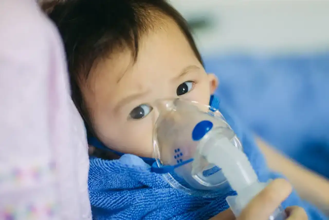
Chronic Respiratory Failure
Overview
The respiratory system is an important aspect of human health since it helps with oxygen absorption and distribution throughout the body. The respiratory system is made up of a variety of tissues and organs. The lungs serve as the respiratory system's focal point. There are various circumstances in which infants may have respiratory failure. In these cases, the juvenile child requires cautious treatment to avoid irreversible harm or deadly consequences.
Chronic respiratory failure in children can be addressed, frequently through a combination of mechanisms and techniques. An early diagnosis of such problems might result in a better prognosis. Parents should be instructed on the signs to look for and when they should take their infant to a doctor for an evaluation. We look at chronic respiratory failure in kids, the signs that may indicate the condition, and current treatments.
What is Chronic Respiratory failure?
Pediatric respiratory failure occurs when the rate of gas exchange between the environment and the blood is insufficient to meet the metabolic demands of the organism. Acute respiratory failure is still a leading cause of morbidity and death in children. Respiratory failure is a common cause of cardiac arrest in children.
The lungs of children with chronic respiratory failure are unable to extract an adequate quantity of oxygen every time the youngster breathes. It is vital to understand that respiratory failure can be acute or chronic. In severe occurrences of the illness, it is frequently regarded as an emergency requiring immediate medical treatment.
When a pediatric patient is diagnosed with chronic respiratory failure, it indicates that the youngster has a long-term condition. This need therapy over time for symptoms to improve. Certain consequences might arise if symptoms are not diagnosed and the ailment is not addressed at an early stage. According to one research, failing to recognize respiratory failure in children is one of the primary causes of cardiopulmonary arrest, which can be a life-threatening consequence.
It is crucial to understand that respiratory failure does not only relate to the influx of oxygen into the circulation. Each breath draws oxygen from the air the youngster breaths in. At the same time, carbon dioxide is evacuated from the lungs during exhalation. In certain situations, the child's lungs are unable to adequately remove the stored carbon dioxide. There are also circumstances in which both of these possibilities exist.
Respiratory failure causes
The following are the anatomic compartments that separate the most prevalent causes of respiratory failure in the pediatric population.
Acquired extrathoracic airway causes include the following:
- Infections (eg, retropharyngeal abscess, Ludwig angina, laryngotracheobronchitis, bacterial tracheitis, peritonsillar abscess)
- Trauma (eg, postextubation croup, thermal burns, foreign-body aspiration)
- Other (eg, hypertrophic tonsils and adenoid)
Congenital extrathoracic airway causes include the following:
- Subglottic stenosis
- Subglottic web or cyst
- Laryngomalacia
- Tracheomalacia
- Vascular ring
- Cystic hygroma
- Craniofacial anomalies
Intrathoracic airway and lung causes include the following:
- Acute respiratory distress syndrome (ARDS)
- Asthma
- Aspiration
- Bronchiolitis
- Bronchomalacia
- Left-sided valvular abnormalities
- Pulmonary contusion
- Near drowning
- Pneumonia
- Pulmonary edema
- Pulmonary embolus
- Sepsis
Respiratory pump causes include the following:
- Diaphragm eventration
- Diaphragmatic hernia
- Flail chest
- Kyphoscoliosis
- Duchenne muscular dystrophy
- Guillain-Barré syndrome
- Infant botulism
- Myasthenia gravis
- Spinal cord trauma
- Spinal muscular atrophy (SMA)
Central control causes include the following:
- CNS infection
- Drug overdose
- Sleep apnea
- Stroke
- Traumatic brain injury
Pathophysiology
Respiratory failure can result from a malfunction in any of the breathing system's components, including the airways, alveoli, central nervous system (CNS), peripheral neural system, respiratory muscles, and chest wall. Patients with hypoperfusion due to cardiogenic, hypovolemic, or septic shock frequently suffer respiratory failure.
The maximum spontaneous ventilation that can be sustained without developing respiratory muscle exhaustion is referred to as ventilation capacity. The spontaneous minute ventilation that results in a steady PaCO2 is referred to as ventilation demand.
Normally, ventilation capacity much outnumbers ventilation demand. Respiratory failure can occur as a result of either a decrease in ventilatory capacity or an increase in ventilatory demand (or both). A disease disorder impacting any of the functioning components of the respiratory system and its controller can reduce ventilation capacity. Ventilatory demand is increased by an increase in minute ventilation and/or labor of breathing.
Respiratory physiology
The act of respiration engages the following three processes:
- Transfer of oxygen across the alveolus
- Transport of oxygen to the tissues
- Removal of carbon dioxide from blood into the alveolus and then into the environment
Any of these processes can fail, resulting in respiratory failure. Understanding pulmonary gas exchange is critical for understanding the pathophysiologic foundation of acute respiratory failure.
Gas exchange
Respiration takes place predominantly at the alveolar capillary units of the lungs, where oxygen and carbon dioxide are exchanged between alveolar gas and blood. After diffusing into the circulation, oxygen molecules bond to hemoglobin in a reversible manner. Each hemoglobin molecule has four sites for combining with molecular oxygen; 1 g of hemoglobin may mix with a maximum of 1.36 mL of oxygen.
The amount of oxygen mixed with hemoglobin is determined by the blood PaO2 level. This connection, known as the oxygen hemoglobin dissociation curve, is not linear, but rather has a sigmoid form with a sharp slope between PaO2s of 10 and 50 mm Hg and a flat part at PaO2s of 70 mm Hg.
The carbon dioxide is transported in 3 main forms:
- In simple solution,
- as bicarbonate, and
- combined with protein of hemoglobin as a carbamino compound.
Blood flow and ventilation would be precisely matched during ideal gas exchange, resulting in no alveolar-arterial oxygen tension (PO2) gradient. However, even in normal lungs, not all alveoli are completely vented and perfused. Some alveoli are underventilated for a given perfusion, whereas others are overventilated. Similarly, when it comes to recognized alveolar ventilation, some units are underperfused while others are overperfused.
Hypoxemic respiratory failure
V/Q mismatch and shunt are the pathophysiologic processes that account for the hypoxemia seen in a wide range of disorders. These two methods cause the alveolar-arterial PO2 gradient, which is generally less than 15 mm Hg, to expand. They can be distinguished by evaluating the response to oxygen supplementation or estimating the shunt fraction after inhaling 100% oxygen. These two processes coexist in the majority of individuals with hypoxemic respiratory failure.
V/Q mismatch
The most prevalent cause of hypoxemia is a V/Q mismatch. In the presence of a disease condition, alveolar units can range from low-V/Q to high-V/Q. Low-V/Q units contribute to hypoxemia and hypercapnia, whereas high-V/Q units waste ventilation but have little effect on gas exchange unless the anomaly is severe.
A low V/Q ratio can result from either a reduction in ventilation due to airway or interstitial lung disease or from overperfusion in the context of normal ventilation. Overperfusion may occur in the event of pulmonary embolism, when blood is redirected from areas of the lungs with blood flow restriction due to embolism to regularly ventilated units.
The administration of 100 percent oxygen removes all of the low-V/Q units, resulting in hypoxemia correction. Hypoxia enhances minute breathing via chemoreceptor activation, although it has little effect on PaCO2.
Shunt
Shunt is described as the continuation of hypoxemia while inhaling 100% oxygen. Deoxygenated blood (mixed venous blood) skips the ventilated alveoli and mixes with oxygenated blood that has already passed through the ventilated alveoli, resulting in a decrease in arterial blood content.
Because of the bronchial and thebesian circulations, which account for 2-3% of shunt, anatomic shunt exists in normal lungs. A normal right-to-left shunt in the lung might result from an atrial septal defect, ventricular septal defect, patent ductus arteriosus, or arteriovenous malformation.
Shunt as a cause of hypoxemia is most commonly seen in pneumonia, atelectasis, and severe pulmonary edema of cardiac or noncardiac origin. Unless the shunt is large (> 60%), hypercapnia does not usually arise. In comparison to V/Q mismatch, hypoxemia caused by shunt is more difficult to treat with oxygen delivery.
Hypercapnic respiratory failure
Hypoventilation is a rare cause of respiratory failure that mainly results from CNS depression caused by medications or neuromuscular illnesses affecting respiratory muscles. Hypercapnia and hypoxemia are symptoms of hypoventilation. The existence of a normal alveolar-arterial PO2 gradient distinguishes hypoventilation from other types of hypoxemia.
Symptoms of Chronic Respiratory Failure in Pediatrics
When pediatric patients acquire chronic respiratory failure, certain symptoms tend to emerge. These indications must be thoroughly understood by parents. When this occurs, parents should speak with a doctor who can do a physical examination on the kid. This guarantees that the problem is detected at an early stage, which can significantly enhance the treatment prospects.
Breathing difficulties are one of the most prevalent symptoms that pediatric patients with chronic respiratory failure may face. Babies may be noticed breathing fast in addition to shortness of breath. Furthermore, toddlers and children with the illness may complain of headaches in addition to breathing issues.
A headache, for example, may be difficult to notice in a baby. Parents should, however, be on the watch for problems like as blue lips or skin. In certain circumstances, fingernails may have a blue hue. Excessive drowsiness is another indicator, since the youngster may feel exhausted all the time owing to a lack of oxygen entering their body.
Seizures may occur in the youngster in extreme circumstances. There have also been cases where a pediatric kid goes into a coma as a result of prolonged respiratory insufficiency. This is more common in extreme situations where the child's body is depleted of oxygen entering the blood circulation system.
Respiratory failure Diagnosis
Respiratory failure can have a number of clinical symptoms. However, these are vague, and severe respiratory failure may exist without obvious signs or symptoms. This underlines the significance of testing arterial blood gases in all critically sick patients or those suspected of having respiratory failure.
Chronic respiratory failure is diagnosed by recognizing symptoms that indicate to this illness. If the parents notice symptoms of the condition in their baby, they should seek medical attention. The doctor will examine the infant physically and use a stethoscope to listen to their lungs and heart. This will help the doctor to discover any anomalies and evaluate whether or not the child's lungs are working normally.
Chest radiography is required. Echocardiography is not regularly performed, although it can be beneficial in rare cases. If possible, pulmonary function tests (PFTs) may be informative, however they are more effective in determining recovery potential. Electrocardiography (ECG) should be conducted to rule out a cardiovascular cause of respiratory failure; it may also reveal dysrhythmias caused by severe hypoxemia or acidosis. Right-sided heart catheterization is debatable.
- Laboratory Studies
Once clinical respiratory failure is suspected, arterial blood gas study should be undertaken to confirm the diagnosis and to distinguish between acute and chronic versions. This aids in determining the degree of respiratory failure and guiding therapy.
A complete blood cell count (CBC) may show anemia, which can lead to tissue hypoxia, whereas polycythemia may show persistent hypoxemic respiratory failure.
A chemical panel may be useful in evaluating and managing a patient who is experiencing respiratory failure. Abnormalities in renal and hepatic function may either give insights to the etiology of respiratory failure or warn the doctor to respiratory failure consequences. Electrolyte imbalances, such as potassium, magnesium, and phosphate, can exacerbate respiratory failure and other organ function.
In a patient with respiratory failure, measuring blood creatine kinase with fractionation and troponin I helps rule out a recent myocardial infarction. A high creatine kinase level combined with a normal troponin I level may suggest myositis, which can occasionally lead to respiratory failure.
Serum thyroid-stimulating hormone (TSH) levels should be tested in persistent hypercapnic respiratory failure to rule out hypothyroidism, a possibly reversible cause of respiratory failure.
- Radiography
Because it typically discloses the etiology of respiratory failure, chest radiography is crucial in the assessment of respiratory failure. It is, however, sometimes difficult to distinguish between cardiogenic and noncardiogenic pulmonary edema. Increased heart size, vascular redistribution, peribronchial cuffing, pleural effusions, septal lines, and perihilar bat-wing infiltration distribution all point to hydrostatic edema; the absence of these characteristics points to acute respiratory distress syndrome (ARDS).
Certain medical tests can be conducted to aid in the diagnosis of persistent respiratory failure in pediatric patients. Prior to initiating a treatment plan, the doctor may conduct these tests. The findings of the tests will provide the doctor a more precise picture of the illness, including its severity. Once the doctor determines the severity of the problem, he or she may devise a treatment approach that will produce more efficient outcomes. The majority of children with the illness survive, however, the underlying problems revealed upon diagnosis may have a role in the outcome. Among the other investigations are:
- Information from a pulmonary artery catheter or echocardiography may also be valuable in determining volume status, ventricular function, cardiac output, and the existence and severity of pulmonary hypertension.
- A serum lipase test can help rule out pancreatitis.
- To rule out pulmonary embolism, a spiral chest CT with a CT angiography may be required. If the reason cannot be diagnosed otherwise, consider a bronchoscopy.
- In a patient with hypoxia and an unexplained reduction in hemoglobin, hemosiderin-laden macrophages from bronchoalveolar lavage imply widespread alveolar hemorrhage.
- A high number of lipid-laden macrophages indicates persistent aspiration. The presence of a substantial number of eosinophils in bronchoalveolar lavage specimens distinguishes idiopathic acute eosinophilic pneumonia from ARDS.
Management
The treatment strategy launched by the healthcare professional must address the underlying issues. It is vital to highlight, however, that treatment measures are also necessary to give more immediate alleviation of the child's symptoms. Several treatment options can improve oxygen transport to tissue in the patient's body. A beta-agonist may be administered in some instances. This drug is intended to aid in the correction of the underlying causes that contribute to persistent respiratory failure. In addition to a beta-agonist, some doctors may give the youngster corticosteroids.
Mechanical ventilation is commonly included in these therapy regimens. This method aids in getting more oxygen into the child's lungs. The goal is to offer the lungs with a higher concentration of oxygen, which can aid in increasing the amount of this gaseous material pushed into the blood circulatory system. As a result, using mechanical ventilation equipment on a frequent basis during the therapy stage is recommended. This may minimize the child's chance of having severe respiratory problems or other consequences. Furthermore, mechanical ventilation can help the kid breathe better, enhancing their quality of life while the underlying etiology of chronic respiratory failure is addressed throughout the therapy program.
Disease monitoring and follow-up
The majority of patients have a fairly traditional history, with severe hypoxemia followed by a lengthy requirement for mechanical ventilation, but the course of each phase and overall illness development is diverse.
The acute or exudative phase is characterized by a sudden onset of respiratory failure with hypoxemia that is resistant to treatment with supplementary oxygen. This can continue anywhere from a few hours to a week and is followed by the subacute or proliferative phase, which is marked by chronic hypoxia and the development of hypercarbia. Some patients move to a fibrotic phase, complicating their clinical course with barotrauma, nosocomial infection, or the development of Multiple Organ System Failure (MOSF).
The recovery phase is distinguished by progressive resolution of hypoxemia and better lung compliance. Approximately one-third of ARDS patients die as a result of the condition. Long-term survivors may have modest pulmonary function problems but are usually asymptomatic unless they proceed to the fibrotic phase.
Conclusion
Chronic respiratory failure in infancy can be deadly if effective therapy is not offered promptly. This disorder has a negative impact on the oxygen supply from the lungs to the blood circulation system. Because of the vital function that oxygen plays in the body, tissue damage and other issues might occur. As a result, the illness is seen as a long-term concern that must be closely watched by both parents and healthcare experts.
